Cell 46: 63-74. 2. Barin, F., M. McLane, J. Allan, T. Lee, and M. Essex. 1985. Virus envelope protein of HTLV-III represents major target antigen for antibodies.
Vol. 62, No. 12
JOURNAL OF VIROLOGY, Dec. 1988, p. 4703-4711
0022-538X188/124703-09$02.00/0 Copyright C 1988, American Society for Microbiology
Epitope Mapping of the Human Immunodeficiency Virus Type gpl20 with Monoclonal Antibodies DONALD DOWBENKO,1 GERALD NAKAMURA,' CHRISTOPHER FENNIE,1 CRAIG SHIMASAKI
1
2
LAVON RIDDLE,2 REED HARRIS,2 TIMOTHY GREGORY,2 AND LAURENCE LASKY'* Departments of Molecular Immunology' and Process Development,2 Genentech, Inc., 460 Point San Bruno Boulevard,
South San Francisco, California 94080 Received 23 May 1988/Accepted 8 August 1988
A soluble form of recombinant gpl20 of human immunodeficiency virus type 1 was used as an immunogen for production of murine monoclonal antibodies. These monoclonal antibodies were characterized for their ability to block the interaction between gpl20 and the acquired immunodeficiency syndrome virus receptor, CD4. Three of the monoclonal antibodies were found to inhibit this interaction, whereas the other antibodies were found to be ineffective at blocking binding. The gpl20 epitopes which are recognized by these monoclonal antibodies were mapped by using a combination of Western blot (immunoblot) analysis of gpl20 proteolytic fragments, immunoaffinity purification of fragments of gpl20, and antibody screening of a random gpl20 gene fragment expression library produced in the lambda gtll expression system. Two monoclonal antibodies which blocked gpl20-CD4 interaction were found to map to adjacent sites in the carboxy-terminal region of the glycoprotein, suggesting that this area is important in the interaction between gpl20 and CD4. One nonblocking antibody was found to map to a position that was C terminal to this CD4 blocking region. Interestingly, the other nonblocking monoclonal antibodies were found to map either to a highly conserved region in the central part of the gpl20 polypeptide or to a highly conserved region near the N terminus of the glycoprotein. N-terminal deletion mutants of the soluble envelope glycoprotein which lack these highly conserved domains but maintain the C-terminal CD4 interaction sites were unable to bind tightly to the CD4 receptor. These results suggest that although the N-terminal and central conserved domains of intact gpl20 do not appear to be directly required for CD4 binding, they may contain information that allows other parts of the molecule to form the appropriate structure for CD4 interaction.
The human immunodeficiency viruses (HIV) are a family of retroviruses which are the probable causative agents of the acquired immunodeficiency syndrome (4, 26). These retroviruses fall into the lentivirus class, and they appear to induce immunodeficiency by infecting a subclass of immune system cells which are responsible for T-cell help. These T helper cells, in addition to other cells which are infectible by these viruses, all bear a common surface protein, the CD4 antigen (23). Several lines of evidence suggest that CD4 functions as the receptor for HIV. These include (i) the demonstration that monoclonal antibodies to CD4 are able to block virus infection (5, 12); (ii) the formation of a highaffinity complex between the external envelope glycoprotein of HIV, gpl20, and the CD4 molecule (16, 18); and (iii) the conversion of noninfectible cells to HIV-infectible cells by gene transfection and surface expression of CD4 (17). The viral protein that is responsible for binding of the virion to the receptor is the large external envelope glycoprotein, gpl20 (16, 18). The most convincing demonstration of this fact is the formation of a high-affinity complex between this viral glycoprotein and the CD4 receptor. A fascinating aspect of the structure of gpl20 is the high degree of variability found between different isolates of HIV (1, 7, 25, 28). One hypothesis for the function of these variable regions suggests that they are the result of immunoselection of neutralization-resistant viruses during the course of infection (9). In agreement with this possibility, cross-neutralization studies have shown that antibodies to a given isolate of HIV gpl20 are incapable of neutralizing many divergent *
Corresponding author.
isolates (11, 14, 27). This variability is confined to clearly delineated regions that are interspersed with domains which are highly conserved between different isolates. The high degree of interisolate conservation suggests a functional role for these interspersed conserved domains. For example, one such domain was found by in vitro mutagenesis to be critical for CD4 binding (16), whereas a mutation in a second conserved domain was found to disrupt virus infection but not to grossly inhibit binding of the glycoprotein to its receptor (29). In addition, mutations in other conserved domains were found to affect envelope processing and gpl2O-gp4l interactions (13). These results suggest that HIV glycoprotein gp120 contains conserved structural domains which have several different functions. In this study, we used gpl20-specific monoclonal antibodies as probes for the investigation of one functional aspect of gpl20, binding of the glycoprotein to CD4. These monoclonal antibodies were used in a gpl2O-CD4-binding assay to determine those that are capable or incapable of inhibiting this interaction. The epitopes recognized by both types of monoclonal antibodies were mapped by using a combination of techniques, and a region critical for receptor binding was found. Interestingly, we also mapped monoclonal antibodybinding sites to two other highly conserved domains that do not appear to be directly involved in CD4 interaction. Production of secreted, soluble deletion mutants that lack these conserved regions of gpl20 results in glycoproteins which are unable to bind tightly to CD4, suggesting that these domains contribute structurally important information to the formation of an appropriately structured carboxyterminal CD4 interaction site. 4703
4704
DOWBENKO ET AL. MATERIALS AND METHODS
Monoclonal antibodies. A soluble form of recombinant gpl20 (rgpl20) was produced and purified to homogeneity essentially as previously described (15). Mice or rats were injected seven times with 30,ug of purified rgpl20 in Freund adjuvant over a period of 7 months. Spleens from immunized mice or rats were disrupted, and the cells were fused with NP3x63.Ag8.653 myeloma cells and selected in hypoxanthine-aminopterin-thymidine medium. Individual wells were analyzed for reactivity with recombinant gpl20 in a solidphase enzyme-linked immunosorbent assay. Cells in positive wells were cloned by limiting dilution, and the monoclonal antibody produced by each cell was purified by staphylococcal protein A column chromatography. Monoclonal antibody isotypes were determined by using anti-immunoglobulin antibodies. Epitope groups were determined by competition between unlabeled antibodies (15,ug/ml) and either 12511 or horseradish peroxidase-labeled antibodies for binding to immobilized recombinant gpl20. gpl20-CD4 receptor-binding assay. The source of CD4 receptor was a Chinese hamster ovary (CHO) cell line which was transfected with an expression plasmid containing the human CD4 cDNA sequence (16). This line was previously shown to produce large amounts of a membrane-bound form of human CD4. Binding experiments with N-terminal deletion mutants of gpl20 (see below) were performed with a highly purified soluble form of CD4 (6, 24). Purified rgpl20 was radioiodinated by the lactoperoxidase method to a specific activity of 0.53 nCi/ng. We incubated 100.0 ng (-100,000 dpm) of 1251I-labeled gpl20 with the CD4-CHO cell line for 20 h at 4°C in the presence of increasing quantities of purified gpl20 monoclonal antibodies, after which the specifically bound radioactivity was quantitated by counting in a gamma counter. Approximately 7.5 to 10% of the total input radioactivity bound specifically to the CD4-expressing CHO cells in the absence of any added monoclonal antibodies. Background binding of radiolabeled rgpl20 to CHO cells not expressing CD4 was found to be 1,000:1
>1,000:1
-
a Animal species from which spleen cells were derived for fusion. M, Mouse; R, rat. All spleen cells were fused to a murine hybridoma line (M). The isotypes, determined with rabbit anti-mouse isotype-specific IgG were all IgGl. b Epitopes were grouped as shown in Table 1. c Increasing quantities of purified monoclonal antibodies were added to the '25I-labeled gpl2O-recombinant CD4 interaction assay, after which the radioactivity specifically bound to the receptor was determined by counting in a gamma counter.
of potential steric hindrance caused by the bound nonblocking monoclonal antibodies did not seem to affect CD4 binding. However, with monoclonal antibodies lDlO and 6D8, a low level of blocking was found at extremely high antibody levels. It is probable that this was an artifact of nonspecific binding, since detectable nonspecific cross-reactivity to a number of other gpl20 epitopes has been found with these monoclonal antibodies at very high antibodyantigen ratios (data not shown). Three of the gpl20 monoclonal antibodies were found to block the interaction between gpl20 and CD4. These antibodies, termed 5C2, 7F11, and 7G11, fell into the same epitope group (group b). Interestingly, blocking by the 5C2 and 7F11 monoclonal antibodies occurred over a wide range of antibody concentrations, with only relatively high antibody-antigen ratios (-30:1) giving significant blocking of receptor binding. This result suggests that these monoclonal antibodies compete directly with the high-affinity binding of rgpl20 with the CD4 receptor and agrees with the finding that the blocking monoclonal antibodies are ineffective at neutralizing virus infectivity (unpublished data). In addition, this result complements earlier experiments with a soluble form of the CD4 receptor which showed that a large excess (-1,000 fold) of the viral receptor was necessary to inhibit the binding and subsequent infectivity of the virus (6, 24). In summary, these results suggest that a relatively limited region of gpl20 is involved in direct CD4 interaction and that there are several epitopes on the molecule which, even with the potential steric hindrance contributed by a bound monoclonal antibody, did not seem to be involved with CD4 binding. Mapping of monoclonal antibody-binding sites on rgpl2O proteolytic fragments. gpl20 monoclonal antibody-binding sites were initially mapped by Western blot analysis with naturally occurring proteolytic fragments of the glycoprotein. Two proteolytic fragments of gpl20, with molecular masses of 75,000 and 55,000 daltons, were routinely obtained during purification (Fig. 2). N-terminal sequencing of the partially proteolyzed rgpl20 preparation demonstrated that the 75,000-dalton fragment was derived from the N terminus of gpl20 and corresponded to amino acids 1 to 295, whereas the 55,000-dalton fragment was derived from the C terminus and corresponded to amino acids 296 to 471 (the numbering of the glycoprotein corresponds to that for the HTLV-3blymphadenopathy-associated virus strain [21] and begins at the mature N terminus of the protein [2]). Western blot analysis of the proteolytic fragments of rgpl2O probed with the various monoclonal antibodies al-
J. VIROL.
DOWBENKO ET AL.
4706
B
A occ
100'
a b c d
Kd
c--0---O
100
1 20-'a
80'
80
60 -
60
40'
40
20
20
C
0
i....i.
75--j^
0
a) a)
55-
a.
O
.01
1
.1
.01
10 100
1
.1
10 100
D
C 100 80
c
-o
m
60
0
c 0
a1)
40
a)
20
0
.01
.1
1
i.
I
I
.01
10 100
.1
10 100
F
E
actually binds to a tryptic fragment corresponding to residues 406 to 417 (16). In a similar experiment, no tryptic fragments of gpl20 were recovered from affinity columns of immobilized 7F11 or 7G11. Thus, even though the competition data shown above suggested that monoclonal antibodies 5C2 and 7F11-7G11 bound to the same epitope, the proteolytic mapping studies shown here, in addition to the previous tryptic mapping of the 5C2-binding site, suggest that these antibodies bind to different epitopes which are in close enough proximity to result in steric hindrance in the competition assay (Table 1).
100-
100 80-
80
C
60-
60
a1)
40-
40-
20-
20
-o 0C
1
a. .01
.1
1
10
mcg/mi
100
.01
.1
1
FIG. 2. Western blot analysis of binding of various gp120 monoclonal antibodies to proteolytic fragments of gpl20. Lane a shows a silver-stained 8 to 15% gradient polyacrylamide gel of gp120 and proteolytic fragments of this glycoprotein produced during isolation. The 75,000- and 55,000-dalton fragments were identified by their apparent molecular masses and the N-terminal sequences observed when the partially degraded gpl20 preparation was sequenced. Lanes: a, silver-stained gel of proteolytically degraded gp120 preparation; b, 9F6 reactivity; c, 5C2 reactivity; d, rabbit polyclonal anti-gpl20 reactivity. Kd, Kilodaltons.
10 100
mcg/mI
FIG. 1. Blocking of the "25I-labeled gpl20-CD4 interaction by gpl20 monoclonal antibodies. Increasing amounts of gpl20 monoclonal antibodies were incubated with 100,000 cpm (-100 ng) of gpl20 and CHO cells expressing CD4 on their surfaces for 20 h at 4°C. The unbound gpl20 was washed with phosphate-buffered saline, and the specifically bound radioactive glycoprotein was determined by counting in a gamma counter. The abscissa shows the amounts of gpl20 monoclonal antibodies added to the binding reactions. Panels: A, 1F9; B, 5G9 and 3E10; C, 5C2; D, 7F11; E, 6D8; F, 1D10. mcg, Micrograms.
TABLE 3. Localization of gpl20 monoclonal antibody epitopes Monoclonal
aontibodyo antibody 1F9 9F6 7F11 7G11
5C2 3E10
5G9 6D8
lDlO 8G4 8D8
lowed for the grouping of these antibodies to various regions of the glycoprotein. An example of these Western blots is shown in Fig. 2. At least one of the monoclonal antibodies bound to each of the proteolytic fragments (Table 3). For example, the 5C2, 7F11, and 7G11 blocking monoclonal antibodies bound to the C-terminal 55,000-dalton fragment. In agreement with earlier work, these results suggested that a critical part of the CD4-binding domain of gp120 is localized to the C-terminal region of gpl20. Immunoaffinity chromatography has previously shown that antibody 5C2
Amino acid position Amino acid position(s) of ofof lambda
proteolyticfragmnt(s)" fragment(s)' proteolytic 1-295 1-295
296-471 296-471 296-471; 406-417' 296-471 296-471 1-295; 18-39; 99-141c 1-295 1-295 1-295
fusion
gtll proteinb
187-276 210-274 393-408 ND 403-457 387-449 409-499 21-85
34-55 ND ND
"Naturally occurring proteolytic fragments were analyzed by N-terminal sequencing of sodium dodecyl sulfate-polyacrylamide gel-purified gpl20 polypeptides. Western blot analysis was done to determine the proteolytic fragment containing a given epitope recognized by each monoclonal antibody. Numbering of amino acids began at the mature N terminus of the HTLV-3b isolate of HIV. b Amino acid positions were determined by DNA sequencing of lambda gtll bacteriophage carrying ,-galactosidase-gpl6O gene fragment fusion proteins which reacted with each monoclonal antibody in immunoscreens. ND, Not done. ' Proteolytic fragments were produced by either tryptic digestion or acetic acid cleavage and purified by immunoaffinity chromatography.
HIV-1 gpl20 EPITOPES
VOL. 62, 1988
0
100
200
1D1O(-) 6D8(-)
300
1F9(-) 9F6(-)
4707
400 AA
3E10(-) 5G9(-) 7F11(+) 5C2(+)
N
Zc
75K
55K
FIG. 3. Alignment of gpl20 monoclonal antibody-binding sites. Antibody-binding sites were determined by Western blot mapping, immunoaffinity purification of gpl20 fragments, lambda gtll mapping, or a combination of these techniques. Although several inserts were sequenced for each positively reacting bacteriophage, only the shortest reactive fragments are shown. The bottom of the figure illustrates the nonconserved (dark), highly conserved (light), and moderately conserved (shaded) regions of gpl20 (1, 7, 25, 28). The pluses in parentheses show monoclonal antibodies which block the gpl2O-CD4 interaction at modest antibody-to-antigen ratios, while the minuses show antibodies which do not block the interaction. The filled area within the lambda gtll-delineated site for antibody 5C2 shows a tryptic fragment which was previously found to bind to this monoclonal antibody (16), while the filled area within the 6D8 region shows the acid-cleaved peptide that bound to the 6D8 immunoaffinity column. Also shown is the proteolytic cleavage site that gives rise to the 75,000- and 55,000-dalton fragments of gpl20 (75K and 55K, respectively). AA, Amino acids.
Three of the four nonblocking monoclonal antibodies were found to map to the N-terminal 75,000-dalton fragment corresponding to amino acids 1 to 295. These included the 1F9-9F6, 6D8, and 1D1O-8G4-8D8 groups. These results suggested that epitopes critical for receptor binding do not reside in the N terminus of gpl20. In addition, one epitope, corresponding to the non-CD4-blocking 3E10-5G9 epitope group, was found to map to the C-terminal 55,000-dalton fragment corresponding to amino acids 296 to 471. This result suggested that there was also a region in the Cterminal domain of gpl20 which is dispensable for the CD4 interaction. These results are summarized in Table 3. In addition to the overlapping tryptic and acetic acid cleavage fragments of rgpl20 that purify on immobilized 5C2 and have been previously described (15), two acetic acid cleavage peptides that were linked by a disulfide bond were recovered from immobilized 6D8 (Table 3). No tryptic fragments were purified on immobilized 6D8. As described below, the lambda gtll mapping studies with this monoclonal antibody suggest that the fragment from amino acids 18 to 39 contains the 6D8 antibody-binding site, whereas the peptide corresponding to amino acids 99 to 141 was copurified on the basis of its disulfide linkage with the peptide from amino acids 18 to 39. Localization of gpl2O monoclonal antibody-binding sites by lambda gtll expression mapping. While the Western blot mapping data localized the binding sites for the various gpl20 monoclonal antibodies to large proteolytic fragments, a second mapping technique was used to map the epitopes recognized by these antibodies to a finer degree. The lambda gtll mapping system allows for production of a large number of random fragments which correspond to small regions throughout the gp120 coding sequence (20, 30). By screening a library of such fragments, the binding site for a given monoclonal antibody could be mapped to a relatively fine level by sequencing DNA fragments encoding fusion proteins which are recognized by the antibodies. A random library of 1.7 x i07 phage was created, one-sixth of which should have the correct reading frame and orientation with
the carrier protein, p-galactosidase, and the inserted gpl20 fragments. All of the monoclonal antibodies tested in the lambda gtll screening system resulted in detection of positively reacting bacteriophage. The fact that all of the anti-gpl20 monoclonal antibodies were able to recognize bacterially produced, presumably denatured proteins suggests that there was an initial bias in monoclonal antibody production or screening, perhaps because of the use of recombinant, yet presumably native, gpl20 for the inoculations. However, it is not clear that any conformational epitopes were demonstrated on the gpl20 molecule. Most of the monoclonal antibodies recognized approximately 0.5% of the phage on a plate, while a rabbit polyclonal antibody to a recombinant gp120 antigen (15) recognized approximately 9% of the bacteriophage (data not shown). This result suggested that a relatively high percentage of bacteriophage correctly expressed gpl20 epitopes which could be recognized by either monoclonal or polyclonal antibodies. While a large number of bacteriophage that reacted with each monoclonal antibody was analyzed, only the shortest reactive insert for each positively reacting virus is discussed. A summary of the lambda gtll mapping experiments is shown in Table 3 and Fig. 3. The lambda gtll mapping agreed completely with the data obtained by using the gpl20 proteolytic fragments on Western blots and immunoaffinity chromatography. For example, the monoclonal antibodies which resulted in blocking of the gpl2O-CD4 interaction were both found to map to a relatively conserved region near the C terminus of the glycoprotein. As expected, monoclonal antibodies 5C2 and 7F11 appeared to map to tightly linked regions of the molecule, in agreement with the competition analysis shown in Table 1. As described above and previously (15), the 5C2 epitope is contained in the tryptic peptide from residues 406 to 415 that is encoded within the gpl20 fragment found in the phage selected by this monoclonal antibody. The 7F11 epitope was contained within a fragment spanning amino acids 393 to 408. Since the 7F11 monoclonal antibody column was unable to bind a tryptic fragment of
gene
4708
DOWBENKO ET AL.
J. VIROL.
cysteines @0
gpl 20
,...
@4
0
NEI
C
CD4 BINDING
WT
0471-711"Mllr
Hinc I1
I
+
AA1 64
Pvu 11
Dra Sca
AA257 AA313
r/////
AA370
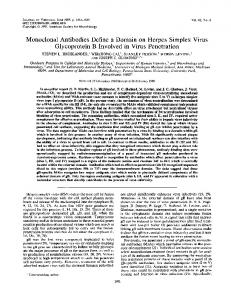
FIG. 4. N-terminal deletion mutants of gpl20. The top of the figure shows the variable and conserved regions of gpl20 as described in the legend to Fig. 3. The 18 conserved cysteine sites located in the glycoprotein are shown at the top. Beneath is illustrated the structure of the HinclI (amino acid [AA] 164), PvuII (amino acid 257), DraI (amino acid 313), and ScaI (amino acid 370) deletion mutants. Also illustrated is the herpes simplex virus signal sequence cassette that was used to express these mutants (shaded). The processed (signal sequence-cleaved) wild-type (WT) and deletion mutant envelope glycoproteins contain nine amino acids from the N terminus of herpes simplex virus glycoprotein D (15; Dowbenko, unpublished data).
rgpl2O, these results suggest that the 7F11 epitope was N terminal to the 5C2 epitope. Thus, these two monoclonal antibodies defined two adjacent epitopes in the C terminus of gp120 that appeared to be contained within a region that was critical for CD4 binding. While the exact delineation of gp120 regions which appeared to be critical for CD4 binding was important, the high-resolution localization of epitopes which could bind monoclonal antibodies without affecting gpl2O-CD4 interaction was also of interest. In this respect, the lambda gtll mapping system provided some important results. For example, the two epitopes recognized by the nonblocking monoclonal antibodies 6D8 and lDlO were found to co-map near the N terminus of gpl20. This region of the glycoprotein is conserved between virus isolates, yet the lack of blocking of the gpl20-CD4 interaction by monoclonal antibodies binding to this region suggests that this conserved region does not directly contribute to this interaction (Fig. 3). In addition, a second highly conserved domain contained within amino acids 180 to 250 of the glycoprotein was found to bind to two other nonblocking monoclonal antibodies corresponding to epitope group a, 9F6 and 1F9. These antibodies appeared to bind to the same highly conserved region in the central part of gp120, and their lack of blocking suggests that this region is involved with functions other than CD4 binding. Thus, these results show that two highly conserved domains of HIV gp120 appear to bind monoclonal antibodies that do not inhibit the interaction between the glycoprotein and its receptor, CD4. The highly conserved nature of these regions suggests that they are involved in other functions that are necessary for the infectivity of the virus.
The final epitope group mapped in this manner was epitope group c, which contained monoclonal antibodies
3E10 and 5G9. These antibodies
were
found to map to
positions in the C terminus of gpl20, in agreement with the proteolytic mapping data described above (Table 3). Since these antibodies neither competed for binding with epitope group b nor blocked gpl20-CD4 interaction, the mapping results suggest that this epitope is C terminal to the epitope group delineated by antibodies 5C2 and 7F11-7G11. The lack of blocking of the glycoprotein-receptor interaction by these antibodies suggests that epitopes C terminal to the site delineated by epitope group b may not be critical for this binding interaction. N-terminal deletion mutants of the envelope glycoprotein. The monoclonal antibody studies shown above, in conjunction with mutagenesis studies by Kowalski et al. (13), suggested that the N-terminal and central conserved domains of gp120 are dispensable for CD4 binding. In addition, mutagenesis studies by Willey et al. (29) revealed that mutations in the central conserved domain of gp120, while resulting in noninfectious virus, had no effect on CD4 binding, also suggesting that this region is dispensable for CD4 interaction. Finally, recent work by Ho et al. (10) has
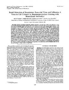
shown that antibodies directed to peptides within the central conserved domain neutralize HIV infectivity but do not block virus-receptor binding. To determine whether Nterminal regions of gp120 are dispensable for binding, Nterminal deletion mutants of gpl20 were constructed and expressed as secreted proteins in transfected mammalian cells (Fig. 4). The secreted glycoproteins were immunoprecipitated with a high-titer rabbit anti-gp120 serum and analyzed by sodium dodecyl sulfate-polyacrylamide gel electrophoresis. All of the deletion mutants were secreted and appeared to react with the antiserum (Fig. 5). The decreased immunoreactivity of the smallest deletion mutant may have been due to a combination of poor expression levels and the
HIV-1 gpl2O EPITOPES
VOL. 62, 1988 a cd4
i
b
c
cd4
cd4
d
i
cd4
e i
cd4
M
200
-97.4
-
68
-
43
N.
_
-25.7
FIG. 5. Lack of CD4 binding of N-terminal deletion mutants. Mammalian cells were transfected with each mutant as well as with wild-type (Fig. 4, legend; 16) gpl20 and labeled with [35S]me-
thionine, and the secreted material was used in the CD4-binding assay as previously described (16). One-tenth of the secreted material was immunoprecipitated with rabbit polyclonal serum directed to gpl20 (15). The rest of the material was reacted with a soluble form of CD4 (24) and immunoprecipitated with monoclonal antibody OKT4. Each pair of lanes shows total immunoprecipitable (i) and CD4-bound (cd4) material. Lanes: a, wild-type gpl20; b, HincIl deletion; c, PvuII deletion; d, DraI deletion; e, Scal deletion; M, molecular weight markers. Kd, Kilodaltons.
maintenance of only
a
few of the original native gpl20
epitopes. Interestingly, the deletion mutants were unable to bind to CD4 with high affinity. Approximately 5 to 10% of the immunoprecipitable wild-type gpl20 was able to coprecipitate with CD4 (Fig. 5), in agreement with previous studies (16). However, none of the N-terminal deletion mutants were able to bind significantly to a soluble form of the CD4 receptor molecule. Lack of glycosylation of the secreted deletion mutants could not explain this lack of receptorbinding activity, since all of these proteins were of higherthan-expected molecular weight, appeared to be heterogeneous in size, and could be glycosylated in an in vitro system (data not shown). These results suggest that sequence information contained within the N-terminal, central, or both domains of gpl20 is necessary to confer a high-affinity CD4-binding phenotype on this glycoprotein. DISCUSSION The external envelope glycoprotein, gpl20, appears to bind HIV to its cellular receptor, CD4. The delineation of a
region of gpl20 which appears to be critical for binding in this report, as well as in previous reports (13, 16), suggests that a defined region of the glycoprotein serves a critical function in binding the virus to its receptor. This region shows a relatively high degree of conservation between diverse HIV isolates, as well as two other CD4-trophic retroviruses, HIV-2 and simian T-lymphotropic virus type 3 (16). However, although the number of domains necessary to accomplish high-affinity binding of gpl20 to CD4 remains unknown, sequence comparisons of the different HIV iso-
4709
lates analyzed to date suggest that there are other regions of the glycoprotein which show a high degree of homology and are, therefore, presumably of functional significance. The data reported here suggest that at least two of these conserved regions have functions other than direct participation in CD4 binding. The mapping of the two gp120-CD4-blocking monoclonal antibodies to adjacent epitopes near the C terminus of the glycoprotein agrees with previously reported results (13, 16). Thus, the localization of the 5C2 epitope by immunoaffinity chromatography of gp120 peptides and subsequent amino acid sequencing demonstrated that the epitope recognized by this blocking monoclonal antibody was localized to a region within a relatively conserved domain of the envelope from residues 406 to 417, in agreement with the localization of this epitope by lambda gtll mapping reported here (residues 403 to 457). The finding that a second blocking monoclonal antibody, 7F11, also binds near this site is further confirmation that this region is critical for binding of the virus to its receptor. The mapping of a nonblocking epitope, corresponding to epitope group c, C terminal to the blocking domain suggests that this critical region does not require C-terminally located sequences for high-affinity binding. C-terminal deletion and random mutagenesis of the gpl20 gene have demonstrated that the CD4 interaction region of this glycoprotein may be conformationally complex, with critical regions from three different areas in the C-terminal region of the protein being important (13). One of the critical mutations that these investigators found was located in a region that mapped near the epitope which bound the nonblocking monoclonal antibodies 3E10-5G9. The nature of this discrepancy is not clear, but it may result from a combination of several variables, including, for example, a conformational change induced by the blocking mutation of Kowalski et al. (13) or only marginally effective blocking by the monoclonal antibodies used by us. In addition, it appears clear from our N-terminal deletion mapping results that deletions (13) may result in the absence of CD4 binding, even in cases in which monoclonal antibodies specific for the deleted regions do not block the interaction. The demonstration of highly conserved domains of gpl20 that do not appear to contribute directly to CD4 binding suggests that these regions have other functions important to the virus. In agreement with the results reported here, previous work (29) has demonstrated that mutations made in the centrally located, highly conserved domain of gpl20 (amino acids 180 to 250) result in mutant viruses that are completely noninfectious but whose envelope glycoproteins appear to bind to the CD4 receptor in both the binding assay described here and a syncitium-forming assay. This result supports the finding shown here and suggests that an important functional domain exists in this central, highly conserved region. Recent work reported by Ho et al. (10) has demonstrated that a peptide from this region was able to induce low levels of neutralizing antibodies that were unable to block virus-receptor interaction. The potential roles for this central conserved region are many and include interaction with the transmembrane protein, gp4l, uncoating of the virus, maintenance of an appropriate overall gp120 conformation, etc. (13, 16). A similar argument could be applied to the N-terminal conserved domain, which contained epitopes for two nonblocking monoclonal antibodies. Interestingly, previous work demonstrated that a mutant gpl20 that lacked the N-terminal 30 amino acids bound with high affinity to the CD4 receptor (15, 16). This suggests that a minor deletion at
4710
DOWBENKO ET AL.
the very N terminus of the protein, in addition to steric inhibition brought on by the binding of monoclonal antibodies to this region, does not seem to grossly affect the ability of gp120 to bind to CD4. The potential of this N-terminal region to act as a structural component of the envelope glycoprotein is suggested by previous work which demonstrated that the noninfectious glycosylation mutant produced in the central highly conserved domain (amino acids 180 to 250) could revert to an infectious virus by a serine-toasparagine change near the C-terminal end of the N-terminal highly conserved domain (amino acids 30 to 130) (29). Thus, it is possible that the N-terminal conserved domain interacts structurally with the central domain, with the result that both of these regions are indispensable for appropriate gp120 structure and function. In support of this possibility, preliminary mapping of disulfide linkages within rgpl20 suggests that there are disulfide interactions between cysteines in the N-terminal and central conserved regions (T. Gregory and M. Spellman, manuscript in preparation). The preliminary data suggest that a complex structural interaction exists between the N-terminal conserved domain and the centrally located conserved domain of gp120. The finding that deletion of the first 164 amino acids of gp120, including the first highly conserved domain as well as the first 6 cysteine residues of the glycoprotein, results in an apparent lack of CD4 binding suggests that there is information, either in the form of direct CD4-binding sites or structure-inducing determinants, that is contained within this region. Previous work using in vitro mutagenesis (13), in addition to work reported here, suggests that direct CD4 interaction sites do not occur in this part of the glycoprotein but are found in the C-terminal region of the molecule. Thus, it appears that the structural aspects of the C-terminally located CD4-binding regions of gpl20 may in part be dependent upon sequences from the N terminus of the protein. Alternatively, removal of several cysteines in the N-terminal deletion mutants may result in aggregated proteins that are unable to bind to CD4, since their removal might result in a number of free cysteine residues remaining in the deletion mutants. In addition, the binding assay that was used here may have been too insensitive to detect deletion mutants with greatly (i.e., -1,000 fold) decreased yet significant binding affinities for CD4, since it is likely that the levels of secreted gpl20 expressed in these transient transfections were quite low (16). In conclusion, the large number of disulfide bonds in gpl20, in addition to the apparent complexity of the disulfide-induced structure (19; T. Gregory and M. Spellman, unpublished data), suggests that appropriate folding of the C-terminal CD4 interaction domain is dependent upon other sequences in the molecule. In summary, the results presented here suggest that although the highly conserved domains in the central and N-terminal regions of gpl20 do not appear to be directly involved in CD4 binding, they may be required for induction of an appropriately folded envelope glycoprotein. Unfortunately, these results also suggest that attempting to produce high-titer virus-blocking antibodies to CD4-binding domains by deletion of other, nonessential regions of the HIV glycoprotein will be difficult, since the shorter gp120 molecules do not appear to retain the correct configuration that might be expected to be necessary for induction of an appropriate antiviral immune response. Finally, these results suggest that development of small-molecule therapeutics based on gp120 for the prevention of HIV binding and infection would be a difficult enterprise (22).
J. VIROL.
ACKNOWLEDGMENTS We thank the many individuals involved in the production of the gpl20 monoclonal antibodies, including P. Berman, M. Renz, D. Vetterlein, N. Hung, F. Chopin, and J. Porter. LITERATURE CITED 1. Alizon, M., S. Wain-Hobson, L. Montagnier, and P. Sonigo. 1986. Genetic variability of the AIDS retrovirus: nucleotide sequence analysis of two isolates from African patients. Cell 46: 63-74. 2. Barin, F., M. McLane, J. Allan, T. Lee, and M. Essex. 1985. Virus envelope protein of HTLV-III represents major target antigen for antibodies. Science 228:1094-1096. 3. Cabradilla, C., J. Groopman, J. Lanigan, M. Renz, L. Lasky, and D. Capon. 1986. Serodiagnosis of antibodies to the human AIDS retrovirus with a bacterially synthesized ENV polypeptide. Bio/Technology 4:128-133. 4. Curran, J., W. Morgan, A. Hardy, H. Jaffe, W. Darrow, and W. Dowdle. 1985. The epidemiology of AIDS: current status and future prospects. Science 229:1352-1357. 5. Dalgleish, A., P. Beverley, P. Clapham, D. Crawford, M. Greaves, and R. Weiss. 1984. The CD4 (T4) antigen is an essential component of the receptor for the AIDS retrovirus. Nature (London) 312:763-766. 6. Deen, K., J. McDougal, R. Inacker, G. Folena-Wasserman, J. Arthos, J. Rosenberg, P. Maddon, R. Axel, and R. Sweet. 1988. A soluble form of CD4 (T4) protein inhibits AIDS virus infection. Nature (London) 331:82-84. 7. Desai, S., V. Kalyanamaran, J. Casy, A. Srinivasan, P. Anderson, and S. Devare. 1986. Molecular cloning and primary nucleotide sequence analysis of a distinct human immunodeficiency virus isolate reveal significant divergence in its genomic sequences. Proc. Natl. Acad. Sci. USA 83:8380-8384. 8. Eaton, D., W. Wood, P. Hass, P. Hollingshead, K. Wion, J. Mather, R. Lawn, G. Vehar, and C. Gorman. 1986. Construction and characterization of an active factor VIII lacking the central one-third of the molecule. Biochemistry 25:8343-8348. 9. Hahn, B., G. Shaw, M. Taylor, R. Redfield, P. Markham, S. Salahuddin, F. Wong-Staal, R. Gallo, E. Parks, and W. Parks. 1986. Genetic variation in HTLVIII/LAV over time in patients with AIDS or at risk for AIDS. Science 232:1548-1553. 10. Ho, D., J. Kaplan, I. Rackhaus, and M. Gurney. 1988. A second conserved domain of gpl20 is important for HIV infectivity and antibody neutralization. Science 239:1021-1024. 11. Ho, D., M. Sarnghadaran, M. Hirsch, R. Schooley, T. Rota, R. Kennedy, T. Chanh, and V. Sato. 1987. Human immunodeficiency virus neutralizing antibodies recognize several conserved domains on the envelope glycoproteins. J. Virol. 61: 2024-2028. 12. Klatzman, D., E. Champagne, S. Chamarat, J. Gruest, D. Guetard, T. Hercend, J. Gluckman, and L. Montaignier. 1984. T-lymphocyte T4 molecule behaves as the receptor for human retrovirus LAV. Nature (London) 312:767-768. 13. Kowalski, M., J. Potz, L. Basiripour, T. Dorfman, W. Chun, E. Terwilliger, A. Dayton, C. Rosen, W. Haseltine, and J. Sodoroski. 1987. Functional regions of the envelope glycoprotein of human immunodeficiency virus type 1. Science 237:1351-1355. 14. Krohn, K., W. Robey, S. Putney, L. Arthur, P. Nara, P. Fischinger, R. Gallo, F. Wong-Staal, and A. Ranki. 1987. Specific cellular immune response and neutralizing antibodies in goats immunized with native or recombinant envelope proteins derived from human T-lymphotrophic virus type IIIb and in human immunodeficiency virus infected men. Proc. Natl. Acad. Sci. USA 84:4994-4998. 15. Lasky, L., G. Groopman, C. Fennie, P. Benz, D. Capon, D. Dowbenko, G. Nakamura, W. Nunes, M. Renz, and P. Berman. 1986. Neutralization of the AIDS retrovirus by antibodies to a recombinant envelope glycoprotein. Science 233:209-212. 16. Lasky, L., G. Nakamura, D. Smith, C. Fennie, C. Shimasaki, E. Patzer, P. Berman, T. Gregory, and D. Capon. 1987. Delineation of a region of the human immunodeficiency virus type 1 gpl20 glycoprotein critical for interaction with the CD4 receptor. Cell 50:975-985.
HIV-1 gpl20 EPITOPES
VOL. 62, 1988
17. Maddon, P., A. Daigleish, J. McDougal, P. Clapham, R. Weiss, and R. Axel. 1986. The T4 gene encodes the AIDS virus receptor and is expressed in the immune system and the brain. Cell 47: 333-348. 18. McDougal, J., M. Kennedy, J. Sligh, S. Cort, A. Mawle, and J. Nicholson. 1986. Binding of HTLVIII/LAV to T4+ T cells by a complex of the 110K viral protein and the T4 molecule. Science 231:382-385. 19. McDougal, J., J. Nicholson, G. Cross, S. Cort, M. Kennedy, and A. Mawle. 1986. Binding of the human retrovirus HTLVIII/ LAV/ARV/HIV to the CD4(T4) molecule: conformation dependence, epitope mapping, antibody inhibition and potential antiidiotype mimicry. J. Immunol. 137:2937-2944. 20. Mehra, V., D. Sweetser, and R. Young. 1986. Efficient mapping of protein antigenic determinants. Proc. Natl. Acad. Sci. USA 83:7013-7017. 21. Muesing, M., D. Smith, C. Cabradilla, C. Benton, L. Lasky, and D. Capon. 1985. Nucleic acid structure and expression of the human AIDS/lymphadenopathy virus. Nature (London) 313: 450-458. 22. Pert, C., J. Hill, M. Ruff, R. Berman, W. Robey, L. Arthur, F. Ruscetti, and W. Farrar. 1986. Octa peptide derived from the neuropeptide receptor-like pattern of antigen T4 in brain potently inhibits human immunodeficiency virus receptor binding and T-cell infectivity. Proc. Natl. Acad. Sci. USA 83:92549258. 23. Reinherz, E., and S. Schlossman. 1980. The differentiation and function of human T lymphocytes. Cell 19:821-827. 24. Smith, D., R. Byrn, S. Marsters, T. Gregory, J. Groopman, and
25.
26.
27.
28.
29.
30.
4711
D. Capon. 1987. Blocking of HIV I infectivity by a soluble, secreted form of the CD4 antigen. Science 238:1704-1707. Starcich, B., B. Hahn, G. Shaw, P. McNeely, S. Modrow, H. Wolf, E. Parks, W. Parks, S. Josephs, R. Gallo, and F. WongStaal. 1986. Identification and characterization of conserved and variable regions in the envelope gene of HTLVIII/LAV, the retrovirus of AIDS. Cell 45:637-648. Weiss, R. 1985. Human T cell retroviruses, p. 405-484. In R. Weiss, N. Teich, and J. Coffin (ed.), RNA tumor viruses: molecular biology of tumor viruses. Cold Spring Harbor Laboratory, Cold Spring Harbor, N.Y. Weiss, R., P. Clapham, J. Weber, A. Dalgleish, L. Lasky, and P. Berman. 1986. Variable and conserved neutralization antigens of the human immunodeficiency virus (HIV-1). Nature (London) 324:572-575. Willey, R., R. Rutledge, S. Dias, T. Folks, T. Theodore, C. Buckler, and M. Martin. 1986. Identification of conserved and divergent domains within the envelope gene of the acquired immunodeficiency retrovirus. Proc. Natl. Acad. Sci. USA 83: 5038-5042. Willey, R., D. Smith, L. Lasky, T. Theodore, P. Earl, B. Moss, D. Capon, and M. Martin. 1988. In vitro mutagenesis identifies a site within the envelope gene of the human immunodeficiency virus that is critical for infectivity. J. Virol. 62:139-147. Young, R., B. Bloom, C. Grosskinsky, J. Ivanyi, D. Thomas, and R. Davis. 1985. Dissection of Mycobacterium tuberculosis antigens using recombinant DNA. Proc. Natl. Acad. Sci. USA 82: 2583-2587.