RESEARCH ARTICLE 4283
Development 134, 4283-4295 (2007) doi:10.1242/dev.005272
Granulosa cells regulate oocyte intracellular pH against acidosis in preantral follicles by multiple mechanisms Greg FitzHarris1,2,*, Violetta Siyanov1,3 and Jay M. Baltz1,2,3 Mammalian oocytes grow within ovarian follicles in which the oocyte is coupled to surrounding granulosa cells by gap junctions. We report here that growing oocytes isolated from mouse preantral follicles are incapable of recovering from an experimentally induced acidosis, and that oocytes acquire the ability to manage acid loads by activating Na+/H+ exchange during growth. By contrast, granulosa cells from similar preantral follicles possess substantial Na+/H+ exchange capacity, which is attributable to the simultaneous action of two Na+/H+ exchanger isoforms: NHE1 and NHE3. Granulosa cells were also found to possess a V-type H+ATPase that drives partial acidosis recovery when Na+/H+ exchange is inactivated. By monitoring intracellular pH (pHi) in small follicle-enclosed oocytes, we found that the oocyte has access to each of these acidosis-correcting activities, such that small follicleenclosed oocytes readily recover from acidosis in a manner resembling granulosa cells. However, follicle-enclosed oocytes are unable to access these activities if gap-junction communication within the follicle is inhibited. Together, these experiments identify the NHE isoforms involved in regulating oocyte pHi, indicate that gap junctions allow granulosa cells to exogenously regulate oocyte pHi against acidosis until the oocyte has acquired endogenous pHi regulation, and reveal that granulosa cells possess multiple mechanisms for carrying out this function.
INTRODUCTION Regulation against unwelcome perturbations in intracellular pH (pHi) is a fundamental and essential process carried out by virtually all cells. The principal pHi regulatory mechanisms in mammalian cells are HCO3–/Cl– exchangers of the AE (anion exchange) family, which export HCO3– in exchange for Cl– thereby correcting pHi increases (Romero et al., 2004; Alper, 1994), and Na+/H+ exchangers of the NHE (also known as Scl9a – Mouse Genome Informatics) gene family which correct pHi decreases by exporting protons in exchange for Na+ (Orlowski and Grinstein, 1997; Orlowski and Grinstein, 2004). Multiple isoforms of the Na+/H+ exchanger (NHE1-9) and the HCO3–/Cl– exchanger (AE1-3, with numerous splice variants) exist, with heterogeneous tissue expression that probably reflects their different kinetic properties and regulation (Romero et al., 2004; Orlowski and Grinstein, 2004). These transport systems may be supplemented by additional components, such as Na+, HCO3–/Cl– exchangers (NBC), which relieve acidosis in some cell types by importing HCO3– (Romero et al., 2004), and V-type H+-ATPases (V-ATPases) which export protons across the plasmalemma in some cells (Nelson and Harvey, 1999; Merzendorfer et al., 1997; Kawasaki-Nishi et al., 2003). The multifaceted array of pHi regulatory mechanisms provides the basis for tight control of intracellular pH in the face of external pH changes and metabolic acid generation. Perhaps unsurprisingly, impairment of pHi regulation can compromise cell function and viability. For example, growth and proliferation is impaired in some pHi regulation-compromised cells when intracellular pHi is 1 Ottawa Health Research Institute, 2Department of Obstetrics and Gynecology (Division of Reproductive Medicine), and 3Department of Cellular and Molecular Medicine, University of Ottawa, Ottawa, ON, K1Y 4E9, Canada.
*Author for correspondence at present address: Institute for Women’s Health, University College London, Gower Street, London WC1E 6BT, UK (e-mail:
[email protected]) Accepted 5 September 2007
disturbed (Grinstein et al., 1989; Kapus et al., 1994), and pHi dysregulation jeopardizes cell survival (Pouyssegur et al., 1984). pHi regulation is of particular importance in early mammalian development, as inhibition of pHi regulatory mechanisms hinders preimplantation development in mouse and hamster embryos (Lane et al., 1998; Zhao et al., 1995). Mammalian oocytes grow within ovarian follicles, discrete microorgans consisting of the oocyte and a surrounding shell of (somatic) granulosa cells. Oocyte growth takes about 15 days in mouse, during which time the oocyte increases in diameter from about 15 to 70-80 m. Throughout growth, the oocyte remains coupled to its granulosa cells by gap junctions, intercellular channels that permit small molecules (65 m) and competent to complete meiosis. Using an assay in which any Na+/H+ exchanger activity could be revealed by an amiloride-sensitive intracellular acidification upon external Na+ removal, there was also evidence that Na+/H+ exchanger activity follows a similar pattern during oocyte growth (Erdogan et al., 2005). Thus, we concluded that small growing oocytes do not possess intrinsic pHi regulatory mechanisms, but develop them when they are nearly fully grown. Subsequently it was found that, in the intact follicle, oocytes have access to significant amounts of alkalosis-correcting HCO3–/Cl– exchange enabling them to recover from alkalosis, and that granulosa cells surrounding growing oocytes had substantial HCO3–/Cl– exchange, which led us to propose that ovarian oocytes may be able to access HCO3–/Cl– exchangers in the granulosa cells (FitzHarris and Baltz, 2006). However, it was difficult to discount the possibility that granulosa cells may activate pHi regulation endogenous to the oocyte (FitzHarris and Baltz, 2006), and the lack of isoform-specific HCO3–/Cl– exchange inhibitors prevented identification of the specific alkalosis-correcting mechanisms present in granulosa cells and fully grown oocytes. The ability of growing oocytes to regulate against acidosis has not yet been addressed, nor have the molecular identities of any pHiregulatory mechanisms in oocytes or surrounding granulosa cells been determined. Our experiments described here indicate that growing oocytes rely upon granulosa cells to regulate against acidosis until the oocyte’s own Na+/H+ activity is activated during growth, and provide strong evidence that ooplasmic pHi is regulated by pHi-regulatory mechanisms within granulosa cells, which possess multiple mechanisms that can raise oocyte pHi, including two Na+/H+ exchanger isoforms and a proton-extruding V-ATPase, some of which are not present even in the fully grown oocyte.
Development 134 (23)
Acidosis correction in ovarian oocytes
RNA extraction was performed using an RNAeasy Micro kit (Qiagen), and reverse transcription performed using a Retroscript kit (Ambion), according to manufacturers’ instructions. RNA extraction and reverse transcription were performed upon groups of no fewer than 60 oocytes or between eight and 25 granulosa shells at a time. PCR was performed using HotStarTaq PCR kit (Qiagen) on an appropriate amount of cDNA template corresponding to three oocytes, or one granulosa shell. Kidney (NHE1-4) and brain (NHE5) cDNA were used as positive controls. The amount of positive control tissue cDNA used was chosen to approximately correspond with that of three oocytes based upon measurement of RNA content of control tissue RNA preparations, and the known RNA content of one oocyte (~0.6 ng) (Sternlicht and Schultz, 1981). The same amount of positive control cDNA was used for comparison with granulosa shells, one granulosa shell having approximately the same volume as three fully grown oocytes. Samples of the final drops of medium in which oocytes were washed were subjected to the same reverse transcription protocol and used as negative controls. PCR was repeated on a minimum of two different oocyte preparations, or three different granulosa cell preparations. Amplicons were visualized on a 1% agarose gel containing 0.13 g/ml ethidium bromide. All products were of the predicted size according to their position on the gel, and the identities of products from all five primer pairs were confirmed by direct sequencing. Primers used for PCR were as follows (5⬘-3⬘) [accession number; predicted amplicon length]. NHE1 (also known as Scl9a1): CACCAGTGGAACTGGACCTT, AAGGTGGTCCAGGAACTGTG [NM_016981.1; 372]. NHE2 (Scl9a2): ATCACGGCTGCTATTGTCGTT, GTGACCCCAGTGTCCACACACA [NM_001033289; 189]. NHE3 (Scl9a3): TCAGCTAAGCTAGGCATCAACC, ATGGTGTCAGGCGGCGGAAGT [AK033564; 412]. NHE4 (Scl9a4): CCGGAGGAACCTGCCAAAATC, TCTTCAGGAGAAAGCCGCTTGA [NM_177084; 162]. NHE5 (Scl9a5): CCTCCCTGTTTGTGGTCAGT, TATGGGAGATGTTGGCTTCC [NM_138858; 235]. Primers for NHE1-4 were designed based upon
mouse mRNA sequences. Primers for NHE5 were generated by ‘blasting’ the sequence for rat NHE5 mRNA (NM_138858) against the mouse genome. Primers were then designed using the matching mouse sequence. PCR products were sequenced and similarity to predicted sequences were as follows: NHE1 98%; NHE2 97%; NHE3 100%; NHE4 96%; NHE5 95%. Data analysis
Where rates of pHi change were calculated, individual oocytes and/or granulosa shells within a given replicate were pooled to provide an average rate, and the mean±s.e.m. of the different replicates presented. Rates were calculated using SigmaPlot 8.0. Data points presented are the mean±s.e.m. of all replicates performed. Means of replicates were compared using t-tests (two groups) or ANOVA (three or more groups). Where ANOVA was used, Tukey-Kramer’s and Dunnett’s post-hoc tests were applied as appropriate (using Instat; GraphPad, San Diego, CA, USA).
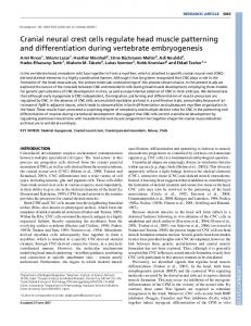
RESULTS Oocytes acquire the capacity to recover from acidosis during growth by activating Na+/H+ exchange We first set out to determine whether denuded growing oocytes are capable of recovering from acidosis. Two separate populations were examined; fully grown oocytes (70-75 m in diameter) obtained from 20-day-old mice, and smaller, mid-growth-phase oocytes (40-60 m) obtained from 10-day-old mice (see Materials and methods). Oocytes were freed of their granulosa cells, and loaded with the pH-sensitive fluorophore SNARF. Acidosis was induced by exposing oocytes to a 10 minute pulse of 25 mM NH4Cl (NH4Cl pulse assay; see Materials and methods for detailed explanation). This protocol caused substantial net acidosis in oocytes of all sizes (~0.5 pHU; Fig. 1).
Fig. 1. Activation of Na+/H+ exchange during oocyte growth. Intracellular pH was monitored in growing oocytes using epifluorescence microscopy. (A) Each panel shows a typical replicate of a given experiment, each individual trace representing a single oocyte. Acidosis was induced in mid-growth phase (left panel) and fully grown (middle panel) oocytes using a 10-minute pulse of NH4Cl (black bar). Note that neither population of oocytes mounts a substantial recovery from acidosis in media devoid of Na+ (gray bar), but resting pH is rapidly restored in fully grown oocytes following Na+ replacement. Insets show examplar fluorescence micrographs from each respective experiment. Scale bar: 50 m. Where used, amiloride (1 mM) was added at t=20 minutes, and remained in the bath thereafter. Note that amiloride retards recovery from acidosis in fully grown oocytes (right panel). (B) Analysis of rate of recovery from acidosis following Na+ replacement. Different letters above the bars indicate significant differences P