tional recovery in patients with chronic coronary artery ... Contractile recovery of an infarct region was .... screen format to improve the detection of dobutamine.
European Heart Journal (2000) 21, 981–991 doi:10.1053/euhj.2000.1946, available online at http://www.idealibrary.com on
Head to head comparison of dobutamine–transoesophageal echocardiography and dobutamine–magnetic resonance imaging for the prediction of left ventricular functional recovery in patients with chronic coronary artery disease F. M. Baer1, P. Theissen2, J. Crnac1, M. Schmidt2, H. J. Deutsch1, U. Sechtem1, H. Schicha2 and E. Erdmann1 1
Klinik III fu¨r Innere Medizin, 2Klinik und Poliklinik fu¨r Nuklearmedizin, der Universita¨t zu Ko¨ln, Ko¨ln, Germany
Aims A substantial number of patients with dysfunctional but potentially viable myocardium cannot be accurately evaluated by transthoracic echocardiography due to a poor acoustic window. This study compares the diagnostic value of alternative functional imaging techniques, such as dobutamine–transoesophageal echocardiography (dobutamine–TEE) and dobutamine magnetic resonance imaging (dobutamine–MRI) for the detection of viable myocardium and the prediction of left ventricular functional recovery in patients with chronic coronary artery disease following successful revascularization procedures. Methods and Results Rest and low-dose (5, 10 �g dobutamine . min �1 . kg �1) multiplane dobutamine–TEE and ultrafast cine–MRI studies were performed in 103 patients. Contractile recovery of an infarct region was predicted if a dobutamine contraction reserve could be assessed visually by TEE or MRI in d50% of infarctrelated a- or dyskinetic segments. Revascularization of the infarct-related vessel was successful in 88 patients, and 4·9�0·7 months later 52 patients still had an angiographically controlled open target vessel. These patients underwent another rest MRI study to assess left ventricular functional recovery. A dobutamine contraction reserve was observed in 27/52 (52%) patients by TEE and in 26/52 (50%) patients by MRI. Functional improvement of the infarct region was diagnosed in 28/52 (54%) patients. The positive
Revision submitted and accepted 1 October 1999. Correspondence: Frank Michael Baer, MD, Klinik III fu¨r Innere Medizin, Universita¨t zu Ko¨ln, Joseph-Stelzmann-Str. 9, D-50924 Ko¨ln, Germany. 0195-668X/00/120981+11 $35.00/0
and negative predictive accuracy of dobutamine–TEE and dobutamine–MRI for the prediction of left ventricular functional recovery was not significantly different (85% vs 92%, ns and 80% vs 85%, ns). Diagnosis of a predominantly viable infarct region by TEE and MRI predicted a significant increase in left ventricular ejection fraction (TEE: 12�8% vs 2�7% P4 months) and left ventricular dysfunction who were on the waiting list for revascularization procedures (Fig. 1). Forty-eight of these patients were also included in a previous MRI study on myocardial viability[12]. Patients were excluded if they had unstable angina, congestive heart failure (NYHA IV), atrial fibrillation, a permanent pacemaker, a history of multiple myocardial infarctions or a history of sustained ventricular tachycardia. Dobutamine–TEE Eur Heart J, Vol. 21, issue 12, June 2000
and dobutamine–MRI studies were performed in random order within 3 days without intervening cardiac events. Beta-blockers were withdrawn 48 h preceding the pharmacological stress tests and the control studies after revascularization. All patients received a long acting nitrate (Isoket Retard 120�) before rest and dobutamine–TEE and dobutamine–MRI studies to allow for constant coronary artery vasodilation. Additionally, premedication with 5 mg midazolam was administered to all patients prior to the TEE studies to prevent an endogeneous release of catecholamines in case of anxiety or difficulties in swallowing the multiplane TEE probe. After 4·9�0·7 months, 65 patients underwent control coronary angiography (Fig. 1). Patients with restenosis/ obstruction (>70% diameter reduction) of the infarctrelated coronary vessel or bypass were excluded from the final evaluation. The subset of 52 patients who fulfilled the inclusion criteria underwent an additional rest–MRI study for quantitative evaluation of left ventricular functional recovery (Fig. 1). The baseline and angiographic characteristics of these patients are presented in Fig. 2. This study was approved by the Hospital Human Rights Committee (Institutional Review Board) and informed consent was obtained from every patient.
Left ventriculography Regional left ventricular wall motion was evaluated from the left ventriculogram (DCI, Philips Einthoven) and graded as normokinetic, hypokinetic, akinetic or dyskinetic by a consensus of two independent, experienced observers. The left ventricular ejection fraction was calculated by the area–length method and multiple views of each coronary artery were obtained (DCI, Philips, Einthoven, Germany) before and after revascularization procedures. Coronary artery or bypass graft narrowing was assessed as % diameter stenosis using electronic calipers. Patients with >70% diameter restenosis of the infarct-related vessel or >70% diameter stenosis of the infarct-related bypass graft at control angiography were excluded from the study because recurrent hibernation or repetitive stunning could not be ruled out.
TEE studies Transoesophageal echocardiography was performed after a 6 h fast by a multiplane probe with a 5 MHz transducer attached to a Vingmed CFM750/System5 (Sonotron) instrument. Rest and dobutamine image sequences were digitized online with the Vingmed EchoPac� system. After the rest–transoesophageal echocardiography studies dobutamine (Dobutrex�) was administered intravenously, at an initial dose of 5 �g . kg �1 . min �1. After an equilibration period of
TEE and MRI in CAD
983
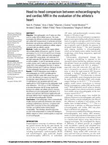
103 patients fulfilled inclusion criteria Inadequate image quality for viability assessment TEE n = 3/MRI n = 2
Dobutamine TEE/MRI studies 98 patients with accurate TEE/MRI studies
Unsuccessful target vessel revascularization Intervention n = 8/ACB n = 2
PTCA/ACB Successful revascularization procedure n = 88
Acute myocardial infarct n = 3 Deaths n = 2 Follow-up Phase (4·9 ± 0·7 months) No angiographic follow-up n = 18 Angiographic follow up = 65 patients Target vessel restenosis n = 13 Successful revascularization and MRI follow-up study n = 52
Figure 1 Flow-chart characterizing the study population, observation periods and related clinical complications, accuracy of TEE and MR image acquisition and frequency of follow-up studies. ACB=aorto coronary bypass graft; MRI=magnetic resonance imaging, PTCA= percutaneous transluminal coronary angioplasty; TEE=transoesophageal echocardiography. 5 min transoesophageal echocardiography scans were repeated. Fine adjustments were made with regard to the size and shape of the papillary muscles to provide an image as close as possible to the baseline image. The dose of dobutamine was then increased to 10 �g . kg �1 . min �1 and transoesophageal echocardiography scans were repeated after another 5 min. There were no serious side effects or complications during the low-dose dobutamine infusion. Control transoesophageal echocardiography studies performed 4–6 months after revascularization followed the same protocol as the rest–transoesophageal echocardiography studies. Total imaging time for rest– and dobutamine–TEE studies ranged between 20–30 min.
TEE image analysis Images were analysed on a segment-by-segment basis by two experienced observers independently and without
knowledge of the findings of the other imaging techniques. Digitized cine-loops were displayed in a quadscreen format to improve the detection of dobutamine contraction reserve. In case of disagreement, a third observer reviewed the study and the subsequent majority judgement was binding. Data on inter- and intraobserver reproducibility of TEE studies and a detailed description of the visual image analysis based on a 28-segment model has been described previously[9]. Akinetic and dyskinetic segments at rest were graded viable if dobutamine-induced wall thickening could be observed. Individual infarct regions were graded viable if 50% of a- or dyskinetic segments showed dobutamine-induced systolic wall thickening.
MRI studies MR images were obtained by using a 1·5-Tesla superconducting magnet (Philips ACS NT). A multislice Eur Heart J, Vol. 21, issue 12, June 2000
984
F. M. Baer et al.
Patients
n = 52
Age (years)
58·2 ± 8·8
Men
Revascularization procedures
n = 52
PTCA
n = 29 (56%)
ACB
n = 23 (44%)
Target vessel: RCA
21 (40%)
Target vessel: LAD
26 (50%)
Target vessel: LCx
5 (10%)
Target vessel occlusion
23 (44%)
n = 48 (92%)
Anterior MI
n = 29 (56%)
n = 23 (44%)
Inferior MI
Infarct age (months)
LVEF < 35%
15 ± 23 n = 15 (29%)
Mean LVEF
41 ± 10·0
Multi-vessel disease
n = 39 (75%)
Figure 2 Baseline and angiographic characteristics of the patients with successful revascularization and MRI follow-up of left ventricular functional recovery. ACB=aorto-coronary bypass graft; LCA=left anterior descending coronary artery; LCx=left circumflex coronary artery; LVEF=left ventricular ejection fraction; MI=myocardial infarction; PTCA=percutaneous transluminal coronary angioplasty; RCA=right coronary artery. ECG-triggered spin–echo sequence was initially acquired to define the cardiac axes. Subsequently, the entire left ventricle was covered by 8–10 short axes and one long axis tomogram using a breathhold turbo field gradient echo technique (flip angle of 30�, echo time (TE) 4 ms and a repetition time (TR) 9 ms, temporal resolution approximately 40 ms)[12]. Slice thickness was 10 mm with no interslice gap and the spatial resolution was 1·7�1·4 mm. Without removing the patient from the magnet, a dose of 5 and 10 �g . kg �1 . min �1 dobutamine (Dobutrex�) was administered intravenously under continuous ECG monitoring and blood pressure recording (Boso Oscillomat) beginning 5 min before and ending after the MR image acquisition. There were no serious side effects or complications during the low-dose dobutamine infusion. The total imaging time for rest and dobutamine MR studies ranged between 20–30 min.
MR image analysis Corresponding to the TEE studies, MRI cineloops were analysed on a visual basis by two experienced observers independently and without knowledge of the findings of the other imaging techniques. MR short axis tomograms covering the entire left ventricle and one transverse tomogram were displayed in a quad-screen format using an Apple� Power PC platform to allow for comparison of rest and dobutamine–stress studies of identical tomograms. Wall motion and systolic wall thickening were Eur Heart J, Vol. 21, issue 12, June 2000
assessed semiquantitatively on a segmental basis using a system where score 1 indicated normal or hyperkinetic myocardium, score 2 hypokinesia (i.e. reduced but not absent systolic wall thickening and inward wall motion), score 3 akinesia (i.e. absent wall thickening and wall motion) and score 4 dyskinesia (i.e. systolic outward movement of the endocardial border and absent systolic wall thickening or systolic wall thinning. Akinetic and dyskinetic segments at rest were graded viable if dobutamine-induced wall thickening could be observed (score improvement from 3 or 4 to 1 or 2). The reference standard ‘contractile recovery after successful revascularization’ was evaluated quantitatively by measuring end-diastolic and end-systolic wall thickness[12]. Improvement of left ventricular function was defined as systolic wall thickening at restd2 mm. This threshold value was chosen based on a spatial resolution of 1·3mm of the MR-machine. However, this implicates that viable regions with a weak contractile recovery are not detected. Functional recovery of the entire infarct region was defined as systolic wall thickening d2 mm in d50% of the dysfunctional segments prior to revascularization.
Statistical analysis All data were expressed as mean values �1SD. A simple regression analysis was performed to assess the correlation of the change in left ventricular ejection
TEE and MRI in CAD
985
Table 1 Infarct region based predictive accuracy of dobutamine–TEE and dobutamine–MRI for the recovery of regional left ventricular function after successful revascularization Imaging technique
Sensitivity (%)
Specificity (%)
Positive predictive value (%)
Negative predictive value (%)
Diagnostic accuracy (%)
Dobutamine–TEE Dobutamine–MRI
82 (23/28) 86 (24/28)
83 (20/24) 92 (22/24)
85 (23/27) 92 (24/26)
80 (20/25) 85 (22/26)
83 (43/52) 88 (46/52)
fraction before and after revascularization and the number of dobutamine responsive segments. ANOVA was performed to evaluate the significance level of mean LVEF for different TEE and MRI viability categories. Sensitivity, specificity and the predictive accuracy of dobutamine–TEE and dobutamine–MRI for the recovery of regional left ventricular function were calculated by applying standard formulas which were then compared by McNemar’s test. The null hypothesis was rejected at the 95% confidence level, in which a P value