Ann Rheum Dis 2000;59:857–863
857
Health impact of pain in the hip region with and without radiographic evidence of osteoarthritis: a study of new attenders to primary care Fraser Birrell, Peter Croft, Cyrus Cooper, Gillian Hosie, Gary Macfarlane, Alan Silman, on behalf of the PCR Hip Study Group
ARC Epidemiology Unit, University of Manchester, M13 9PT, UK F Birrell G Macfarlane A Silman Centre for Primary Care Sciences, University of Keele, Stoke-on-Trent, ST4 7QB, UK P Croft MRC Environmental Epidemiology Unit, University of Southampton, SO9 4XY, UK C Cooper Great Western Health Centre, Glasgow, G13 2SW, UK G Hosie Unit of Chronic Disease Epidemiology, University of Manchester, M13 9PT, UK G Macfarlane Correspondence to: Professor Silman
[email protected] Accepted for publication 7 March 2000
Abstract Objectives—To assess the health impact of hip pain at the time of first presentation to primary care, and the influence on this of radiographic evidence of osteoarthritis. Subjects and methods—Cross sectional survey of 195 patients (63 male, 132 female), aged 40 years and over, presenting with a new episode of hip pain, recruited from 35 general practices across the UK. Health status at presentation was determined by a structured questionnaire on symptoms, healthcare use, and health related quality of life (SF-36). Pelvic radiographs were assessed blindly for hip osteoarthritis using standard scoring systems. Results—The overall impact on health was substantial. Before their first consultation, three quarters of patients needed analgesics, half used topical creams or ointments, and one in eight used a walking stick. Most of these impact measures were, however, unrelated to the degree of radiographic change, though use of a walking stick was increased in those with the most severe damage. Health status, as judged by the SF-36, was also impaired for measures of physical function and pain, but the impact on the “mental health”, “general health”, and “vitality” dimensions was small. There was a weak relation between the SF-36 scores and radiographic change, with many domains unrelated to the severity of radiographic damage. Conclusions—This study is the first to show the therapeutic impact and pattern of impairment in health status resulting from hip pain at the time of first presentation to the healthcare services. Unlike many regional pain syndromes seen in primary care, such as back pain, hip pain does not impact on wider aspects of quality of life, such as general health status, mental health, or vitality. Furthermore, any impact of hip pain in this group is not markedly influenced by the degree of structural damage. Further follow up is required to determine whether such damage influences the persistence of any adverse impact. (Ann Rheum Dis 2000;59:857–863)
Although there have been studies investigating the health impact of hip osteoarthritis (OA) at the time of total hip arthroplasty,1 the decision to perform total hip arthroplasty in patients
www.annrheumdis.com
with OA of the hip is based largely on the patient’s report of pain and disability, usually with demonstrable structural change.2–4 There has, however, been little research examining the impact of hip disease at the time of first presentation to primary care. This may be important for the following reasons. Health status of early disease can oVer insight into the need for arthroplasty at this stage. Secondly, at the time of hospital referral, most patients have severe disease and the natural history before referral is unknown.5 Thirdly, the inherent selection bias of hospital based studies does not permit any analysis of the relative contributions of pain and structural (that is, radiographic) damage on impact. The objective of this study was to answer two questions: firstly, what is the impact on health status of hip pain in patients newly presenting in primary care and, secondly, is impact increased in those with radiographic hip OA compared with those with other causes for their hip pain? Subjects and methods SUBJECTS
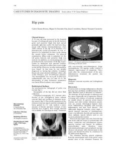
This was a multicentre study of patients presenting with new episodes of hip pain in primary care. In all, 36 general practitioner (GP) members of the Primary Care Rheumatology Society, a group of doctors who have a particular interest in musculoskeletal pain, were recruited from all over the United Kingdom. They were asked to enrol consecutive new attenders aged 40 years or older with hip pain. The practices were divided between urban and rural areas and covered a broad socioeconomic spectrum. Ethical permission was secured locally for each practice before the study started. As no standardised definition for hip pain exists, a working case definition was derived by a consensus group including the study coordinators and GPs and validated by pilot testing. This was defined as pain within a preshaded area on a standardised pain drawing (fig 1) which, on GP clinical assessment, was not thought to originate from structures outside the hip (for example, low back, trochanteric bursa). Use of a pictorial instrument of the area to be regarded as “hip pain” enhanced the standardisation of case definition. Patients presenting with pain in a hip with prior arthroplasty were excluded. The aim was to study early disease severe enough to prompt a visit to the doctor. As hip pain may be a recurrent problem throughout
858
Birrell, Croft, Cooper, et al
Back
Front
Left
Right
(A)
Right
(A)
Left
(C)
(C)
(D) (B)
(E)
(B)
(E)
Landmarks:
Figure 1
(E)
(E)
A - Iliac crest B - Ischial tuberosity C - Anterior superior iliac spine D - Pubic tubercle E - 1/3 way down thigh
Standardised drawing for hip pain.
life, the definition of a new episode presented obvious diYculties and thus a pragmatic approach was adopted. Patients were excluded if they either had consulted at any time in the past for their current pain; or had consulted with any hip pain in the previous 12 months, even if not related to their current pain. A subset of the medical records of the patients recruited was reviewed to validate the case entry criteria and to provide an estimate of the proportion of the study group who were first-ever consulters.
METHODS
The patient completed a structured questionnaire at presentation. Pain severity was measured using a numerical rating scale (1–10). This approach is well validated, performing comparably or better than other pain rating instruments.6 Duration of hip pain was measured by recalled interval since the current episode started. There were closed questions on current physical activities, including walking, and treatments or assistance sought for the hip pain leading to presentation. Health related quality of life was assessed using the Short Form 36 Health Questionnaire (SF-36),7 to which responses were coded and transformed according to standard algorithms.8 An anteroposterior pelvis radiograph was requested on each subject, with a set of standard instructions given to the local x ray department. As there is no consensus definition for radiographic OA,9 x rays were graded for both Croft’s modification of the Kellgren and Law-
www.annrheumdis.com
rence grade (“Croft grade”) and minimum joint space.10 Gradings were made by two independent observers, unaware of the clinical status of the subject, with adjudication of any discrepancies made by a third observer.
ANALYSIS
The impact of hip pain was expressed in terms of pain severity, walking, treatments, and health professionals seen, stratified by age group and sex. The influence of radiographic hip OA was assessed after stratification into three groups for each of the Croft grades (0–1, 2–3, 4–5) and for minimum joint space (>2.5 mm, 1.5–2.5 mm, 2.5 136 (70) >1.5, 1) and a substantial minority had advanced x ray changes (table 1). Figure 3 shows the relation between SF-36 Z scores and radiographic severity stratified by Croft grade (fig 3A) and minimum joint space (fig 3B). There was no overall consistent pattern of increasing impact with increasing radiographic severity. The pain scale scores were not related to the degree of radiographic damage. By contrast, the functional scale scores did show the greatest
860
Birrell, Croft, Cooper, et al
impairment in those with the most damage. No other domains showed this pattern. Tables 4 and 5 show the relation between the other impact measures and radiographic change by Croft grade and minimum joint space, respectively. These data show, as with the SF-36, that independent of grading scheme, the impact measures studied were broadly similar across the grades of radiographic damage. However, both minimum joint space of 1.5 mm or less and Croft grades of 4–5 are associated with a threefold increased odds of using a walking stick, after adjustment for age and sex. There was also a doubling of
the odds of pain severity score above the median, though this was not statistically significant. Discussion We have shown that there is a substantial impact from hip pain, even at first presentation to primary care with this problem. The most striking impact on health related quality of life was seen for physical measures of the SF-36— physical functioning, physical role, and pain, but the impact on mental health, general health, and vitality, when compared with normative data, was small. Further, one in
A 0.2
Emotional role
Mental health Mental health
Social functioning
Vitality
Bodily pain
Emotional role
Z score
–0.4
Physical role
Physical functioning
–0.2
General health
SF-36 measure
0
–0.6
Croft 0 and 1
–0.8
Croft 2 and 3 Croft 4 and 5 –1
–1.2
–1.4
–1.6
B
Vitality
General health
Bodily pain
Social functioning
–0.4
Physical role
–0.2
SF-36 measure Physical functioning
0
Z score
–0.6
>2.5 mm 1.6–2.5 mm –30 min/day Used a walking stick Taken analgesics Used topical treatment Consulted physiotherapist Consulted osteopath/chiropractor
Sex
All (n=195)
40–63 (n=98)
64+ (n=97)
Male (n=63)
Female (n=132)
5 (3–7) 130 (68) 23 (12) 141 (73) 94 (46) 15 (8) 15 (8)
5 (3–7) 71 (74) 6 (6) 72 (75) 44 (46) 6 (6) 10 (10)
5 (3–7) 59 (61) 17 (18) 69 (71) 50 (52) 9 (9) 5 (5)
4 (3–7) 49 (78) 13 (21) 38 (60) 38 (60) 8 (13) 3 (5)
5 (3–7) 81 (63) 10 (8) 103 (79) 56 (43) 7 (5) 12 (9)
*IQR = interquartile range. † Percentages reflect subjects with full data for each category
Table 4
Logistic regression of the impact of symptoms by grade of osteoarthritis‡ *Croft grade group 0–1 (n=109)
Pain severity >4 Walk >30 min/day Use a walking stick Take analgesics Use topical treatments See physiotherapist See osteopath/chiropractor
2–3 (n=53)
4–5 (n=33)
No (%)
OR† Reference
No (%)
OR† (95% CI)
No (%)
OR† (95% CI)
÷2 trend p
2.5 (n=136)
Pain severity >4 Walk >30 min/day Use a walking stick Take analgesics Use topical treatments See physiotherapist See osteopath/chiropractor
>1.5,