Journal of the Formosan Medical Association (2015) 114, 569e576
Available online at www.sciencedirect.com
ScienceDirect journal homepage: www.jfma-online.com
REVIEW ARTICLE
Hedgehog signaling: From basic research to clinical applications Erica Yao, Pao-Tien Chuang* Cardiovascular Research Institute, University of California, San Francisco, CA, USA Received 6 November 2014; accepted 1 January 2015
KEYWORDS cancer; cilium; development; Hedgehog; mechanism; signaling
Studies of the major signaling pathways have revealed a connection between development, regeneration, and cancer, highlighting common signaling networks in these processes. The Hedgehog (Hh) pathway plays a central role in the development of most tissues and organs in mammals. Hh signaling is also required for tissue homeostasis and regeneration in adults, while perturbed Hh signaling is associated with human cancers. A fundamental understanding of Hh signaling will not only enhance our knowledge of how the embryos are patterned but also provide tools to treat diseases related to aberrant Hh signaling. Studies have yielded a basic framework of Hh signaling, which establishes the foundation for addressing unresolved issues of Hh signaling. A detailed characterization of the biochemical interactions between Hh components will help explain the production of graded Hh responses required for tissue patterning. Additional cell biological and genetic studies will offer new insight into the role of Hh signaling in homeostasis and regeneration. Finally, drugs that are capable of manipulating the Hh pathway can be used to treat human diseases caused by disrupted Hh signaling. These investigations will serve as a paradigm for studying signal transduction/integration in homeostasis and disease, and for translating discovery from bench to bedside. Copyright ª 2015, Elsevier Taiwan LLC & Formosan Medical Association. All rights reserved.
Introduction The Hedgehog (Hh) pathway is one of several major signaling pathways that control key steps of embryonic
Conflicts of interest: The authors have no conflicts of interest relevant to this article. * Corresponding author. Cardiovascular Research Institute, University of California, 555 Mission Bay Blvd. South, San Francisco, CA 94158, USA. E-mail address:
[email protected] (P.-T. Chuang).
development.1e3 Hh signaling also participates in tissue homeostasis and regeneration4 while perturbation of Hh signaling is associated with several human cancers.5e7 This highlights the versatility of conserved signaling pathways in multiple signaling contexts during the life span of an organism from embryonic development to postnatal physiology and pathology. Elucidating the molecular mechanisms of Hh signaling will increase our fundamental understanding of Hh signaling and serve as a paradigm to illuminate pathway design and evolution. These studies also hold the promise to provide tools for detecting and eventually treating human diseases caused by aberrant Hh signaling.
http://dx.doi.org/10.1016/j.jfma.2015.01.005 0929-6646/Copyright ª 2015, Elsevier Taiwan LLC & Formosan Medical Association. All rights reserved.
570
The current model of mammalian Hh signal transduction The basic framework of Hh signaling has been established through extensive studies in cell-based assays and model organisms. Hh signaling adopts multiple control steps at different subcellular locales that engage various regulatory mechanisms, some of which seem to be unique to Hh signaling. This important property ensures the production of graded responses in Hh-responsive cells. Moreover, such a pathway design enables tight regulation of Hh signaling in a temporally and spatially specific manner, a key requirement for tissue patterning and homeostasis.
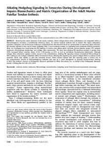
Hedgehog ligand biogenesis and release Among the three mammalian Hh ligands, Sonic hedgehog (Shh), Indian hedgehog (Ihh), and Desert hedgehog (Dhh), Shh exhibits the widest tissue expression and is also the best characterized. Hh proteins elicit dose-dependent responses and, in several tissues, fulfill the definition of a classical morphogen, for instance, in the developing neural tube and the limb. Thus, elucidating the molecular basis of Hh protein distribution and action will reveal a fundamental mechanism of tissue patterning (see Fig. 1). The Hh ligand undergoes proteolytic cleavage to produce an N-terminal signaling fragment with dual lipid modification.8,9 The mature form has a cholesterol adduct at its N-terminus and a palmitoyl moiety at its C-terminus. Cholesterol modification is mediated by the C-terminal fragment of the Hh protein while Skinny hedgehog (Skn/Hhat), an acyltransferase, catalyzes the addition of a palmitoyl group.10 Lipid modification enhances local Hh protein levels and activity but also restricts its long-range distribution.11 The lipidated Hh ligand is released from Hh-producing cells, a process that requires the transmembrane protein Dispatched (Disp), and the secreted protein SCUBE2, and is subsequently delivered to Hh-responding cells. Several models have been proposed for Hh distribution, including a soluble Hh protein complex,12 a lipoprotein particle,13 exosomes (membrane vesicles), and actin-based specialized filopodia (cytonemes).14 An important task in this active area of research is to uncover the in vivo relevance of the proposed modes of Hh distribution. This could be challenging because many of these processes involve machinery that also functions in other processes and the machinery that executes each mode of transport has not been fully characterized. Moreover, whether Hh-producing cells utilize a unique method or a combination of mechanisms to distribute its Hh ligand and whether different tissues preferentially rely on a particular mechanism require further studies. Finally, it is unclear if these are universal mechanisms in distributing other signaling molecules.
Hedgehog signal transduction Hh signaling is initiated through binding of the Hh ligand to its receptor, Patched 1 (Ptch1), a 12-pass transmembrane protein with topological similarity to transporters (see Fig. 2). This process is facilitated by several accessory proteins, including Boc (bioregional Cdon-binding protein),
E. Yao, P.-T. Chuang Cdon (cell-adhesion-molecule-related), and Gas1 (growth arrest specific 1).15e19 Distinct complexes that are comprised of Ptch1 and one of its coreceptors have been reported, indicating the overlapping and distinct functions of these accessory proteins.20,21 However, the molecular basis of how Hh coreceptors enhance Hh signaling remains unknown. Hh binding to Ptch1 relieves Ptch1 repression of the seven-pass transmembrane protein, Smoothened (Smo), and triggers a cascade of signaling events downstream from Smo.22e24 Lipidated Hh protein has increased signaling activity and it is unclear how lipid modification confers such an important property. Similarly, despite extensive studies, the molecular mechanisms by which Ptch1 regulates Smo activity remain an enigma. Because Ptch1 does not directly interact with Smo,25 it is generally believed that a small molecule (or molecules) regulated by Ptch1 mediates the interaction between Ptch and Smo. The identity of the mediator between Ptch and Smo remains elusive. Such molecules may not be amenable to genetic manipulations and their discovery would be likely to rely on cell-based assays and proteomic approaches. Interestingly, oxysterols bind Smo and promote Smo activity,22e24,26 adding another layer of regulation to Hh signaling and strengthening a link between Hh signaling and lipid metabolism. The purported small molecule mediator of Ptch can possibly influence oxysterols in addition to inhibiting Smo. As discussed below, mutations in Ptch1 or Smo underlie several human tumors, and a mechanistic understanding of how Hh signal is transduced via Ptch/co-receptor/Smo will have a major impact on our fundamental understanding of tumor development and treatment.
Control of Hh target gene expression Three transcription factors, Gli1e3, mediate Hh target gene expression in the nucleus.27,28 Gli329 and to some extent Gli230,31 undergo limited proteolysis to generate a transcriptional repressor that inhibits Hh target gene expression in the absence of Hh signaling.32 Hh signaling not only inhibits Gli repressor formation but also promotes the production of Gli activators (derived from full-length Gli proteins) to activate nuclear Hh targets. Unlike Gli2/3, Gli1 does not undergo limited proteolysis and only functions as an activator. Gli1 transcription is induced by Hh signaling and serves to amplify the Hh signal. Hh target gene expression is controlled by varying levels and combinations of Gli activators and repressors in a given tissue, thus generating Hh outputs appropriate for tissue development.29,33e36 The overlapping function of Gli proteins and the variable expression and requirements of individual Gli proteins in diverse tissues render predictions of Hh responses and phenotypes a challenging task.35,37 An important question is to identify and characterize Gli targets in diverse tissues in order to further understand how Hh signaling controls various aspects of tissue development and homeostasis.27,38,39 In this regard, significant progress has been made in the neural tube and the limb. A combination of chromatin immunoprecipitation sequencing (ChIPSeq), transgenesis and modeling has yielded a network of signaling that serves as a blueprint for further
Hedgehog signaling in development and disease
571
Hh-producing cell Nucleus
Hh transcription Endoplasmic reticulum Autoproteolysis
Hh
Hhat
Hh
Hh
b
Disp
a
Scube2
c
d
Hh
Hh
Hh Hh Soluble multimer
Hh Hh Lipoprotein
Cytoneme
Exovesicle
Hh-receiving cell
Figure 1 Production and secretion of Hedgehog (Hh) protein. The Hh protein is synthesized as an w45kDa precursor, which is then transported to the endoplasmic reticulum for processing. The Hh protein undergoes autocatalytic cleavage, facilitating the addition of a cholesterol moiety to its C-terminus. Hh acyltransferase (Hhat) catalyzes the addition of palmitate to the N-terminus. The processed Hh protein is released from the Hh-producing cell through the action of the 12-pass transmembrane protein, Dispatched (Disp) in combination with the soluble protein, Scube2. The Hh protein is delivered to Hh-responsive cells by one of four possible mechanisms: (a) Hh is transported to the Hh-receiving cell via the cytoneme; (b) the Hh protein is released from the plasma membrane as a soluble multimer; (c) Hh proteins assemble with lipophorin apolipoproteins to form lipoprotein particles; (d) Hh proteins are released and delivered through exovesicles.
refinement.40e42 Extension of these studies to all three Gli proteins and to other tissues would enable a better description of how a combinatorial use of the three Gli proteins can produce graded Hh responses. A key negative regulator in Hh signaling downstream from Smo is Suppressor of Fused (Sufu).43,44 Sufu controls the protein levels, activity and distribution of the three Gli
transcription factors and Sufu/Gli constitute a major regulatory hub in Hh signaling downstream from Smo. Sufu can sequester Gli proteins,45e48 regulate Gli protein levels,49e51 promote the production of Gli repressor and control Gli protein activity.52,53 This perhaps endows Hh-responsive cells with the ability to produce complex responses and offers focal points for integrating inputs. Loss of Sufu leads
572
E. Yao, P.-T. Chuang
Figure 2 Mammalian Hedgehog (Hh) signal transduction. (A) In the absence of Hh ligand, the 12-pass transmembrane protein, Patched (Ptch), inhibits the activity of the seven-pass transmembrane protein Smoothened (Smo). Suppressor of Fused (Sufu) sequesters Gli2/3 in the cytosol and perhaps also in other subcellular locales such as the cilium and the nucleus. Phosphorylation of Gli by the kinases, PKA, CK1 and GSK3b, promotes the formation of Gli repressor (GliR), a process that is also facilitated by Sufu. GliR enters the nucleus and inhibits the expression of target genes (e.g., Hip1, Gli1, and Ptch1). (B) Hh ligand binds to its receptor, Ptch, and its coreceptors (e.g., Boc, Cdon, and Gas1), relieving the inhibition of Smo by Ptch. Ptch is internalized and degraded, and Smo is enriched on the primary cilium. Smo is activated through phosphorylation of its C-terminal tail. This is associated recruitment of the Sufu-Gli2/3 complexes to the primary cilium and translocation of Kif7 to the cilium tip. Full-length Gli2/3 protein is processed into its activator form (GliA), which enters the nucleus and promotes the expression of target genes.
to upregulation of Hh signaling that is associated with reduced Gli protein levels. This is consistent with Sufu’s role as a major negative regulator of Hh signaling. Interestingly, maximal Hh signaling in Sufu mutants is not achieved,37,49 suggesting that Sufu also exerts positive effects on Hh signaling. This likely results from Sufu regulation of full-length Gli protein levels (and consequently Gli activators). In fact, simultaneous perturbation of Gli activators and repressors in other settings also increases Hh signaling but compromises maximal Hh signaling. Studies on Sufu regulation and function will continue to yield key insight into Hh pathway regulation.
Feedback control of Hh signaling In addition to signal amplification using a positive feedback loop, Hh signal transduction also employs negative feedback to modulate the Hh signal. Both Ptch1 and Hhip are Hhbinding proteins and are induced by Hh signaling. They can function to sequester the Hh ligand and alter the dynamics
of Hh signaling.54,55 Interestingly, Boc and Cdon do not participate in Ptch-mediated feedback regulation since their expression is downregulated by Hh signaling56 although Boc/Cdon facilitate Ptch-mediated Hh reception. Because both signaling strength and duration are important for Hhresponsive cells,57 feedback control of Hh signaling provides an important means to fine-tune signaling outputs. It is conceivable that feedback control could be utilized to regulate the levels or activity of other Hh pathway components. This would confer new properties to Hh signaling. Modeling would facilitate predictions of properties of Hh responses but these computational models need to be tested in an experimental setting both in vitro and in vivo. Hh pathway components exhibit transcriptional and post-transcriptional regulations. They are utilized in different subcellular locales and control different steps of Hh signaling. A key issue is to assess the contribution of each type of regulation to essential properties of Hh signaling such as signal amplification, signal output, and signal modulation.
Hedgehog signaling in development and disease
573
Evolutionary divergence of Hh signaling
Noncanonical Hh signaling
While the basic design of the Hh pathway is largely conserved among diverse species, evolutionary divergence has occurred, in particular from fly to mammal.58 Gene duplication has yielded three Hh ligands, two Ptch receptors, two Kif kinesin proteins, and three Gli transcription factors in mammals. This could have allowed paralogs to diverge in function and trigger corresponding changes in their interacting partners. Consistent with this, Sufu subsumes a central role in negatively regulating Gli activity while the Fu kinase is dispensable in mammals. This is in contrast to a central role of Fu in Drosophila Hh signaling and lack of overt phenotypes in Sufu mutant flies. The powerful technology of next-generation sequencing and genome editing (such as CRISPR/Cas) can be readily applied to diverse organisms. This would open up a new avenue to test experimentally how various Hh pathway components and their interactions emerged, evolved and were even replaced in different species during evolution. The origin of the Hh pathway is unknown. It has been speculated that it may have evolved from machinery that senses nutrients.59 This is based on the connection between lipid metabolism and Hh signaling and the function of homologs of Hh pathway components in organisms lacking active Hh signaling and thus predating the Hh pathway. Again, the ability to manipulate the genome with greater ease would allow testing of these hypotheses.
Interestingly, several Hh-dependent functions, such as chemotaxis and pathfinding, appear to be independent of the primary cilium and Gli-mediated transcription.81 Smo at different subcellular locales was proposed to account for eliciting distinct Hh outputs. Smo on the primary cilium relays the Hh signal to Gli proteins, culminating in transcriptional responses whereas Smo outside the cilium regulates chemotactic responses to Hh. Further studies will shed light on the adaptation of the primary cilium in Hh signaling.
The primary cilium and vertebrate Hedgehog signaling Another unique aspect of vertebrate Hh signaling is the utilization of the primary cilium for transducing the Hh signal.60e71 The primary cilium is a microtubule-based, evolutionarily conserved organelle. Construction of the cilium relies on intraflagellar transport powered by the anterograde kinesin and the retrograde dynein motor. Most vertebrate cells have a single primary cilium. Genetic analysis of mutants defective in ciliogenesis or ciliary function provides strong evidence to support an essential role of the cilium in Hh signal transduction. Consistent with this model, most core Hh components localize to the cilium in a dynamic manner.49,72e77 How primary cilia control various steps of Hh signal transduction is under intensive investigation. A current model suggests that Sufu is recruited to the cilium via Gli proteins. Sufu, Gli, and kinesin Kif7 are enriched at the cilium tip. Sufu-Gli association is proposed to be disrupted upon Hh stimulation perhaps at the cilium tip, allowing the production of Gli activators.52,53 Kif7 controls cilium architecture at the tip and this could affect Sufu-Gli activity.78 New tools are needed in order to establish the function of ciliary localization of Hh pathway components. The connection between Hh signaling and lipid metabolism has long fueled the speculation that changes in membrane lipid contents have an important impact on Hh signaling. Interestingly, the ciliary membrane seems to contain specialized lipid composition and may provide a conducive environment for Hh signal transduction.79,80
Hh signaling and congenital anomalies Studies in model organisms indicate that the Hh pathway controls key steps of the developmental processes in many tissues. It is expected that mutations in Hh pathway components will lead to human congenital anomalies. Indeed, mutations in Shh have been detected in human patients with holoprosencephaly while mutations in Ihh are associated with brachydactyly and Dhh mutations are found in gonadal dysgenesis. Disruption of the human Hh acyltransferase that catalyzes the transfer of a palmitate moiety to Hh proteins results in 46, XY Disorder of Sex Development.82 As next-generation sequencing becomes more routinely used in clinical diagnosis, it is anticipated that additional cases of birth defects related to the Hh pathway will be discovered. Severe loss-of-function mutations in Hh pathway components are likely to cause serious developmental defects incompatible with life and the fetuses die in utero. In this case, whole genome sequencing would allow us to determine whether missense mutations produce milder or unexpected phenotypes in living patients, which are in general under-diagnosed. These studies will provide important information on phenotype-genotype correlations. They will not only assist clinical diagnosis but also inform us of the function of Hh signaling in human development. With induced pluripotent stem cell and genome editing technology, mutations corresponding to human mutations can be readily created in cell lines and model organisms to facilitate phenotypic analysis and mechanistic studies. Mutations that disrupt ciliary function (ciliopathies) are associated with a wide range of human syndromes. A number of disease-causing genes have been identified in ciliopathy patients. Developmental abnormalities with hallmarks of Hh phenotypes in these patients are probably caused by defective Hh signaling. By contrast, it has been difficult to pinpoint the underlying signaling defects in other anomalies, particularly dysfunction of postnatal physiology. In many instances, it is unclear whether and how disruption of Hh signaling contributes to the phenotypic consequences.
Hh signaling and tissue regeneration and repair Hh signaling has been studied in many adult tissues in the context of progenitor cells and tissue regeneration and repair.4 In a number of tissues, active Hh signaling can be detected in a small population that possesses progenitor potential, and in many cases Hh signaling is activated upon
574 tissue injury. It is postulated that activation of the Hh pathway serves to stimulate cell proliferation essential for tissue repair. For instance, Gli-expressing cells contribute to the neural stem cell population located in the subventricular zone of the lateral ventricles and in the subgranular zone of the hippocampal dentate gyrus in the murine nervous system.83 A major challenge is to establish the functional role of Hh signaling in tissue regeneration in a given tissue. In many instances, it is difficult to unambiguously identify Hhproducing and -responsive cells in adult tissues in homeostasis and following injury. Tools are generally not available to eliminate these cell populations exclusively and efficiently in adults in order to evaluate the effects of perturbed Hh signaling. Moreover, Hh signaling probably interacts with other pathways in the process of tissue repair. Dissecting the signaling cascade and crosstalk and assessing the contribution of Hh signaling in a given tissue is a major effort.
Hh signaling and tumor development Perturbation of Hh signaling is associated with several human cancers, notably basal cell carcinoma (BCC) and medulloblastoma. Mutations in Ptch1 are found in most human BCCs, suggesting that enhanced Hh signaling probably causes BCC. While it is not possible to establish definitively a causal relationship between mutations in Hh components and human cancers, studies in mice and cell lines have provided strong evidence to support Hh pathway disruption as a major factor in developing human BCC and a subset of medulloblastomas. Several other types of human cancers such as digestive tract tumors have also been associated with perturbed Hh signaling.84,85 Even if mutations in Hh components are not required for tumor initiation, it remains possible that these tumors utilize the Hh pathway for growth or survival at certain stages of tumor development. Next-generation sequencing will continue to provide important information on Hh signaling and human cancers, in particular, in uncovering the spectrum of cancers that relies on Hh signaling. Strikingly, molecules that inhibit Smo activity have been shown to be effective in treating patients with invasive BCC or medulloblastoma related to Hh pathway overactivity.86,87 Smo appears to be a very druggable target in the Hh pathway and various Smo inhibitors (such as GDC-0449) have been developed. This illustrates the feasibility of targeting the Hh pathway for treating diseases caused by aberrant Hh signaling. Patients who received GDC-0449 (Vismodegib, Genentech Inc., South San Francisco, CA, USA) treatment initially responded but eventually developed resistance. Genomic analysis, in some cases, revealed mutations in Smo that conferred resistance to GDC-0449.88 This problem is common in targeted therapies and effective approaches that overcome resistance will serve as a paradigm for understanding drug resistance in targeted therapies. Apart from its immediate side effects, Smo antagonists can inhibit bone growth in animal studies89 presumably related to Hh function in tissue homeostasis and regeneration/repair. This raises the important question of how long-term use of Hh inhibitors can disturb normal physiology. Molecules that target other Hh pathway components such as Gli have also been reported.90 Efforts to develop
E. Yao, P.-T. Chuang drugs against other Hh players are underway. In addition to offering tools to probe the mechanisms of Hh signaling, they could provide potential therapies for Hh-related tumors or diseases. As discussed above, Hh signaling may be utilized at one stage of tumor development for their growth in tumor types other than the well-characterized, Hh-related tumors. In this scenario, the blockade of Hh signaling in combination with chemotherapeutic drugs would still confer therapeutic benefits. Given tissue complexity, tumors derived from different germ layers seem to respond differently to small molecules that perturb Hh signaling. The aforementioned Smo antagonists reduce the growth of basal cell carcinoma and medulloblastoma, indicating a critical role of Hh signaling in promoting tumor growth. By contrast, Hh agonists cause stromal hyperplasia in the pancreas and subsequently inhibit epithelial growth.91 Moreover, genetic ablation of Hh responses in murine models can accelerate tumor development in the pancreas or bladder.91,92 This suggests that Hh pathway activation could slow tumor development in these cases. These seemingly paradoxical outcomes stem from complex tissue interactions. Nevertheless, they can be understood on the basis of the proliferative effect of Hh signaling and activation of other signaling pathways in a particular tissue.
Acknowledgments We thank Chuwen Lin and Audrey Liu for critical reading of the manuscript. In this cursory review of the Hh pathway, the selection of topics is somewhat arbitrary due to space constraints. We sincerely apologize to those whose work was not discussed or cited. This work was supported by grants from the National Institutes of Health (U01 HL111054) to P.-T.C.
References 1. Chen Y, Jiang J. Decoding the phosphorylation code in Hedgehog signal transduction. Cell Res 2013;23:186e200. 2. Ingham PW, Nakano Y, Seger C. Mechanisms and functions of Hedgehog signalling across the metazoa. Nat Rev Genet 2011; 12:393e406. 3. Briscoe J, The ´rond PP. The mechanisms of Hedgehog signalling and its roles in development and disease. Nat Rev Mol Cell Biol 2013;14:416e29. 4. Petrova R, Joyner AL. Roles for Hedgehog signaling in adult organ homeostasis and repair. Development 2014;141: 3445e57. 5. Scales SJ, de Sauvage FJ. Mechanisms of Hedgehog pathway activation in cancer and implications for therapy. Trends Pharmacol Sci 2009;30:303e12. 6. Barakat MT, Humke EW, Scott MP. Learning from Jekyll to control Hyde: hedgehog signaling in development and cancer. Trends Mol Med 2010;16:337e48. 7. Bijlsma MF, Roelink H. Non-cell-autonomous signaling by Shh in tumors: challenges and opportunities for therapeutic targets. Expert Opin Ther Targets 2010;14:693e702. 8. Porter JA, Young KE, Beachy PA. Cholesterol modification of hedgehog signaling proteins in animal development. Science 1996;274:255e9.
Hedgehog signaling in development and disease 9. Pepinsky RB, Zeng C, Wen D, Rayhorn P, Baker DP, Williams KP, et al. Identification of a palmitic acid-modified form of human Sonic hedgehog. J Biol Chem 1998;273:14037e45. 10. Buglino JA, Resh MD. Palmitoylation of Hedgehog proteins. Vitam Horm 2012;88:229e52. 11. Li Y, Zhang H, Litingtung Y, Chiang C. Cholesterol modification restricts the spread of Shh gradient in the limb bud. Proc Natl Acad Sci U S A 2006;103:6548e53. 12. Zeng X, Goetz JA, Suber LM, Scott Jr WJ, Schreiner CM, Robbins DJ. A freely diffusible form of sonic hedgehog mediates long-range signalling. Nature 2001;411:716e20. 13. Pana ´kova ´ D, Sprong H, Marois E, Thiele C, Eaton S. Lipoprotein particles are required for Hedgehog and Wingless signalling. Nature 2005;435:58e65. 14. Ramı´rez-Weber FA, Kornberg TB. Cytonemes: cellular processes that project to the principal signaling center in Drosophila imaginal discs. Cell 1999;97:599e607. 15. Wang Y, McMahon AP, Allen BL. Shifting paradigms in Hedgehog signaling. Curr Opin Cell Biol 2007;19:159e65. 16. Ryan KE, Chiang C. Hedgehog secretion and signal transduction in vertebrates. J Biol Chem 2012;287:17905e13. 17. Farzan SF, Singh S, Schilling NS, Robbins DJ. The adventures of sonic hedgehog in development and repair. III. Hedgehog processing and biological activity. Am J Physiol Gastrointest Liver Physiol 2008;294:G844e9. 18. Beachy PA, Hymowitz SG, Lazarus RA, Leahy DJ, Siebold C. Interactions between Hedgehog proteins and their binding partners come into view. Genes Dev 2010;24:2001e12. 19. Filmus J, Capurro M. The role of glypicans in Hedgehog signaling. Matrix Biol 2014;35:248e52. 20. Izzi L, Le ´vesque M, Morin S, Laniel D, Wilkes BC, Mille F, et al. Boc and Gas1 each form distinct Shh receptor complexes with Ptch1 and are required for Shh-mediated cell proliferation. Dev Cell 2011;20:788e801. 21. Allen BL, Song JY, Izzi L, Althaus IW, Kang JS, Charron F, et al. Overlapping roles and collective requirement for the coreceptors GAS1, CDO, and BOC in SHH pathway function. Dev Cell 2011;20:775e87. 22. Myers BR, Sever N, Chong YC, Kim J, Belani JD, Rychnovsky S, et al. Hedgehog pathway modulation by multiple lipid binding sites on the smoothened effector of signal response. Dev Cell 2013;26:346e57. 23. Nachtergaele S, Whalen DM, Mydock LK, Zhao Z, Malinauskas T, Krishnan K, et al. Structure and function of the Smoothened extracellular domain in vertebrate Hedgehog signaling. Elife 2013;2:e01340. 24. Nedelcu D, Liu J, Xu Y, Jao C, Salic A. Oxysterol binding to the extracellular domain of Smoothened in Hedgehog signaling. Nat Chem Biol 2013;9:557e64. 25. Taipale J, Cooper MK, Maiti T, Beachy PA. Patched acts catalytically to suppress the activity of Smoothened. Nature 2002;418:892e7. 26. Corcoran RB, Scott MP. Oxysterols stimulate Sonic hedgehog signal transduction and proliferation of medulloblastoma cells. Proc Natl Acad Sci U S A 2006;103:8408e13. 27. Hui CC, Angers S. Gli proteins in development and disease. Annu Rev Cell Dev Biol 2011;27:513e37. 28. Aberger F, Ruiz I, Atalba A. Context-dependent signal integration by the GLI code: the oncogenic load, pathways, modifiers and implications for cancer therapy. Semin Cell Dev Biol 2014;33:93e104. 29. Bai CB, Stephen D, Joyner AL. All mouse ventral spinal cord patterning by hedgehog is Gli dependent and involves an activator function of Gli3. Dev Cell 2004;6:103e15. 30. Pan Y, Bai CB, Joyner AL, Wang B. Sonic hedgehog signaling regulates Gli2 transcriptional activity by suppressing its processing and degradation. Mol Cell Biol 2006;26: 3365e77.
575 31. Li J, Wang C, Pan Y, Bai Z, Wang B. Increased proteolytic processing of full-length Gli2 transcription factor reduces the hedgehog pathway activity in vivo. Dev Dyn 2011;240: 766e74. 32. Wang B, Fallon JF, Beachy PA. Hedgehog-regulated processing of Gli3 produces an anterior/posterior repressor gradient in the developing vertebrate limb. Cell 2000;100:423e34. 33. Ding Q, Motoyama J, Gasca S, Mo R, Sasaki H, Rossant J, et al. Diminished Sonic hedgehog signaling and lack of floor plate differentiation in Gli2 mutant mice. Development 1998;125: 2533e43. 34. Matise MP, Epstein DJ, Park HL, Platt KA, Joyner AL. Gli2 is required for induction of floor plate and adjacent cells, but not most ventral neurons in the mouse central nervous system. Development 1998;125:2759e70. 35. Bowers M, Eng L, Lao Z, Turnbull RK, Bao X, Riedel E, et al. Limb anterior-posterior polarity integrates activator and repressor functions of GLI2 as well as GLI3. Dev Biol 2012;370: 110e24. 36. Cao T, Wang C, Yang M, Wu C, Wang B. Mouse limbs expressing only the Gli3 repressor resemble those of sonic hedgehog mutants. Dev Biol 2013;379:221e8. 37. Liu J, Heydeck W, Zeng H, Liu A. Dual function of suppressor of fused in Hh pathway activation and mouse spinal cord patterning. Dev Biol 2012;362:141e53. 38. Rabinowitz AH, Vokes SA. Integration of the transcriptional networks regulating limb morphogenesis. Dev Biol 2012;368: 165e80. 39. Falkenstein KN, Vokes SA. Transcriptional regulation of graded Hedgehog signaling. Semin Cell Dev Biol 2014;33: 73e80. 40. Vokes SA, Ji H, McCuine S, Tenzen T, Giles S, Zhong S, et al. Genomic characterization of Gli-activator targets in sonic hedgehog-mediated neural patterning. Development 2007; 134:1977e89. 41. Vokes SA, Ji H, Wong WH, McMahon AP. A genome-scale analysis of the cis-regulatory circuitry underlying Sonic hedgehog-mediated patterning of the mammalian limb. Genes Dev 2008;22:2651e63. 42. Balaskas N, Ribeiro A, Panovska J, Dessaud E, Sasai N, Page KM, et al. Gene regulatory logic for reading the Sonic Hedgehog signaling gradient in the vertebrate neural tube. Cell 2012;148:273e84. 43. Cooper AF, Yu KP, Brueckner M, Brailey LL, Johnson L, McGrath JM, et al. Cardiac and CNS defects in a mouse with targeted disruption of suppressor of fused. Development 2005;132:4407e17. 44. Sva ¨rd J, Heby-Henricson K, Persson-Lek M, Rozell B, Lauth M, et al. Genetic elimination of suppressor of fused reveals an essential repressor function in the mammalian Hedgehog signaling pathway. Dev Cell 2006;10:187e97. 45. Ding Q, Fukami Si, Meng X, Nishizaki Y, Zhang X, Sasaki H, et al. Mouse suppressor of fused is a negative regulator of sonic hedgehog signaling and alters the subcellular distribution of Gli1. Curr Biol 1999;9:1119e22. 46. Kogerman P, Grimm T, Kogerman L, Krause D, Unde ´n AB, Sandstedt B, et al. Mammalian suppressor-of-fused modulates nuclear-cytoplasmic shuttling of Gli-1. Nat Cell Biol 1999;1: 312e9. 47. Murone M, Luoh SM, Stone D, Li W, Gurney A, Armanini M, et al. Gli regulation by the opposing activities of fused and suppressor of fused. Nat Cell Biol 2000;2:310e2. 48. Barnfield PC, Zhang X, Thanabalasingham V, Yoshida M, Hui CC. Negative regulation of Gli1 and Gli2 activator function by Suppressor of fused through multiple mechanisms. Differentiation 2005;73:397e405. 49. Chen MH, Wilson CW, Li YJ, Law KK, Lu CS, Gacayan R, et al. Cilium-independent regulation of Gli protein function by Sufu
576
50.
51.
52.
53.
54.
55.
56.
57.
58.
59.
60.
61. 62.
63. 64. 65. 66.
67. 68. 69.
70.
71. 72.
E. Yao, P.-T. Chuang in Hedgehog signaling is evolutionarily conserved. Genes Dev 2009;23:1910e28. Jia J, Kolterud A, Zeng H, Hoover A, Teglund S, Toftga ˚rd R, et al. Suppressor of Fused inhibits mammalian Hedgehog signaling in the absence of cilia. Dev Biol 2009;330:452e60. Wang C, Pan Y, Wang B. Suppressor of fused and Spop regulate the stability, processing and function of Gli2 and Gli3 fulllength activators but not their repressors. Development 2010;137:2001e9. Tukachinsky H, Lopez LV, Salic A. A mechanism for vertebrate Hedgehog signaling: recruitment to cilia and dissociation of SuFu-Gli protein complexes. J Cell Biol 2010;191:415e28. Humke EW, Dorn KV, Milenkovic L, Scott MP, Rohatgi R. The output of Hedgehog signaling is controlled by the dynamic association between Suppressor of Fused and the Gli proteins. Genes Dev 2010;24:670e82. Goodrich LV, Johnson RL, Milenkovic L, McMahon JA, Scott MP. Conservation of the hedgehog/patched signaling pathway from flies to mice: induction of a mouse patched gene by Hedgehog. Genes Dev 1996;10:301e12. Chuang PT, McMahon AP. Vertebrate Hedgehog signalling modulated by induction of a Hedgehog-binding protein. Nature 1999;397:617e21. Tenzen T, Allen BL, Cole F, Kang JS, Krauss RS, McMahon AP. The cell surface membrane proteins Cdo and Boc are components and targets of the Hedgehog signaling pathway and feedback network in mice. Dev Cell 2006;10:647e56. Harfe BD, Scherz PJ, Nissim S, Tian H, McMahon AP, Tabin CJ. Evidence for an expansion-based temporal Shh gradient in specifying vertebrate digit identities. Cell 2004;118:517e28. Wilson CW, Chuang PT. Mechanism and evolution of cytosolic Hedgehog signal transduction. Development 2010;137: 2079e94. Hausmann G, von Mering C, Basler K. The hedgehog signaling pathway: where did it come from? PLoS Biol 2009;7: e1000146. Nozawa YI, Lin C, Chuang PT. Hedgehog signaling from the primary cilium to the nucleus: an emerging picture of ciliary localization, trafficking and transduction. Curr Opin Genet Dev 2013;23:429e37. Kim S, Dynlacht BD. Assembling a primary cilium. Curr Opin Cell Biol 2013;25:506e11. Goetz SC, Anderson KV. The primary cilium: a signalling centre during vertebrate development. Nat Rev Genet 2010; 11:331e44. Drummond IA. Cilia functions in development. Curr Opin Cell Biol 2012;24:24e30. Eggenschwiler JT, Anderson KV. Cilia and developmental signaling. Annu Rev Cell Dev Biol 2007;23:345e73. Bay SN, Caspary T. What are those cilia doing in the neural tube? Cilia 2012;1:19. Berbari NF, O’Connor AK, Haycraft CJ, Yoder BK. The primary cilium as a complex signaling center. Curr Biol 2009;19: R526e35. DeRouen MC, Oro AE. The primary cilium: a small yet mighty organelle. J Invest Dermatol 2009;129:264e5. Oh EC, Katsanis N. Cilia in vertebrate development and disease. Development 2012;139:443e8. Wong SY, Reiter JF. The primary cilium at the crossroads of mammalian hedgehog signaling. Curr Top Dev Biol 2008;85: 225e60. Mukhopadhyay S, Rohatgi R. G-protein-coupled receptors, Hedgehog signaling and primary cilia. Semin Cell Dev Biol 2014;33:63e72. Nachury MV. How do cilia organize signalling cascades? Philos Trans R Soc Lond B Biol Sci 2014;369:20130465. Haycraft CJ, Banizs B, Aydin-Son Y, Zhang Q, Michaud EJ, Yoder BK. Gli2 and Gli3 localize to cilia and require the
73.
74. 75.
76.
77.
78.
79. 80. 81.
82.
83.
84.
85.
86.
87.
88.
89.
90.
91.
92.
intraflagellar transport protein polaris for processing and function. PLoS Genet 2005;1:e53. Corbit KC, Aanstad P, Singla V, Norman AR, Stainier DY, Reiter JF. Vertebrate Smoothened functions at the primary cilium. Nature 2005;437:1018e21. Rohatgi R, Milenkovic L, Scott MP. Patched1 regulates hedgehog signaling at the primary cilium. Science 2007;317:372e6. Liem Jr KF, He M, Ocbina PJ, Anderson KV. Mouse Kif7/Costal2 is a cilia-associated protein that regulates Sonic hedgehog signaling. Proc Natl Acad Sci U S A 2009;106:13377e82. Endoh-Yamagami S, Evangelista M, Wilson D, Wen X, Theunissen JW, Phamluong K, et al. The mammalian Cos2 homolog Kif7 plays an essential role in modulating Hh signal transduction during development. Curr Biol 2009;19:1320e6. Kim J, Kato M, Beachy PA. Gli2 trafficking links Hedgehogdependent activation of Smoothened in the primary cilium to transcriptional activation in the nucleus. Proc Natl Acad Sci U S A 2009;106:21666e71. He M, Subramanian R, Bangs F, Omelchenko T, Liem Jr KF, Kapoor TM, et al. The kinesin-4 protein Kif7 regulates mammalian Hedgehog signalling by organizing the cilium tip compartment. Nat Cell Biol 2014;16:663e72. Kuzhandaivel A, Schultz SW, Alkhori L, Alenius M. Cilia-mediated hedgehog signaling in Drosophila. Cell Rep 2014;7:672e80. Kornberg TB. The contrasting roles of primary cilia and cytonemes in Hh signaling. Dev Biol 2014;394:1e5. Bijlsma MF, Damhofer H, Roelink H. Hedgehog-stimulated chemotaxis is mediated by smoothened located outside the primary cilium. Sci Signal 2012;5:ra60. Callier P, Calvel P, Matevossian A, Makrythanasis P, Bernard P, Kurosaka H, et al. Loss of function mutation in the palmitoyltransferase HHAT leads to syndromic 46,XY disorder of sex development by impeding Hedgehog protein palmitoylation and signaling. PLoS Genet 2014;10:e1004340. Ahn S, Joyner AL. In vivo analysis of quiescent adult neural stem cells responding to Sonic hedgehog. Nature 2005;437: 894e7. Berman DM, Karhadkar SS, Maitra A, Montes De Oca R, Gerstenblith MR, Briggs K, et al. Widespread requirement for Hedgehog ligand stimulation in growth of digestive tract tumours. Nature 2003;425:846e51. Thayer SP, di Magliano MP, Heiser PW, Nielsen CM, Roberts DJ, Lauwers GY, et al. Hedgehog is an early and late mediator of pancreatic cancer tumorigenesis. Nature 2003;425:851e6. Von Hoff DD, LoRusso PM, Rudin CM, Reddy JC, Yauch RL, Tibes R, et al. Inhibition of the hedgehog pathway in advanced basal-cell carcinoma. New Engl J Med 2009;361:1164e72. Rudin CM, Hann CL, Laterra J, Yauch RL, Callahan CA, Fu L, et al. Treatment of medulloblastoma with hedgehog pathway inhibitor GDC-0449. New Engl J Med 2009;361:1173e8. Yauch RL, Dijkgraaf GJ, Alicke B, Januario T, Ahn CP, Holcomb T, et al. Smoothened mutation confers resistance to a Hedgehog pathway inhibitor in medulloblastoma. Science 2009;326:572e4. Kimura H, Ng JM, Curran T. Transient inhibition of the Hedgehog pathway in young mice causes permanent defects in bone structure. Cancer Cell 2008;13:249e60. Lauth M, Bergstro ¨m A, Shimokawa T, Toftga ˚rd R. Inhibition of GLI-mediated transcription and tumor cell growth by smallmolecule antagonists. Proc Natl Acad Sci U S A 2007;104: 8455e60. Lee JJ, Perera RM, Wang H, Wu DC, Liu XS, Han S, et al. Stromal response to Hedgehog signaling restrains pancreatic cancer progression. Proc Natl Acad Sci U S A 2014;111:E3091e100. Shin K, Lim A, Zhao C, Sahoo D, Pan Y, Spiekerkoetter E, et al. Hedgehog signaling restrains bladder cancer progression by eliciting stromal production of urothelial differentiation factors. Cancer Cell 2014;26:521e33.