Page 1 of 18 in PresS. Am J Physiol Gastrointest Liver Physiol (December 6, 2007). doi:10.1152/ajpgi.00457.2007 Articles
Parkin & Ingham
1
Hedgehog Signalling in Gastrointestinal Development and Disease
Caroline A. Parkin and Philip W. Ingham1
Institute of Molecular and Cell Biology 61 Biopolis Drive Proteos Singapore 138673
(1) Corresponding author Philip Ingham, e-mail:
[email protected]
Copyright © 2007 by the American Physiological Society.
Page 2 of 18
Parkin & Ingham
2
Abstract Hedgehog (Hh) proteins are members of a family of secreted signalling factors that orchestrate the development of many organs and tissues including those of the gastrointestinal (GI) tract. The requirement for Hh activity is not limited to early development but underlies the homeostsasis of a number of tissues and abnormal activity of the Hh pathway is associated with several GI malignancies. Understanding the roles and mechanisms of action of Hh signalling both in development and post-natally should thus give novel insights into potential treatments for these diseases. Here we focus on the Hh signalling pathway and its role in GI tract development and maintenance, and consider the diseases resulting from aberrant Hh activity.
Page 3 of 18
Parkin & Ingham
3
A Brief Introduction to the Hh Pathway Originally identified in Drosophila through its role in controlling the pattern and polarity of the larval body segments, homologues of the Hh gene have been identified in a wide variety of metozoa ranging from sponges to human. The latter, like all vertebrates, possess multiple Hh genes; two of these, designated Dhh (Desert hh) and Ihh (Indian hh) are named after species of the spiny mammal, whilst the third, Shh (Sonic hedgehog) is named after a computer game. All known Hh family genes encode pro-proteins that undergo autoproteolytic cleavage to yield a lipid modified N-terminal secreted polypeptide in which all the signalling activity resides. The transcription of Hh genes is under complex spatio-temporal control, reflecting their involvement in multiple processes both during embryonic development and post embryonically. The genes encoding the core components of the Hh signal transduction pathway depicted in Figure 1 were also first characterised in Drosophila and, like the Hh genes themselves, have been highly conserved through evolution. Reception of the Hh signal is mediated by Patched (Ptc: Ptch1 in vertebrates), a multipass transmembrane protein that belongs to the RND family of transporter proteins. Unusually for a receptor, binding of Hh to the extracellular loops of the Ptc protein results in its inactivation, releasing a second transmembrane protein, Smoothened (Smo) from Ptc mediated inhibition. The transcriptional response to Hh signalling is mediated by members of the Gli family of zinc finger transcription factors. In Drosophila, a single Gli protein, known as Cubitus interruptus (ci), with both activator and repressor activities is responsible for the vast majority of Hh transcriptional responses, whereas in vertebrates, this function is distributed between three Gli proteins, Gli1, Gli2 and Gli3. Activation of Smo in response to Hh leads to a shift in the relative levels of the opposing Gli activities resulting in the activation of Hh target genes. Exactly how Smo modulates the activity of the Gli proteins is still not fully understood. Despite its serpentine structure and homology to members of the GPCR superfamily, there is little evidence that Smo acts via heterotrimeric Gproteins. In Drosophila, Smo has been shown to interact physically with an intracellular protein complex that modulates the activity of the Ci transcription factor. Although at least some of the components of this complex have orthologues in vertebrates, the regions of Smo critical for this interaction are not well conserved. Moreover, the function of some of the intracellular components appears to have diverged during evolution: the Suppressor of fused (Sufu for short) protein, for instance, is essentially dispensable in Drosophila but is essential in mammals for suppression of Hh pathway activity. Other differences between Drosophila and vertebrates have emerged with the identification of vertebrate specific components of the pathway. The Hh interacting protein (Hhip), for
Page 4 of 18
Parkin & Ingham
4
example is a vertebrate specific transmembrane protein that regulates Hh signalling by binding and sequestering the ligand, while members of the Intraflagellar Transport (IFT) family of proteins are essential for Hh signalling in mice but not in flies. More detailed accounts of the Hh signalling pathway and its deployment in animal development can be found in a number of recent reviews (6; 7) Hh Signalling in the GI Tract The GI tract can be divided into three domains, the foregut, midgut, and hindgut, each of which develops specialised functions or accessory organs. The patterning of the gut along its A-P axis depends on various, complex interactions between the endoderm and the surrounding mesoderm. Signalling between the different germ layers via the Hh, Bone Morphogenetic Protein (BMP), Wnt/Beta catenin, and Notch pathways - along with positional cues generated by the differential expression of the Hox genes - controls much of the patterning and specification events during early gut development. In the mouse embryo, Shh and Ihh are expressed in distinct though overlapping regions of the developing endoderm whilst the Hh receptor and effectors, Ptc1 and Gli1-3, respectively, are upregulated in the surrounding mesoderm. The distinct domains of expression of Ihh and Shh are reflected in the differing requirements for each during normal development of the GI tract; Analysis of the phenotypes of mice mutant for either gene, combined with studies in chick, fish and frog, have revealed that both genes are required for the radial patterning of the gut and for specifying various regional identities along the GI tract. Additionally Shh is necessary for the correct development of the hindgut and Ihh is implicated in the maintenance of stem cell proliferation in the small intestine. The different domains of expression of Ihh and Shh may generate varying concentration gradients in the gut in line with Hh proteins acting as concentration dependent morphogens. Hedgehog Function in Fore and Hindgut Development Animal mutant for Hh pathway genes have greatly advanced our understanding of the roles of Hh in the normal development of the gut. GI phenotypes that resemble a number of human congenital abnormalities have been observed. The aetiology of foregut and hindgut malformations that cause fistulas and atresia are poorly understood, but it is now believed that aberrant Hh activity could be a major cause, an important revelation, since these gastrointestinal malformations are associated with several congenital syndromes, including Pallister-Hall, Feingold and Currarino syndromes, and the fairly common VACTERL association (vertebral,
Page 5 of 18
Parkin & Ingham
5
anal, cardiac, tracheosophageal fistula, radial, renal and limb abnormalities). Many aspects of the VACTERL association, including oesophageal fistula/tracheo-oesophageal fistulas and anorectal malformations of varying degrees of severity, such as stenosis, imperforate anus (either due to fistula or atresia) and persistent cloaca are manifest in Shh -/-, Gli2 -/- and Gli3 -/- mutant mice. Partitioning of the cloaca into the gastrointestinal, urinary and genital openings is still poorly understood, but the presence of various cloacal defects in Hh mutant mice, together with strong expression of Hh signals and receptors in the hindgut endoderm and mesoderm suggests that Hh signalling is at least partially responsible for the normal patterning of the hindgut (16). How Hh may regulate cloacal partitioning is not known, although examination of Shh and Gli mutants in heterozygous and homozygous combinations suggests a dose dependent effect, as the severity of malformation increases with decreasing levels of Hh activity (16). Despite strong expression of Shh in the hindgut endoderm, the chick cloaca, by contrast to its mammalian and teleost counterparts, does not partition during normal development. Notably, Gli2 is only weakly expressed in the surrounding mesoderm, in contrast to the situation in the mouse and since Gli2 -/- mice exhibit a persistent cloaca, reminiscent of the chick, it was speculated that Gli2 is required for the partitioning. Expression of a constitutively active Gli2, however, fails to cause cloacal separation in the chick (12). Although the majority of cases of anorectal malformations appear to be sporadic, suggesting they are caused by epigenetic modulation of Hh signalling during development, at least two Mendelain syndromes that give rise to hindgut defects are associated with mutations in Hh pathway genes or its targets. Pallister-Hall syndrome, which includes imperforate anus, as well as hypothalamic haematomas, polydactyly and syndactyly is associated with a frame shift mutation in Gli3, Cterminal to the zinc finger domain, resulting in a truncated protein with an intact DNA binding domain. Mice with a similar mutation show Pallister-Hall-like defects, including the polydactyly and anorectal abnormalities (5) . Autosomally dominant Feingold syndrome results in a range of GI abnormalities including oesophageal, duodenal and anal atresia as well as annular pancreas in addition to microcephaly and syndactally. It is caused by heterozygosity for mutation of the N-MYC gene, a known downstream target of Shh, at least in the cerebellum where it regulates cell cycle progression (26). Interestingly, the autosomal dominant Currarino syndrome, which results in anorectal abnormalities as well as presarcal teratoma and sacral agenesis, was mapped to chromosome 7q36, close to Shh, suggesting it to be a good candidate for the mutated gene. Analysis of the Shh gene in several patients with Currarino syndrome, however, failed to identify any mutations in the open reading frame whilst another study found several mutations in the neighbouring homeobox gene, Hlxb9. The role of Hlxb9 in the aetiology of
Page 6 of 18
Parkin & Ingham
6
Currarino syndrome is not understood, and interpretation is further complicated by the failure of Hlxb9 knock out mice to show any malformation in the anus or sacral vertebrae although this may be partially explained by the different expression patterns of Hlxb9 in human and mouse (3). Several sporadic cases of Currarino syndrome have also been described, which have no mutations or microdeletions in Hlxb9. It thus remains possible that long-range regulatory elements of Shh are located in the Hlxb9 coding region, mutations of which result in Currarino syndrome. Another aspect of the VACTERL association is the presence of foregut malformations, which are also observed in Hh mutants. Similar to the hindgut, expression of Shh in the foregut endoderm is high, but is restricted to the prospective trachea and excluded from the putative oesophagus. Shortly following the split of the trachea and oesophagus, this pattern of expression is reversed, the oesophagus but not the trachea now exhibiting high levels of Shh transcription. Hh activity is essential for the patterning of the foregut endoderm; in mice mutant for Shh or Gli the trachea and oesophagus may not divide, resulting in a fistula between them. In zebrafish, Hh pathway mutants also exhibit foregut malformations and it may be that reduced Hh activity in the mutants inhibits the separation (11). The VACTERL-like abnormalities characteristic of Hh mutant mice suggests that alterations in Hh activity may be responsible for the malformations in human. Models of VACTERL, such as the Adriamycin rat, have focused on the possibility that abnormal expression of Shh in the notochord may be responsible for the GI phenotypes. Foregut Shh expression is reduced and eventually lost when rats are treated with Adriamycin, while expression of Shh in the notochord is maintained (2). The notochord is, however, abnormal in these rats and may remain attached to the foregut, or bifurcate producing ectopic notochord. Interestingly, expression of Shh in the duplicated notochord has been reported to be higher than in control rats (2). It has been speculated that the altered notochord in the treated rats is responsible for some of the VACTERL anomalies, including the anorectal malformations, either due to changes in Shh expression or unknown changes in factors normally secreted from the notochord. Notably, in contrast to the consistent anorectal malformations seen in foetal rats exposed to Adriamycin, no VACTERL defects were detectable in similarly treated chick embryos (17). The development of many VACTERL like abnormalities in Shh and Gli mutant mice suggests that a reduction in Hh pathway activity in the endoderm may underlie the phenotypes observed in both Adriamycin rats and human patients with VACTERL association. Consistent with this, zebrafish embryos mutant for shh or smo display anorectal malformations similar to those seen in the VACTERL association, whereas mutations that disrupt development of the notochord, show little if any effect on the development of the cloaca (Parkin and Ingham, in preparation).
Page 7 of 18
Parkin & Ingham
7
Radial and AP patterning of the GI tract Hh signalling is required for radial patterning of the GI tract, its activity specifying the ratio of endodermal lamina propria and submucosa to smooth muscle and enteric neurons (21; 23). In mice null for either Shh or Ihh, the circular smooth muscle along the small intestine is thinner compared to wildtype siblings; such mutants also have abnormal innervation of the intestine. In Shh -/- mice, the neurons differentiate abnormally and migrate into the villi, an effect that is mimicked in the chick by the application of cyclopamine, a small molecule antagonist of the Hh pathway that blocks Smo activity and therefore all signal transduction (21; 23). In the normal intestine, Shh expression in the epithelium inhibits proliferation of enteric neurons and differentiation of smooth muscle and instead promotes the differentiation of the lamina propria and submucosa in the most proximal tissues. Loss of Ihh and/or expression of dominant negative Gli3 in mice results in a different phenotype, whereby neurons are absent from areas of the small intestine and colon, resulting in a marked intestinal dilation. Non-dilated colon occurs in other areas where the neuronal number is not affected; the pattern of normal and abnormal innervation resulting in intestinal dilation is a phenotype that resembles Hirschsprung disease, although linkage to Ihh has not been established. Mice that ectopically express the hedgehog antagonist, Hhip, in the intestinal epithelium have expanded smooth muscle, although innervation is normal; crypt-villus patterning is also affected via a paracrine mechanism, whereby the surrounding mesenchyme is affected by loss of Hh signalling and in turn it signals back and stimulates increased epithelial proliferation, which was not detected in the Ihh or Shh null mice (13) Hedgehog signalling in the adult GI tract Notably, Hh genes and their products have also been found to be expressed in specific domains of the adult GI tract: Shh is expressed in the acid secreting fundic glands (primarily the parietal cells) of the human and mouse stomach as well as in the base of crypts of the small intestine and colon, but not the oesophagous or antrum of the stomach. In the glands, Shh expression correlates with fundic gland differentiation and its activity can stimulate expression of the acid pump, H+/ K+ATPase (22). Gastrin appears to be required for Shh expression in the parietal cells since gastrin null mice have much lower levels of Shh (27). Intriguingly the cleavage of Shh into its active form within the parietal cells is also stimulated by gastrin activity through the induction of pepsin A (27). Pepsin A expression is reduced in gastric tumours, concomitant with a near complete loss of processed Shh protein and in line with this, the gastric glands develop abnormally and are described as either hyperplastic or metaplastic in Shh null mice (21). On the other hand, however, Shh levels have been found to be elevated in human gastric adenomas and intestinal metaplasias suggesting that over activation of the Hh
Page 8 of 18
Parkin & Ingham
8
pathway may be involved in early gastric carcinomas. Ectopic Shh (and H+ /K+ATPase) expression is also detected in chronic gastritis when there are fundic gland metaplasias, indicating that the switch from squamous to gastric epithelial tissue is accompanied by induction of Shh expression (11). This process may occur during development if intestinal epithelium is the default type; during early development Hh expression is down regulated in the small intestine, whilst the putative stomach may require Shh expression to switch it to a gastric identity. Ihh transcripts and protein have been detected in surface absorptive enterocytes in the human colon and may also be expressed elsewhere in the gut. There have been significant problems with elucidating the spatial distribution of the Hh transcripts and proteins in the adult GI tract due to cross reactivity of proteins and overall low levels of mRNA expression. Therefore dissecting the distinct roles of each gene, which have so far been mostly determined by broader inhibition of the pathway and interpreted based on their supposed domains of expression, will require further analysis using new techniques (e.g. RNAi) to manipulate the individual components of the pathway. Hh Signalling in Pancreatic Development and Disease Although Ihh and Shh are widely expressed along the A-P axis of the developing gut, expression of both is notably excluded from the pancreatic anlage. Various studies have demonstrated that the exclusion of Shh expression from the prospective pancreatic endoderm is critical for normal pancreatic development. The pancreas originates as two domains; a ventral and dorsal bud which later fuse to form the definitive pancreas, itself composed of two main cell types: hormonesecreting endocrine cells and enzyme-secreting exocrine cells and the associated ducts. Development of the pancreas from the dorsal domain requires signals from the notochord; in the chick Activin betaB and FGF2 from the notochord specifically repress Shh transcription in the future dorsal domain, while expression is maintained elsewhere in the dorsal endoderm (1; 4). The importance of excluding Shh expression is demonstrated by the effects of its ectopic expression in the early pancreatic epithelium using the insulin promoting factor1 promoter (Pdx1; an early marker of the pancreatic primordium). Early expression of Shh leads to the loss of pancreatic marker genes (1; 4), while at later stages it results in a disruption of morphological development, including transdifferentiation of putative pancreatic tissue into duodenal mesenchyme (1; 4). Shh also inhibits differentiation and growth of insulin-producing beta cells in embryonic stem cell culture as well as in pancreatic explants, where epithelial branching is reduced; both effects can be rescued by application of cyclopamine (15; 20). Consistent with this suppressive effect of Hh signalling on pancreatic specification, mice homozygous for null alleles of Shh or Ihh have increased pancreatic mass and an annular pancreas phenotype that resembles the human congenital malformation, in
Page 9 of 18
Parkin & Ingham
9
which the pancreas encircles and may constrict the duodenum, sometimes resulting in complete obstruction of the intestines and the flow of food (4; 21). Mice lacking Hhip display a range of morphogenetic abnormalities of the pancreas; in severe cases the ventral and dorsal pancreas fuse to form a small mass of tissue, though this retains pancreatic character, consistent with an effect on proliferation, but not differentiation, of pancreatic endocrine cells (9). Additionally, ectopic pancreatic tissue can be found in the stomach and duodenum while the spleen is also reduced in size (9). Functional redundancy between Ptc1 and Hhip in limiting Hh diffusion is indicated by the increase in severity of pancreatic defects in Hhip mutants that also lack one Ptc1 allele (9). By contrast to the exclusion of Shh in the early pancreas, Ihh and Dhh, as well as Ptc1 and Smo, are expressed in adult exocrine cells in distinct puncta. Expression is also detected in the insulin-secreting cell line, INS-1 (25). Treatment of these cells with cyclopamine decreased insulin production as well as insulin promoter activity, whilst ectopic misexpression of Shh increased promoter activity and insulin secretion levels (25). These findings have led to speculation that Hh activity may be a factor in Type II diabetes mellitus. Interestingly, a number of congenital abnormalities seen in the children of diabetic mothers, such as caudal regression syndrome are also associated with altered Hh activity. Pancreatic Cancer The importance of precise regulation of Hh activity in the adult pancreas is highlighted by the finding that Shh, Ihh, Ptc1, Smo and Gli1 are overexpressed in exocrine derived pancreatic cancer tissues and cell lines. Examination of various pancreatic cancer cell lines showed that at least two Hh pathway components are aberrantly expressed in each line, while subsequent analysis suggested that high levels of Shh enhance the proliferation of pancreatic duct epithelial cells, possibly through the regulation of cyclin D1 and p21 transcription (18; 24). Shh also appears to protect duct cells from apoptosis by activation of phosphatidylinositol 3-kinase signalling and the stabilisation of Bcl-2 and Bcl-X(L) (18; 24) .Pancreatic adenocarcinoma (PDA) is one of the leading causes of cancer death in the United States and UK. PDA arises from exocrine duct cells, which go through morphologically distinct stages, beginning with characteristic pancreatic intraepithelial lesions (PanINs). Aberrant Hh activity was found to increase dramatically during the progression from PanINs to metastatic adenocarcinomas in human samples (18; 24). Forced expression of Shh in the mouse pancreas (using the Pdx1 promoter) leads to the development of abnormal tubular structures that resemble early PanINs, suggestive of an increase in neoplastic potential (18; 24). Misexpression of Shh in the pancreas is able to induce oncogenic mutations in KRas, which is mutated in 99% of human PDA, and in some of the PanIn-like
Page 10 of 18
Parkin & Ingham
10
pancreata Her2/neu overexpression, another PanIn genetic marker, is also found (18; 24). However, initial formation of the pancreas in the Pdx-Shh mice is affected and the mice have a shortened lifespan (less than a month), preventing long term analysis of the effects of Hh deregulation and cancer development. The creation of a mouse expressing a dominant active form of Gli2 only in the pancreatic epithelium obviates some of the problems that may be caused by epithelial-mesenchymal signalling by overexpression of the Hh ligands (19). Although these mice survive longer than the Pdx-Shh mice, 30% developed pancreatic tumours between 6-20 weeks post-natally; however, the tumours are different from those seen in Pdx-Shh mice. The tumours appear to be undifferentiated carcinomas and progression through PanIn lesions does not occur, which differs from the normal development of PDA; additionally, mutations in KRas are not induced in the pancreata. Introducing the oncogenic K-Ras mutation in combination with the dominantly active Gli2 results in extensive PanIn formation, rapid deterioration and increased mortality, but the tumours are still undifferentiated. A significant finding is that Shh and Ihh expression is greatly increased in the double mutant mice, which is not observed in the single mutants, perhaps indicating that the K-Ras mutation is able to induce Hh expression in early Human PanIn lesions. This is important, as increased activity of Hh in pancreatic cancers may not be due to mutations in downstream components of the Hh pathway, but rather in factors that act upstream of Hh to activate its expression, a hypothesis supported by the finding that oncogenic K-Ras activates Hh signalling by increasing Gli1 activity in pancreatic adenocarcinoma cells via the RAF/MEK/MAPK pathway (8). In a number of PDA cell lines examined, inhibition of Hh signal transduction by the application of cyclopamine is able to induce apoptosis and block proliferation and can also reduce tumour growth in xenograft mouse models (24). Hh neutralising antibodies or recombinant Hhip protein are also able to reduce pancreatic cancer cell growth (10; 10; 24). Loss of Hhip activity may be a contributing factor to pancreatic cancer; expression is reduced or absent in many cell lines due to hypermethylation of the Hip promoter (14). Identification of aberrant Hh activity in pancreatic secretions could provide an opportunity for the early detection of premalignant cells, a possibility of great clinical significance as early diagnosis of pancreatic cancers is currently very difficult and contributes significantly to the poor survival rate (2-3% beyond 5 years1). The identification of Hh as an important contributing factor in the development Office for National Statistics. Mortality Statistics: Cause. England and Wales 2005 London TSO 2006 1
Page 11 of 18
Parkin & Ingham
11
and progression of pancreatic cancer (and numerous other cancers including areas of the digestive tract: oesophagus, bilary tract, stomach and liver), may open new doors to therapeutic treatments. Cyclopamine has been found to decrease tumour formation in many different types of cancer in experimental models - even when delivered orally - providing a promising lead for the development of new dugs based upon this molecule and its derivatives. One caveat to this approach is that activation of the pathway downstream of Smo, for instance, through the loss of Su(fu) (eg in Gliomas), will be immune to cyclopamine derivatives: in such cases, agents that target the Gli transcription factors themselves will be required. Conclusion The complete range of requirements for Hh activity in the GI tract is out of the scope of this review. However, forthcoming reviews will cover further aspects of Hh signalling in the specification, development and maintenance of the GI tract. It is clear that tight regulation of Hh activity is crucial for the normal development of the gut. It is also apparent that the loss of control, and perhaps a reversion to a developmental pattern of expression is a major cause of disease in the digestive system. Understanding the interactions between the Hh pathway and the other pathways involved in GI development may lead to an improved knowledge of the disease state and the development of better diagnostic and therapeutic agents; a reality that may be close if cyclopamine derived drugs perform as well in clinical applications as they do in the laboratory.
Page 12 of 18
Parkin & Ingham
12
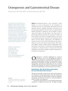
Figure 1 A simplified schematic of the Hh signalling pathway, depicting the key core components that are conserved between vertebrates and Drosophila. This highly stylised representation shows only the functional interactions between the various components: their dynamic subcellular distributions, that are key to their activity, are not depicted. In unstimulated cells (left), the activity of the transmembrane protein Smo is suppressed by the Hh receptor Ptc and the majority of the Gli transcription factors are present in their repressive (R) form. In stimulated cells (right) binding of Hh to Ptc activates Smo which in turn shifts the Gli balance in favour of the activator (A) forms, resulting in the transcription of Hh target genes.
Page 13 of 18
Parkin & Ingham
13
REFERENCES 1. Apelqvist A, Ahlgren U and Edlund H. Sonic hedgehog directs specialised mesoderm differentiation in the intestine and pancreas. Curr Biol 7: 801-4, 1997.
2. Arsic D, Cameron V, Ellmers L, Quan QB, Keenan J and Beasley S. Adriamycin disruption of the Shh-Gli pathway is associated with abnormalities of foregut development. J Pediatr Surg 39: 1747-53, 2004.
3. Hagan DM, Ross AJ, Strachan T, Lynch SA, Ruiz-Perez V, Wang YM, Scambler P, Custard E, Reardon W, Hassan S, Nixon P, Papapetrou C, Winter RM, Edwards Y, Morrison K, Barrow M, Cordier-Alex MP, Correia P, Galvin-Parton PA, Gaskill S, Gaskin KJ, Garcia-Minaur S, Gereige R, Hayward R and Homfray T. Mutation analysis and embryonic expression of the HLXB9 Currarino syndrome gene. Am J Hum Genet 66: 1504-1515, 2000.
4. Hebrok M, Kim SK and Melton DA. Notochord repression of endodermal Sonic hedgehog permits pancreas development. Genes Dev 12: 1705-13, 1998.
5. Hill P, Wang B and Ruther U. The molecular basis of Pallister Hall associated polydactyly. Hum Mol Genet 16: 2089-2096, 2007.
6. Hooper JE and Scott MP. Communicating with Hedgehogs. Nat Rev Mol Cell Biol 6: 306-317, 2005.
Page 14 of 18
Parkin & Ingham
14
7. Huangfu D and Anderson KV. Signaling from Smo to Ci/Gli: conservation and divergence of Hedgehog pathways from Drosophila to vertebrates. Development 133: 3-14, 2006.
8. Ji Z, Mei FC, Xie J and Cheng X. Oncogenic KRAS activates hedgehog signaling pathway in pancreatic cancer cells. J Biol Chem 282: 14048-14055, 2007.
9. Kawahira H, Ma NH, Tzanakakis ES, McMahon AP, Chuang PT and Hebrok M. Combined activities of hedgehog signaling inhibitors regulate pancreas development. Development 130: 4871-4879, 2003.
10. Kayed H, Kleeff J, Esposito I, Giese T, Keleg S, Giese N, Buchler MW and Friess H. Localization of the human hedgehog-interacting protein (Hip) in the normal and diseased pancreas. Mol Carcinog 42: 183-192, 2005.
11. Lees C, Howie S, Sartor RB and Satsangi J. The hedgehog signalling pathway in the gastrointestinal tract: implications for development, homeostasis, and disease. Gastroenterology 129: 1696-1710, 2005.
12. Liu G, Moro A, Zhang JJ, Cheng W, Qiu W and Kim PC. The role of Shh transcription activator Gli2 in chick cloacal development. Dev Biol 303: 448-460, 2007.
Page 15 of 18
Parkin & Ingham
15
13. Madison BB, Braunstein K, Kuizon E, Portman K, Qiao XT and Gumucio DL. Epithelial hedgehog signals pattern the intestinal crypt-villus axis. Development 132: 279-289, 2005.
14. Martin ST, Sato N, Dhara S, Chang R, Hustinx SR, Abe T, Maitra A and Goggins M. Aberrant methylation of the Human Hedgehog interacting protein (HHIP) gene in pancreatic neoplasms. Cancer Biol Ther 4: 728-733, 2005.
15. Mfopou JK, De G, V, Xu X, Heimberg H and Bouwens L. Sonic hedgehog and other soluble factors from differentiating embryoid bodies inhibit pancreas development. Stem Cells 25: 1156-1165, 2007.
16. Mo R, Kim JH, Zhang J, Chiang C, Hui CC and Kim PC. Anorectal malformations caused by defects in sonic hedgehog signaling. Am J Pathol 159: 765-74, 2001.
17. Mortell A, Giles J, Bannigan J and Puri P. Adriamycin effects on the chick embryo. Pediatr Surg Int 19: 359-364, 2003.
18. Morton JP, Mongeau ME, Klimstra DS, Morris JP, Lee YC, Kawaguchi Y, Wright CV, Hebrok M and Lewis BC. Sonic hedgehog acts at multiple stages during pancreatic tumorigenesis. Proc Natl Acad Sci U S A 104: 5103-5108, 2007.
Page 16 of 18
Parkin & Ingham
16
19. Pasca dM, Sekine S, Ermilov A, Ferris J, Dlugosz AA and Hebrok M. Hedgehog/Ras interactions regulate early stages of pancreatic cancer. Genes Dev 20: 3161-3173, 2006.
20. Puri S and Hebrok M. Dynamics of embryonic pancreas development using realtime imaging. Dev Biol 306: 82-93, 2007.
21. Ramalho-Santos M, Melton DA and McMahon AP. Hedgehog signals regulate multiple aspects of gastrointestinal development. Development 127: 2763-72, 2000.
22. Stepan V, Ramamoorthy S, Nitsche H, Zavros Y, Merchant JL and Todisco A. Regulation and function of the sonic hedgehog signal transduction pathway in isolated gastric parietal cells. J Biol Chem 280: 15700-15708, 2005.
23. Sukegawa A, Narita T, Kameda T, Saitoh K, Nohno T, Iba H, Yasugi S and Fukuda K. The concentric structure of the developing gut is regulated by Sonic hedgehog derived from endodermal epithelium. Development 127: 1971-80, 2000.
24. Thayer SP, di Magliano MP, Heiser PW, Nielsen CM, Roberts DJ, Lauwers GY, Qi YP, Gysin S, Fernandez-del Castillo C, Yajnik V, Antoniu B, McMahon M, Warshaw AL and Hebrok M. Hedgehog is an early and late mediator of pancreatic cancer tumorigenesis. Nature 425: 851-856, 2003.
Page 17 of 18
Parkin & Ingham
17
25. Thomas MK, Rastalsky N, Lee JH and Habener JF. Hedgehog signaling regulation of insulin production by pancreatic beta-cells. Diabetes 49: 2039-47, 2000.
26. van Bokhoven H, Celli J, van Reeuwijk J, Rinne T, Glaudemans B, van Beusekom E, Rieu P, Newbury-Ecob RA, Chiang C and Brunner HG. MYCN haploinsufficiency is associated with reduced brain size and intestinal atresias in Feingold syndrome. Nat Genet 37: 465-467, 2005.
27. Zavros Y, Waghray M, Tessier A, Bai L, Todisco A, Gumucio DL, Samuelson LC, Dlugosz A and Merchant JL. Reduced pepsin a processing of sonic hedgehog in parietal cells precedes gastric atrophy and transformation. J Biol Chem 2007.
Page 18 of 18
A simplified schematic of the Hh signalling pathway, depicting the key core components that are conserved between vertebrates and Drosophila. This highly stylised representation shows only the functional interactions between the various components: their dynamic subcellular distributions, that are key to their activity, are not depicted. In unstimulated cells (left), the activity of the transmembrane protein Smo is suppressed by the Hh receptor Ptc and the majority of the Gli transcription factors are present in their repressive (R) form. In stimulated cells (right) binding of Hh to Ptc activates Smo which in turn shifts the Gli balance in favour of the activator (A) forms, resulting in the transcription of Hh target genes. 254x190mm (96 x 96 DPI)