Newton, C. R., A. Graham, L. E. Heptinstall, S. J. Powell, C. Summers, N. ... Feray, C., M. Gigou, D. Samuel, M. Reynes, M. Bismuth, and C. Brechot. 1992.
Hepatitis B Virus Precore Mutation and Fulminant Hepatitis in the United States A Polymerase Chain Reaction-based Assay for the Detection of Specific Mutation T. Jake Liang, * Kiyoshi Hasegawa, * Santiago J. Munoz,$ Craig N. Shapiro,' Boris Yoffe,11 Brian J. McMahon,' Chengyin Feng, * Hucheng Bei, * Miriam J. Alter,' and Jules L. Dienstag *
*Gastrointestinal Unit, Medical Services, Massachusetts General Hospital, and Department ofMedicine, Harvard Medical School, Boston, Massachusetts 02114; *Division of Gastroenterology and Hepatology, Department of Medicine, Jefferson Medical College, Philadelphia, Pennsylvania 19107; §Hepatitis Branch, Center for Disease Control and Prevention, Atlanta, Georgia 30333; IlDivision of GI, Veterans Affairs Medical Center and Baylor College ofMedicine, Houston, Texas 77030; and 'Alaska Native Medical Center and Centerfor Disease Control and Prevention, Anchorage, Alaska 99510
Abstract Hepatitis B virus (HBV) variants with precore mutation(s) resulting in the absence of HBeAg production have been associated with the occurrence of fulminant hepatitis in Japan, Israel, and southern Europe, where the prevalence of this HBV strain appears common. In areas such as United States, where HBV infection is not endemic, the role of this mutant virus in fulminant hepatitis is unknown. We developed an amplification refractory mutation detection system to detect specifically the presence of the G to A mutation at nucleotide position 1898, which is the most frequently observed mutation resulting in a precore stop codon. In addition, this method provided a quantitative measurement of the relative ratio of one strain to the other. Using this system, we tested HBV strains for the presence of the stop codon mutation in sera from 40 cases of fulminant hepatitis B occurring in the United States. Serum HBV DNAs from 28 patients were analyzed successfully. A mixture of wild-type and mutant strains in various ratios were observed in 15 patients, wild type exclusively in 11, and mutant exclusively in 2. Four of these patients had undergone liver transplantation for HBV-associated cirrhosis and developed fulminant HBV-associated hepatitis after transplantation. Pre- and posttransplant serum samples from one patient were analyzed: a mixture of wild-type and mutant HBV strains was detected in both samples. Our study demonstrated that both wild-type and mutant HBV strains are associated with fulminant hepatitis, and that in some patients in the United States, factors other than precore mutations contribute to the development of fulminant hepatitis. (J. Clin. Invest. 1994. 93:550-555.) Key words: hepatitis B virus * fulminant hepatitis,, precore mutation * polymerase chain reaction * hepatitis C virus
Address correspondence to T. Jake Liang, M.D., Gastrointestinal Unit, Massachusetts General Hospital, Jackson 812, Fruit Street, Boston, MA 02114. Received for publication 17 May 1993 and in revised form 7 October 1993.
1. Abbreviations used in this paper: ARMDS, amplification refractory mutation detection system; HBV, hepatitis B virus; IVDA, intravenous drug addict; nt, nucleotide. The Journal of Clinical Investigation, Inc. Volume 93, February 1994, 550-555
550
Liang et al.
Introduction Infection with hepatitis B virus (HBV)' is associated with a broad spectrum of liver injury, ranging from acute self-limited infection to fulminant hepatitis, chronic hepatitis with or without progression to cirrhosis and liver failure, as well as to an asymptomatic chronic carrier state. Evidence has been accumulating to indicate that HBV mutants unable to synthesize HBeAg may influence the course ofinfection and clinical manifestations of disease ( 1-3). Many mutations inactivating the precore open reading frame necessary for the synthesis of HBeAg have been described (4, 5). These mutations have been observed to arise de novo in patients chronically infected with wild-type HBV (6), and acute infection with such mutants appears to be associated with fulminant hepatitis (7-9). We have previously described the association of an HBV variant containing two precore mutations with an epidemic of fulminant hepatitis B in Israel (7). The HBV strain responsible for this epidemic contained two G to A mutations in the precore open reading frame (nucleotides [nt] 1898 and 1901), one of them resulting in a stop codon. Other laboratories have also reported similar findings in patients with sporadic fulminant hepatitis B in Japan (8, 9). Using an amplification refractory mutation detection system (ARMDS) (10) to detect specifically the presence of the G to A mutation at nt position 1898, we studied 40 cases of sporadic fulminant hepatitis B that had occurred in various regions of United States. The system is based on the absolute inability of oligonucleotides with mismatched 3' residues to function as PCR primers under appropriate conditions. Methods Patients. Serum samples were collected from 40 patients with sporadic fulminant hepatitis B that had occurred in various regions of the United States: New England (seven patients), mid-Atlantic coast ( 19 patients), Colorado (two patients), Alabama (two patients), Florida (two patients), Washington (two patients), Alaska (three patients), and Texas (three patients). Most of the serum samples were collected at the Massachusetts General Hospital, Jefferson Medical College, the Baylor College of Medicine-affiliated hospitals, and Alaska Native Medical Center. The eight cases from Colorado, Alabama, Florida, and Washington were collected through an epidemiological surveillance program established by the Center for Disease Control and Prevention (Atlanta, GA). Since 1981, intensive surveillance of acute viral hepatitis has been conducted in four "sentinel counties" in the United States (Denver, CO; Jefferson, AL, Pinellas, FL; and Pierce, WA). 16 cases of acute, self-limited HBV hepatitis were included for comparative studies: four from New England; two from Denver, CO; one from Jefferson, AL; two from Pinellas, FL; one from Pierce, WA; and six from Texas.
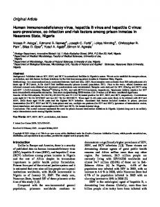
Serum samples obtained during initial hospitalization within 6 wks from the onset of illness were stored frozen at -80'C except for the cases from CDC (Atlanta), where they were stored at -20'C. The clinical characteristics of 28 patients whose serum samples we were able to analyze successfully are summarized in Table I. All patients had circulating IgM anti-HBc, except for patients 20-23, who had endstage HBV-associated cirrhosis, and in whom fulminant hepatitis B developed after transplantation. These four patients developed acute hepatic failure histologically and serologically consistent with fulminant hepatitis B, within 3 mo after transplant. Serum samples from the preoperative stage, as well as during the occurrence offulminant hepatitis were available from one such patient (patient 20). For the other three patients, we were only able to obtain serum samples during the fulminant hepatitis phase. Amplification refractory mutation detection system. Serum DNA was isolated as described previously (11). Extracted DNA was subjected to ARMDS for specific detection of the G to A mutation at nt 1898 (10). This system is depicted schematically in Fig. 1. Two HBV primers (primers 1 and 2) spanning nt 1605-2432 of the HBV genome were used in the first PCR amplification. An aliquot of reaction product was then amplified further in a second reaction with primers 3 and 4, which are contained within the region spanned by the first two primers. Primer 3 had either a G (primer 3W) or A (primer 3M) at the 3' end, corresponding to nt position 1898. Therefore, primers 3W and 3M were referred to as the "detection primers," one specific for the wild-type G and the other for the mutant A sequence, respectively. In addition, a mutation of T to G was introduced at the third position from the 3' end of the detection primer to further enhance the specificity of the amplification reaction (10). The primer sequences were: primer 1, 5' GTTGCATGGAGACCACCGTGAAC 3' (sense, nt 1605-1627); primer 2,5' GCTTCTGCGACGCGGCGATTGAGA 3' (antisense, nt 2432-2410); primer 4, 5' CGAGGGAGTT CTTCTTCTAG 3' (antisense, nt 2394-2375). The sequences for primers 3W and 3M are shown in Fig. 1. The first PCR amplification continued for 35 cycles (940C for 1 min, 50'C for 1 min, 720C for 2.5 min), and the second PCR was designed for 30 cycles with the annealing step at 62°C for 1 min and the elongation step at 72°C for 1.5 min. The reaction volume was 25 Al in each of the PCR reactions, and the standard PCR reaction was performed for the first PCR amplification. 10,ul of the first PCR reaction was subjected to Southern blot hybridization with a 32p_ HBV-specific probe. The samples with a positive PCR signal were nt 1605
-Primer
nt 1879
Primer 3WT Primer 3MT
Core
Precore
Primer 2
1
nt 1898