concepts of the epidemiology, molecular virology, pathogenesis, natural history, diagnosis, therapy, and prevention of hepatitis C. Keywords: hepatitis C virus; ...
Review article
S W I S S M E D W K LY 2 0 0 1 ; 1 3 1 : 2 9 1 – 2 9 8 · w w w . s m w . c h
291
Peer reviewed article
Hepatitis C: an update Darius Moradpour1, Andreas Cerny2, Markus H. Heim3, Hubert E. Blum1 1
Department of Medicine II, University of Freiburg, Freiburg i. Br., Germany Clinica Medica, Ospedale Civico, Lugano, Switzerland 3 Department of Gastroenterology, University Hospital Basel, Switzerland 2
Summary Hepatitis C virus (HCV) infection is a leading cause of chronic hepatitis, liver cirrhosis, and hepatocellular carcinoma worldwide. While current therapeutic options for hepatitis C are limited, recent progress in the understanding of the biology of HCV led to the identification of novel targets for antiviral intervention. In addition, molecular and immunotherapeutic strategies to inhibit HCV replication or gene expression and to enhance the cellular immune response against HCV are being
explored. These and other novel antiviral strategies may eventually complement existing therapeutic modalities. Here, we briefly review current concepts of the epidemiology, molecular virology, pathogenesis, natural history, diagnosis, therapy, and prevention of hepatitis C. Keywords: hepatitis C virus; protease; helicase; polymerase; interferon-a; ribavirin; immunopathogenesis
Introduction Since the identification of the hepatitis C virus (HCV) as the most common etiologic agent of posttransfusion and sporadic non-A, non-B hepatitis [1, 2], important aspects of the epidemiology, molecular virology, and natural course of hepatitis C have been elucidated. In addition, specific and sensitive assays for the diagnosis of HCV infection have been developed. However, important aspects
of the molecular biology of HCV, the pathogenesis of HCV-induced liver disease as well as many therapeutic and preventive issues are still unresolved. In the following, we will briefly review current understanding of the epidemiology, molecular biology, pathogenesis, and natural history of HCV infection as well as give an update of the diagnosis, therapy, and prevention of hepatitis C.
Epidemiology It is estimated that about 170 million people worldwide are infected with HCV [3]. The seroprevalence rate is about 1% in Western Europe and North America, 3–4% in some Mediterranean and Asian countries and up to 10–20% in parts of Central Africa and Egypt [4, 5]. In Switzerland an estimated 50,000 to 70,000 individuals are HCVinfected [6]. HCV is parenterally transmitted. With the introduction of anti-HCV screening of blood and blood products in 1990 new cases of posttransfusion hepatitis C have virtually disappeared and intravenous drug use has become the major identifiable mode of transmission in many countries. Unfortunately, the lack of systematic screening of blood donors continues to result in HCV transmission in countries with developing or transitional economies. In addition, large-scale
immunization and parenteral therapy programs as well as surgical and dental procedures with inadequately sterilized equipment have been important routes of transmission in these countries [7]. HCV transmission has been described in nosocomial settings, including organ transplantation [8]. Occupational needle-stick injuries from anti-HCV positive sources result in seroconversion in about 3% of recipients; thus representing a transmission risk between that of HIV (about 0.3%) and hepatitis B virus (HBV) (about 30% in unvaccinated recipients). Intranasal cocaine use has been identified as a possible mode of transmission (“straw sharing”) [9]. Sexual transmission is rare and correlates with high-risk sexual practices. Mother-to-infant transmission has been observed, but the risk is probably less than 5% unless the mother is co-infected
292
Hepatitis C: an update
with HIV. There is no proven association between transmission and the type of delivery or maternal breast-feeding. Household transmission is uncom-
mon. Intriguingly, in clinical practice no epidemiologic risk factor can be identified in up to 40% of patients with hepatitis C (“sporadic hepatitis C”).
Molecular virology HCV has been classified in the Hepacivirus genus within the Flaviviridae family of viruses which includes the classical flaviviruses, such as yellow fever virus and the animal pestiviruses [10]. The structure and replication cycle of HCV are incompletely understood due to the low viral titers found in sera and livers of HCV-infected individuals and the lack of an efficient cell culture system or small animal model permissive for HCV infection. Nevertheless, considerable progress has been made using heterologous expression systems, functional cDNA clones, and most recently, selectable subgenomic replicons [11–15]. HCV contains a single-stranded RNA genome of positive polarity and approximately 9,600 nucleotides. As in flavi- and pestiviruses, the viral genome is composed of a 5’ noncoding region (5’ NCR), a long open reading frame encoding a polyprotein precursor of about 3,000 amino acids,
Figure 1 Genetic organization and polyprotein processing of HCV. Asterisks in the E1 and E2 region indicate glycosylation of the envelope proteins. Diamonds denote cleavages of the HCV polyprotein precursor by the endoplasmic reticulum signal peptidase, and arrows indicate cleavages by HCV NS2-3 and NS3 proteases.
Figure 2 Structure of the HCV RNA-dependent RNA polymerase. A model for a polymerase-RNA template: primer-rNTP complex is shown. (A) The completely encircled pathway for access of the rNTP to the active site is shown. The molecular surface is colored from blue (positive) to red (negative) according to the local electrostatic surface potential. (B) Ribbon diagram with the fingers subdomain drawn in blue, the palm subdomain in red and the thumb subdomain in green (from Lesburg CA et al. Nat Struct Biol 1999;6:937–943 with permission from Patricia C. Weber, Ph.D. and the Nature Publishing Group, New York, NY, USA).
and a 3’ NCR (figure 1). The 5’ NCR functions as an internal ribosomal entry site (IRES) essential for cap-independent translation of the viral RNA. The HCV polyprotein precursor is co- and posttranslationally processed by cellular and viral proteases to yield the mature structural and non-structural proteins. The structural proteins include the core protein, which forms the viral nucleocapsid, and the envelope glycoproteins E1 and E2. These are released from the polyprotein precursor by the endoplasmic reticulum signal peptidase. CD81 and the low density lipoprotein receptor have been proposed as HCV receptor candidates or potential components of a receptor complex [16, 17]. Presently, however, these molecules do not fulfill the criteria of a bona fide viral receptor and the search for other candidates is going on. E2 has recently been reported to interfere with the catalytic activity of the interferon (IFN)-induced
S W I S S M E D W K LY 2 0 0 1 ; 1 3 1 : 2 9 1 – 2 9 8 · w w w . s m w . c h
double-stranded RNA-activated protein kinase PKR [18]. The significance of this observation, however, for the IFN resistance commonly observed in clinical practice is unclear [19]. The non-structural proteins NS2 through NS5 include the NS2–3 autoprotease and the NS3 serine protease, which are essential for processing of the polyprotein precursor, a RNA helicase located in the carboxy-terminal region of NS3, the NS4A polypeptide, the NS4B and NS5A proteins, and a RNA-dependent RNA polymerase represented by NS5B (figure 1). The crystal structures of the serine protease and RNA helicase domains of NS3, and more recently, of the entire NS3 protein [20] as well as the RNA-dependent RNA polymerase have been elucidated [21] (figure 2). These enzymes are essential for viral replication and have emerged as major targets for the design of novel antiviral agents. NS4A is a cofactor for the NS3 serine protease. The functions of NS4B and NS5A, a serine phosphoprotein, remain to be established. Similar to E2, NS5A has been reported to interfere with PKR function [22]. In this context, a role for NS5A in modulating the IFN response was first suggested by studies performed in Japan, which described a correlation between mutations within a discrete region of NS5A, termed interferon sensitivity determining region (ISDR), and a favorable response to IFN-α therapy [23]. These findings were largely confirmed in Japan,
Figure 3 HCV life cycle. (1) Virus binding and internalization, (2) cytoplasmic release and uncoating, (3) IRESmediated translation, (4) polyprotein processing, (5) RNA replication, (6) packaging and assembly, (7) virion maturation, (8) virion release.
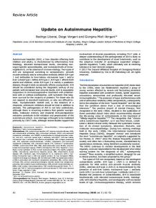
Figure 4
Acute hepatitis
Chronic hepatitis
but not in Europe and North America. The reasons for this discrepancy are not understood but may involve both differences in IFN-α regimens and the low prevalence of ‘mutant type’ HCV genotype 1b isolates in Western countries. Taken together, the presumed life cycle of HCV includes (1) binding to an as yet unidentified cell surface receptor and internalization into the host cell, (2) cytoplasmic release and uncoating of the viral RNA genome, (3) IRES-mediated translation, (4) polyprotein processing by cellular and viral proteases, (5) RNA replication, (6) packaging and assembly, (7) virion maturation, and (8) release from the host cell (figure 3). Recent studies have suggested that HCV infection is a highly dynamic process with a viral half-life of only a few hours and average daily virion production and clearance rates of up to more than 1012 [24]. These findings are similar to the dynamics of HIV infection and provide a rationale for the development and implementation of multidrug combination therapies against hepatitis C. In addition, together with the lack of a proof-reading function of the viral RNA-dependent RNA polymerase, this high replicative activity provides the basis for the genetic variability of HCV. HCV isolates fall into 3 major categories, depending on the degree of sequence divergence: genotypes, subtypes, and isolates [25]. The term quasispecies refers to the genetic heterogeneity of the population of HCV genomes coexisting in an infected individual. Genotypes 1–3 have a worldwide distribution, genotypes 4 and 5 are found principally in Africa and genotype 6 is distributed primarily in Asia. Several studies have shown that infection with genotype 1, with a prevalence of 70–80% in most Western countries, is associated with a poor response to IFN-α therapy [26, 27]. More recently, it was proposed to divide HCV isolates into 6 phylogenetically distinct groups, termed clades 1 to 6 [28]. Clades 1, 2, 4, and 5 correspond to genotypes 1, 2, 4, and 5 while clade 3 comprises genotypes 3 and 10, and clade 6 comprises genotypes 6, 7, 8, 9, and 11.
Cirrhosis
HCC
Natural course of hepatitis C.
70–80%
20%
293
1–4% per year
294
Hepatitis C: an update
Pathogenesis The mechanisms responsible for liver injury in acute and chronic HCV infection are poorly understood [29]. In primary HCV infection, liver cell damage coincides with the development of the host immune response and not with infection and viral replication. In addition, persistent viral replication often occurs without evidence of liver cell damage, suggesting that HCV is not directly cytopathic. The immune response against HCV, therefore, plays a central role in HCV pathogenesis. HCV infection does not induce protective immunity. Re- and superinfection with homologous and heterologous isolates has been shown in both experimental and clinical studies [30, 31]. The role of antibodies in immunopathology has not been studied in much detail so far. HCV-specific major histocompatibility complex (MHC) class I-restricted CD8+ cytotoxic T lymphocyte (CTL) [32, 33] and MHC class II-restricted CD4+ helper T cell responses [34, 35] have been identified in patients with acute and chronic HCV infection. CTL-mediated lysis of virus-infected host cells may lead to clearance of the virus or, if incomplete, to viral persistence and eventually chronic hepatitis. Based on these observations and parallels in other viral diseases, viral persistence and immunologically mediated liver cell injury are important mechanisms leading to chronic hepatitis C [29]. Patients who clear HCV infection have a more vigorous CD4+ [34, 35] and CD8+ T cell response early on [33]. Despite the presence of an immune response, however, HCV is rarely eliminated. For a noncytopathic virus to persist it must overwhelm, not induce, or evade an antiviral immune response. Perhaps the simplest explanation is quantitative, based on the kinetics of infection relative to the in-
duction of a CTL response during the early phase of an infection [36]. According to this model, viral persistence would be predicted if the size of the inoculum or the replication rate of the virus exceeds the kinetics of the immune response. However, to be consistent with the repeated observation that the CTL response is less vigorous in chronically infected patients than it is during acute self-limited infection, additional mechanisms must be involved. These may include the induction of peripheral tolerance or exhaustion of the T cell response, infection of immunologically privileged sites, inhibition of antigen presentation, down regulation of viral gene expression and viral mutations that abrogate, anergize or antagonize antigen recognition by virus-specific T cells [29]. There is some evidence that privileged sites may play a role since HCV may infect extrahepatic cells and tissues. Inhibition of antigen presentation is for some viruses a mechanism to establish a persistent infection. Thus far, however, there is no evidence for these processes in HCV infection [37]. HCV, however, may interfere with the IFN system [18, 22, 38]. As mentioned above, the role of viral escape mutations and the quasispecies nature of HCV as a cause of viral persistence has attracted considerable interest. Molecular mimicry represents another potential mechanism of viral persistence and pathogenesis. In this context, we have recently found that CTL induced by an HCV core-derived synthetic peptide in some cases also recognized cytochrome P450; an observation that lends support for the hypothesis that HCV infection may be causally linked to autoimmune hepatitis in some patients [39].
Natural history After an incubation period of 3 to 12 weeks HCV infection is usually followed by a clinically unapparent hepatitis [40]. Only about 25% of patients are symptomatic. Fulminant hepatitis C is very uncommon. One of the most important clinical features of hepatitis C is its progression to chronicity in about 70–80% (figure 4). Typically, patients with chronic hepatitis C have few symptoms, if any, and these are usually nonspecific, intermittent, and mild. The most common symptom is fatigue. The natural history of chronic hepatitis C has been analyzed in several retro- and prospective studies [41]. While no increased mortality was found in the retrospective Veterans Administration study [42], other studies indicated that chronic hepatitis C frequently progresses to cirrhosis and hepatocellular carcinoma (HCC) [43]. On the other hand, recent studies on highly selected co-
horts have again shown a more benign course [44, 45]. Overall, about 20% of patients with chronic hepatitis C will develop liver cirrhosis within approximately 20 years. Once cirrhosis is established the rate of HCC development is 1–4% per year. HCV infection appears to be responsible for a substantial proportion of the recently observed increase in HCC incidence and mortality [46]. Although recent experimental evidence raises the possibility that HCV might operate through direct pathways in promoting malignant transformation of hepatocytes, it is generally believed that HCC associated with chronic hepatitis C develops through a general pathway of increased liver cell turnover, induced by chronic liver injury and regeneration, resulting in multiple and stepwise genetic alterations [47]. The highly variable natural course of chronic hepatitis C may depend on viral (e.g., inoculum size, genotype, and quasispecies
S W I S S M E D W K LY 2 0 0 1 ; 1 3 1 : 2 9 1 – 2 9 8 · w w w . s m w . c h
complexity) and host factors (e.g., age at the time of infection, gender, MHC haplotype, coinfection with HBV or HIV, and, probably most important, alcohol consumption). Chronic HCV infection has been associated with a number of extrahepatic manifestations, including mixed cryoglobulinemia, glomerulonephritis, lichen planus, and porphyria cutanea
295
tarda [48]. Cryoglobulins are detectable in up to one third of patients with chronic hepatitis C, while the clinical syndrome of mixed cryoglobulinemia occurs in only 1–2% of patients. Recently, chronic hepatitis C has been associated with nonHodgkin’s lymphoma and type 2 diabetes mellitus. However, these associations are still debated.
Diagnosis Diagnosis of hepatitis C is based on serological assays which detect HCV-specific antibodies (anti-HCV) and on molecular assays which detect HCV RNA. The molecular assays currently available are reverse transcriptase (RT)-PCR and the branched DNA (bDNA) assay. By RT-PCR the viral RNA is reverse transcribed into complementary DNA (cDNA) that is then amplified by PCR. In the bDNA assay the signal resulting from a specific hybridization of capture and detection probes with the viral RNA is amplified. Third-generation anti-HCV enzyme-linked immunosorbent assays (ELISAs) are highly sensitive as well as specific and represent the primary diagnostic assay. Anti-HCV tests are less sensitive, however, in immunocompromised individuals or hemodialysis patients. For these patients, a negative ELISA does not exclude HCV infection, and RT-PCR should be performed. The recombinant immunoblot assay (RIBA) is a supplemental assay that can be used to confirm a positive ELISA, particularly in low-risk populations. In this setting, a negative RIBA will make further testing unnecessary. No routine serological test for a viral antigen is available as yet. However, immunoassays based on the use of high-affinity monoclonal antibodies against core protein to detect and quantitate HCV in serum are currently being evaluated [49]. HCV becomes positive by RT-PCR as early as 1–2 weeks after infection and 4-6 weeks before anti-HCV seroconversion. The determination of
HCV RNA is, in principle, important for the selection of patients for antiviral therapy and for the assessment of its efficacy [27]. Optimal RT-PCR assays at present have a sensitivity of less than 100 copies of HCV RNA per ml of plasma or serum. Standardization of assays and proficiency testing of diagnostic laboratories has recently improved the previously high rate of false-positive and -negative PCR results. In the case of a positive ELISA, RTPCR allows to discriminate between patients with chronic hepatitis C and those with resolved HCV infection that can remain anti-HCV positive for years or decades. Intriguingly, recent studies have shown that the antibody response to HCV fades over time in many patients after successful elimination of the virus [50]. Discrimination of genotype 1 from genotypes 2 and 3 as well as quantitative determination of viremia levels has become important for the selection of the optimal treatment regimen [27]. In general, however, genotyping and quantitative RTPCR tests should be used only in the context of a defined therapy protocol and not for the initial diagnosis of HCV infection. Liver biopsy allows to determine the necro-inflammatory activity (grading) and the degree of fibrosis (staging), as well as to recognize or exclude coexisting liver pathology (such as alcoholic liver disease or hemochromatosis). It is strongly recommended before the initiation of antiviral therapy.
Current therapy The decision to treat is based on the analysis of numerous variables and should take into account the specific situation of each patient. In general, treatment is currently recommended for patients with persistently (>6 months) elevated aminotransferase levels, anti-HCV and RNA positivity in serum, and findings of fibrosis and at least moderate degrees of inflammation and necrosis on liver biopsy [26, 27]. For other patients the indications are less clear and decisions will have to be made on an individual basis. Additional factors that come into consideration are, among others, the age and general condition of the patient, the
duration of HCV infection, the risk of developing cirrhosis, the likelihood of response to therapy, comorbidity, and the patient’s personal and professional plans. Importantly, impairment of quality of life during treatment should be taken into account and discussed with the patient. Contraindications to therapy with IFN and ribavirin include decompensated liver cirrhosis, autoimmune hepatitis or other autoimmune diseases, a history of depression or psychosis, pregnancy, the lack of a reliable method of contraception (teratogenicity of ribavirin), cardiopulmonary diseases, leukopenia (