Kidney International. Vol. ... The glomeruhi were obtained from human kidneys removed ... The dishes were placed in an incubator at 37°C and cultured in.
Kidney International. Vol. 35 (1989), pp. 1245—1248
TECHNICAL NOTES
Hepatoma G2 conditioned medium facilitates early outgrowth of endothelial cells from isolated glomeruli SHARON ELLIOT, LILIANE J. STRIKER, TosHlo Doi, W. MARSTON LINEHAN, and GARY E. STRIKER Metabolic Diseases Branch, National Institute of Diabetes and Digestive and Kidney Diseases; Surgery Branch, Division of Cancer Treatment, National Cancer Institute; and Division of Kidney, Urologic and Hematologic Diseases, National Institute of Diabetes and Digestive and Kidney Diseases; National Institutes of Health, Bethesda, Maryland, USA
Since it was first shown that glomerular cells could be peared to settle faster than non-glomerular contaminants, the propagated in culture [1], this technique has proven to be an invaluable tool for studying the physiology of individual gbmerular cells. Epithelial, mesangial, and endothelial cells have been isolated and propagated in several species [2], however, the population of cells obtained consisted of more than one of these glomerular cell types. The limited availability of normal human kidney specimens for tissue culture has led to attempts to optimize culture conditions which could selectively enhance the growth of one specific cell type. Hoshi and McKeehan [3] have demonstrated that medium conditioned by HepG2 cells, a hepatoblastoma line, contained one or more soluble growth factors which favored the growth of human umbilical vein endothelial cells in serum free media. This laboratory has previously reported the growth of human glomerular endothelial cells in the presence of fetal bovine serum supplemented with platelet-derived growth factor [41. In
preparations were resuspended several times in fresh medium and allowed to settle a minimum of five times. The supernatant was aspirated each time, effectively removing non-glomerular contaminants. This procedure was repeated until the preparation was free of contaminants. This relatively gentle method of isolating glomeruli was found to require a short period of time and resulted in isolates with better plating efficiency. Encapsulated glomeruhi were not observed visually.
Cell culture Primary cultures. Glomeruli were plated in 100 mm dishes (Nunclon) coated with human fibronectin (200 sg/ml, Collaborative Research, Lexington, Massachusetts, USA). The gbmerular pellet was divided and plated in either Waymouth's medium (Gibco, Grand Island, New York, USA) supplemented view of our continued interest in these cells, and the variation in with 20% fetal bovine serum (FBS) (Gibco), and 2 ng/ml of the ability of different lots of fetal bovine serum to support platelet-derived growth factor (PDGF) (BRL, Gaithersburg, endothelial cell growth, we investigated the use of hepatoma Maryland, USA) (Medium A), a 1:1 mixture of hepatoma G2 conditioned medium to further optimize the growth of endothe- conditioned medium (CM) and Waymouth's medium with 20% hal cells. FBS (Medium B), or unconditioned MDCB medium with 15% FBS (Medium C). All media contained penicillin (100 U/mI), Methods streptomycin (100 sg/ml, Gibco) and glutamine (1 msi, Gibco). The dishes were placed in an incubator at 37°C and cultured in Isolation of glomeruli The glomeruhi were obtained from human kidneys removed a 5% CO2 and 95% air mixture. for renal cancer. Cortical tissue, far from the tumor zone were Hepatoma conditioned medium (CM) stripped of capsule, and the fragments diced into approximately 2 mm3. The pieces were gently forced through a single stainless Confluent human hepatoblastoma cells, HepG2 (ATCC steel mesh (80, E.C. Apparatus, St. Petersburg, Florida, USA) #HB8065, Rockville, Maryland, USA) were maintained in T175 with a glass pestle. Only material passing easily through this flasks (Nuncbon), in medium MDCB 107 supplemented with single mesh size was collected in Hanks media at 37°C (Quality 10% fetal bovine serum as described previously [3]. CondiBiological, Inc., Gaithersburg, Maryland, USA) and allowed to tioned medium was collected every three days from confluent settle in 50 ml conical tubes (Falcon). Generally, recognizable monolayers, centrifuged at 3000 rpm for 15 minutes to remove glomeruli represented more than 80% of the structures in the cell debris, and frozen at —80°C for periods not exceeding one initial preparation of sieved material. As intact glomeruli ap- month. Outgrowth. Dishes were examined at day 7 after the initial plating, and glomeruhi which had not attached and non-gbomerReceived for publication September 25, 1988 and in revised form November 16, 1988 Accepted for publication November 29, 1988
© 1989 by the International Society of Nephrology
ular fragments were aspirated. At day 7 fresh medium was added to the dish irrespective of the cell number. Once a week thereafter the medium was replaced. At confluence, the cells were passaged and counted. 1245
Fig. I. Primary outgrowth in medium A (xlOO).
Fig. 2. 4. Primary outgrowth in medium B ix l). B. Confluent monolayer of cobblestone endothelial cells in medium B (X250). C. lmmunofluorescence staining of human endothelial cells with antibody against vWF x630).
1247
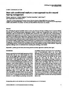
Elliot et al: Hepatoma G2 conditioned medium A Culture #1
B Culture #2
C"
20
x 15
a U
U C
a .0
E
10
E
z
z
5
18 Time, days
Time, days
C Culture #3
15 >