Journal of Translational Medicine
Xue et al. J Transl Med (2015) 13:379 DOI 10.1186/s12967-015-0743-2
Open Access
RESEARCH
HERC5 is a prognostic biomarker for post‑liver transplant recurrent human hepatocellular carcinoma Feng Xue1†, Brandon W. Higgs2*†, Jiaqi Huang2†, Chris Morehouse2, Wei Zhu2, Xin Yao2, Philip Brohawn2, Zhan Xiao2, Yinong Sebastian2, Zheng Liu2, Yun Xia1, Dong Shen2, Mike Kuziora2, Zhengwei Dong3, Hulin Han3, Yi Gu3, Jianren Gu4, Qiang Xia1* and Yihong Yao2
Abstract Background and aims: Orthotopic liver transplantation (OLT) can be an effective treatment option for certain patients with early stage hepatocellular carcinoma (HCC) meeting Milan, UCSF, or Hangzhou criteria. However, HCC recurrence rates post-OLT range from 20 to 40 %, with limited follow-up options. Elucidating genetic drivers common to primary and post-OLT recurrent tumors may further our understanding and help identify predictive biomarkers of recurrence—both to ultimately help manage clinical decisions for patients undergoing OLT. Methods: Whole exome and RNA sequencing in matched primary and recurrent tumors, normal adjacent tissues, and blood from four Chinese HCC patients was conducted. SiRNA knockdown and both qRT-PCR and Western assays were performed on PLCPRF5, SNU449 and HEPG2 cell lines; immunohistochemistry and RNA Sequencing were conducted on the primary tumors of Chinese HCC patients who experienced tumor recurrence post-OLT (n = 9) or did not experience tumor recurrence (n = 12). Results: In three independent HCC studies of patients undergoing transplantation (n = 21) or surgical resection (n = 242, n = 44) of primary tumors (total n = 307), HERC5 mRNA under-expression correlated with shorter: time to tumor recurrence (p = 0.007 and 0.02) and overall survival (p = 0.0063 and 0.023), even after adjustment for relevant clinical variables. HERC5 loss drives CCL20 mRNA and protein over-expression and associates with regulatory T cell infiltration as measured by FOXP3 expression. Further, matched primary and recurrent tumors from the 4 HCC patients indicated clonal selection advantage of Wnt signaling activation and CDKN2A inactivation. Conclusions: HERC5 plays a crucial role in HCC immune evasion and has clinical relevance as a reproducible prognostic marker for risk of tumor recurrence and survival in patients. Keywords: Hepatocellular carcinoma, Orthotopic liver transplantation, Whole exome, HERC5 Background Hepatocellular carcinoma (HCC) is the most common primary liver malignancy and third leading cause of *Correspondence:
[email protected];
[email protected] † Feng Xue, Brandon W. Higgs and Jiaqi Huang contributed equally to this work 1 Department of Liver Surgery and Liver Transplantation, Renji Hospital, Shanghai Jiaotong University School of Medicine, Shanghai 201203, China 2 Translational Bioinformatics, MedImmune, Gaithersburg, MD 20878, USA Full list of author information is available at the end of the article
cancer deaths worldwide, with Hepatitis B virus a major etiological factor [1, 2]. Beyond sorafenib (Bayer HealthCare Pharmaceuticals, Inc.; Onyx Pharmaceuticals, Inc, Germany) which is only effective in a small patient population, there is no approved treatment for HCC. Patients have limited options, and orthotopic liver transplantation (OLT) is viable for certain early stage HCC cases, though it is only efficacious in a subset meeting Milan, Toronto, or UCSF clinical criteria [3–5]. The HCC recurrence rates after OLT range from 20 to 40 %, and treatment options after recurrence are limited [3–5]. To date,
© 2015 Xue et al. This article is distributed under the terms of the Creative Commons Attribution 4.0 International License (http:// creativecommons.org/licenses/by/4.0/), which permits unrestricted use, distribution, and reproduction in any medium, provided you give appropriate credit to the original author(s) and the source, provide a link to the Creative Commons license, and indicate if changes were made. The Creative Commons Public Domain Dedication waiver (http://creativecommons.org/publicdomain/ zero/1.0/) applies to the data made available in this article, unless otherwise stated.
Xue et al. J Transl Med (2015) 13:379
certain clinicopathologic variables such as tumor size and absence of macroscopic vascular invasion are used to predict risk of recurrence, though success of these factors vary from study-to-study [6]. In an effort to improve the prediction of HCC recurrence, molecular profiling has been applied in many studies. Multiple transcriptomic and proteomic studies have been conducted to help understand the link between molecular mediators and factors of etiology, tumorigenesis, disease course, and/or other variables related to survival and recurrence in HCC. In fact, from 2003 to 2010, over 14 studies have identified gene signatures from a minimum of 12 genes or proteins to 186, totally more than 934 genes for purposes of predicting survival and/or recurrence in HCC patients [7–21]. More recently, Kim et al., developed a 233 gene signature to discern early from late tumor recurrence in primarily HBV-positive HCC [22], while Zheng et al., combined a 122 gene hepatic stellate cell signature with clinical variables for a prognostic index to predict overall survival in HCV-positive cirrhosis or HCC patients [23]. In contrast to gene or protein signatures, single analyte prognostic gene expression markers such as TNF-related apoptosisinducing ligand (TRAIL) mRNA was shown to associate with tumor growth and survival, though the latter result did not show statistical significance, and melanoma-associated antigen-D2 (MAGE-D2) mRNA was identified by Hashimoto et al., as a prognostic factor for disease-specific survival following curative hepatectomy [24, 25] The commonality of genes across these studies is low, primarily due to factors of: heterogeneity within HCC populations, degraded RNA isolated from formalin-fixed tissues, differences in clinical stages and etiologies, small sample sizes, lack of independent validation, and basic analytical strategy used to identify predictive genes. Beyond gene or protein expression patterns shown in these studies, the underlying genetic role in HCC recurrence and how it influences pathway modulation has not been explored—something that can greatly enhance our ability to accurately predict tumor recurrence in HCC. Recent sequencing studies have advanced our knowledge of genetic oncodrivers in HCC, identifying the most recurrent functional impacting mutations in genes and frequently modulated pathways such as Wnt signaling, G1/S cell cycle signaling, apoptosis, and JAK/STAT signaling [26–29]. Additional work has helped elucidate both sites and functional effects of the viral-host genome integration for HBV within HCC patients [30, 31]. These studies have provided a foundation for the genetic landscape of primary tumors in HCC patients though the genetic basis leading to tumor recurrence remains poorly understood, particularly somatic variation shared
Page 2 of 15
between the primary and recurring tumors and mechanisms supporting certain predictors of survival or disease recurrence, beyond statistical correlates. In this study, we used an integrated omics strategy to identify a hemizygous DNA deletion and concordant mRNA under-expression of HERC5, an IFN-induced HECT-type E3 protein ligase gene associated with shorter: time to tumor recurrence and overall survival in HCC patients. The downstream immune-pathological impact from loss of HERC5 was also determined. Additionally, this study indicates a clonal selection advantage in the genetic changes in Wnt signaling in the recurrent tumors, relative to primary tumors of HCC patients. Assessing risk of such outcomes in HCC is a significant unmet need and a predictive biomarker to help manage clinical decisions has high relevance for patients potentially undergoing OLT.
Patients and methods Patients and samples
To adhere to REporting recommendations for tumour MARKer prognostic studies (REMARK) reporting of clinical specimens, 21 patients within the Hangzhou criteria [4] who underwent OLT at Renji Hospital from 2008 to 2012 were retrospectively included in this study. No stratification or matching was used for patient inclusion in this study. Informed written consent was obtained from each patient and the study protocol conformed to the ethical guidelines of the 1975 Declaration of Helsinki as reflected in a priori approval by the Ethics Committee of Renji Hospital. No donor organs were obtained from executed prisoners or other institutionalized persons. Within 24 months after OLT, 9 patients had recurrent liver tumors or remote metastasis following OLT, while the remaining 12 patients were tumor free. All clinical variables considered are provided in Table 1 and further detailed in Additional file 1: Table S1. The primary tumor (PT) and normal adjacent tissue (PNAT) were collected from all patients (n = 21). The recurrent tumor (RT), normal adjacent tissue from donor (RNAT), and recipient blood (PB) were collected from 4 of 9 recurrent patients. DNA sequence, read mapping and variant calling
DNA exome sequencing (WES) was generated by Beijing Genomics Institute (BGI) using the Illumina standard library preparation and sequencing protocols [30]. Paired-end 90mer sequence FASTQs for both data types were provided to MedImmune. WES data was available from four patients, all of whom experienced tumor recurrence post-OLT with the following specimens: PT, RT, PNAT, RNAT, and PB. QC and both patient-level and summarized variants results are provided in Additional file 1: Tables S2–S5. Detailed explanations of somatic and
1
1
1
1
1
1
1
1
1
0
0
0
0
0
0
0
0
0
0
0
0
HCC1*
HCC4*
HCC5*
HCC11*
HCC2
HCC3
HCC6
HCC8
HCC10
HCC-C2
HCC-C6
HCC-C8
HCC-C9
HCC-C11
HCC-C12
HCC-C13
HCC-C14
HCC-C15
HCC-C16
HCC-C18
HCC-C19
M
M
M
M
M
M
M
M
M
M
M
M
M
M
M
M
M
F
M
M
M
Sex
41
57
49
49
59
49
40
62
40
64
51
55
59
42
41
42
43
57
67
59
48
Age
II–III
I
II–III
II
II
I–II
II–III
I
II
I
II
I–II
III
III
III
III
II
II–III
II
II
III
Primary tumor grade
A
A
B
A
B
A
A
A
B
B
B
C
A
A
A
B
A
A
A
A
A
Child
B
A2
A3
B
B
C
A2
A3
A3
A3
A3
D
A3
B
B
B
B
B
B
B
A3
BCLC
1
3
3
3
3
1
1
3
1
1
1
1
3
1
1
4*2.5*2*, 2 3*2*1.8*
5*4*4*
5*5*4*
5*4*2*; 3 5*4.5*3*
7*5*5*
3*2.5*2.5*; 1 2*2*2*
2.5*1.5*1* 1
3*3*2*
1*1*1*
5*1.5*1.5*, 3 3*2*2*
3*2.5*2*
5.5*5*4.5* 2
4*3.5*3.5
10*10*7
7*5*4
6*6*2.5, 10*8*4
9*8*6
0.5–2.5
5*4*3
8*6*5.5
5*5*3
220.9
6.6
128.9
NA
240.7
4.3
210.8
5.4
9.2
62.5
137.5
5.2
1888.2
>3000
126
>3000
1461.1
1581.6
>3000
2.5
2935.9
Primary Criteria AFP tumor (Milan = 1; (ng/mL) size (cm) UCSF = 2; Exceed = 3)
2
2
11
4
20
4
4
9
3
13
13
4
15
5
4
5
21
2
2
5
8
MELD
N/A
N/A
N/A
N/A
N/A
N/A
N/A
N/A
N/A
N/A
N/A
N/A
6
11
1.5
14.5
19
16
18
6.5
8
N/A
N/A
N/A
N/A
N/A
N/A
N/A
N/A
N/A
N/A
N/A
N/A
Liver
Liver
Lung
Liver
Liver
Lung
Liver
Liver
Liver
Positive
Positive
Positive
Positive
Positive
Positive
Positive
Positive
Positive
Positive
Positive
Positive
Positive
Positive
Positive
Positive
Positive
Negative
Negative
Positive
Positive
Time of Recurrent HBV recurrent tumor (preafter OLT organ OLT) (month)
Positive
Negative
N/A
Negative
N/A
Negative
Positive
Negative
N/A
Positive
Negative
Negative
Negative
Negative
Negative
Negative
Negative
Negative
Negative
Positive
Negative
HBV (postOLT)
1
0
0
0
0
1
0
0
0
1
0
0
0
0
0
1
0
1
1
1
0
MULT±
0
0
1
1
0
0
0
0
0
0
0
0
0
1
0
0
1
0
0
1
0
SAT±
0
0
0
0
0
0
0
0
0
0
0
0
0
0
0
0
N/A
0
0
1
0
1
1
1
1
1
1
1
1
1
1
1
1
1
1
1
1
1
1
1
0
1
ENCAP± CIRR±
0
0
0
0
0
1
0
0
0
0
0
0
0
N/A
0
0
0
0
0
0
0
VES±
0
0
0
0
0
0
0
0
0
0
0
0
1
1
0
1
0
0
1
0
1
THROMB±
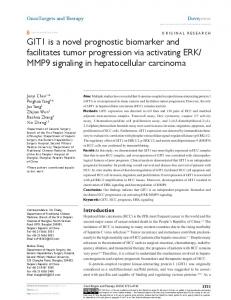
Italicized rows indicate patients who did not experience tumor recurrence post-OLT (n = 12), while non-italicized rows indicate patients who did experience tumor recurrence post-OLT (n = 9)
* These 4 patients have matched primary and recurrent tumor specimens, normal adjacent tissue from the primary and recurrent tumors, and recipient blood specimens (n = 20 total specimens)
AFP alpha-fetoprotein, HBV hepatitis B virus, MULT multiplicity, ENCAP encapsulated, CIRR cirrhosis, THROMB thrombosis, OLT orthotopic liver transplantation, BCLC barcelona Clinical Liver Cancer staging, SAT satellite, VES vessel, Child Child-Pugh score, MELD Model for End-Stage Liver Disease, ±1 Yes, 0 No
Patient OLT inclusion criteria included: ECOG (Eastern Cooperative Oncology Group performance status) score 0–2, tumor within Hangzhou criteria and no major vascular invasion or extra hepatic metastases from imaging studies. Exclusion criteria included absolute contradictions of: involvement of the surrounding tissue or distant metastasis, co-current non-curable extra hepatic malignancies, active infection; relative contraindications included: pulmonary hypertension, symptomatic ischemic heart disease, severe renal insufficiency, and mental disorders
Recurrent with 24 months±
Patient
Table 1 The summary of clinical information for 21 Chinese HCC patients
Xue et al. J Transl Med (2015) 13:379 Page 3 of 15
Xue et al. J Transl Med (2015) 13:379
germline variant and indel calling is provided in Additional file 2, in addition to the following methods: Patient identity, Clonal relationship value derivation, Donor tissue presence in recurrent tumors, Somatic copy number variation (CNV) analysis, Specificity of HERC5 prognostic correlation among genes in chr4q, and Integrated pathway analysis (Additional file 1: Tables S6–S8). RNASeq read mapping and differential expression analysis
RNASeq data was generated by BGI using the Illumina standard library preparation and sequencing protocols [30]. Paired end 90mer sequence FASTQs were provided to MedImmune. Sequence data was QCd using FastQC (v0.10.1), with average read count per mate 50 million. Reads were mapped to reference (UCSC hg19; Feb 2009 release; Genome Reference Consortium GRCh37) using TopHat2 (v2.0.9; 32) using the human reference gtf annotation file (GRCh37.68). Transcript counts were calculated/normalized using htseq-count and DESeq (v1.12.1; 33). DESeq’s negative binomial distribution was used to calculate p-values and fold changes between PT and PNAT as well as RT and RNAT using p 2 as a threshold for the four patients. These results were used in the pathway analyses and combined with the same genes harboring copy number (CN) amplified or deleted regions (see Additional file 2 for CN calling methods). Unadjusted p-values were utilized to simply identify the most differential transcripts within a single patient (PT vs. PNAT) using the fold change magnitude as a primary gene ranking. Since p value calculations were conducted within each patient, there was no replication and statistical power was not adequate to warrant multiple testing adjustment. Tumor cell prevalence was evaluated using ABSOLUTE [34] and verified against pathology assessments for each tumor. RNASeq data was available from 21 patients (9 experienced tumor recurrence post-OLT and 12 did not) with the following specimens: PT and PNAT. Time‑to‑event analyses
Time-to-event analyses were used to correlate the expression of the four genes (NAA11, HERC5, DDX60, and HERC6) identified with tumor recurrence among the 21 Chinese patients’ primary tumors from this study (n = 9 experienced tumor recurrence; n = 12 did not experience tumor recurrence). The expression of each gene in the normal adjacent tissue was subtracted from the tumor expression for each patient (PT-PNAT) individually, then each gene was cut at the median into high or low expression groups. In alignment with REMARK criteria, Kaplan–Meier (KM) analysis, univariate Cox proportional hazards (PH) regression, and multivariate Cox PH
Page 4 of 15
regression analyses were conducted adjusting for HBV status post-OLT, age (binary), gender, and tumor grade. These four variables were the most relevant for potential confounding factors with a molecular prognostic. Cirrhosis status was positive for all but one patient, so this variable was not used in the analysis of these 21 patients. HERC5 was the only significant correlate (p