Material Report
ANTHROPOLOGICAL SCIENCE Vol. 119(1), 87–93, 2011
Hip fractures in the Portuguese archaeological record Francisco CURATE1*, Sandra ASSIS1, Célia LOPES1, Ana Maria SILVA1 1
Research Centre for Anthropology and Health, University of Coimbra, Coimbra 3000, Portugal Received 11 February 2010; accepted 30 March 2010
Abstract Fractures are ubiquitous in the archaeological record but the majority of these are the consequence of a traumatic incident and do not reflect any loss of strength inherent to the bone. So-called fragility fractures, particularly hip fractures, are considered uncommon occurrences in skeletal populations from the past. Nevertheless, evidence of this type of fracture in the archaeological record is increasing. A methodical search for possible hip fractures in the excavation reports, theses and monographs housed in the Department of Anthropology of the University of Coimbra presented an occasion to describe six hip fractures, previously unpublished, from different Portuguese archaeological sites and to challenge the widespread assumption that hip fractures were nearly non-existent in the past. Key words: hip fracture, proximal femur, palaeopathology, Portugal (Cummings et al., 1985; Nurmi et al., 2003; Roche et al., 2005), but the excess mortality due to hip fracture varies with age and sex (Melton, 1995). Increasingly, people who experience hip fracture become institutionalized after acute treatment in hospital (Radley et al., 2008). Hip fractures were clinically recognized in the 16th century by Ambroise Paré (1575). His account of a patient with an intracapsular fracture of the neck of the femur was the earliest case ever to be published in a medical textbook. Although Paré was the first to describe fractures of the hip, it was Sir Astley Cooper (1822) who added the classic discussion of its main problem, the circulation of the femoral head and the condition of what subsequently became recognized as its vascular necrosis. During the 19th century, textbooks by Malgaigne (1842), Smith (1847), Hamilton (1860), and Stimson (1883) made significant advances in the description of the pathological appearance of hip fractures and provided the first statistical data with reference to this type of fracture in the clinical literature. Fractures are omnipresent in paleopathological reports (e.g. Lovejoy and Heiple, 1981; Grauer and Roberts, 1996; Lovell, 1997; Judd and Roberts, 1998; Alvrus, 1999; Jurmain, 1999; Neves et al., 1999; Domett and Tayles, 2006; Djuric et al., 2006; Mitchell, 2006) but the majority of them are the result of a traumatic episode and do not reflect any frailty intrinsic to the bone itself (Dequeker et al., 1997). Fragility fractures, specifically hip fractures, are regarded as unusual occurrences in archaeological skeletal material (Ortner, 2003; Agarwal et al., 2004). A few years ago, Brickley (2002) stated that there were only two published cases of hip fracture in archaeologically derived bone, one from Roman Britain (Roberts and Manchester, 1995) and another from 12th dynasty Egypt (Dequeker et al., 1997), but evidence of fracture of the proximal femur in the archaeological record is growing (e.g. Stroud and Kemp, 1993; Bartonícek and Vlcek, 2001; Campillo, 2001; Ibáñez, 2001; Ferreira and Silva, 2002; Mays, 2006; Buzon and Richman, 2007; Salter-Pedersen, 2007; Ives, 2007; Curate et al.,
Introduction The overall fracture pattern in the population is bimodal, with peaks in the younger and geriatric groups. The fractures responsible for the late peak are often considered ‘osteoporotic fractures,’ ‘fragility fractures,’ or ‘J-Type fractures’ (Melton, 1995; Strømsøe, 2004). These fractures are distinguished epidemiologically by representative characteristics, specifically: connection with moderate trauma at sites containing significant quantities of trabecular bone; incidence rates that increase with age; and prevalence is higher among females than males (Melton, 1995). Osteoporotic fractures are usually associated with a fall to the floor from a standing height (Kannus et al., 1996). It is widely accepted that osteoporosis is the main cause of reduced bone strength, and epidemiological data indicate that low bone mineral density (BMD) values are related with an increased risk of fraction at the population level (Melton, 1995; Marcus, 1996; Grynpas, 2003; Strømsøe, 2004; Sievänen et al., 2007). The worst complications resulting from osteoporosis are fractures of the hip, or fractures of the proximal femur, which contribute disproportionately to the cost, morbidity and mortality attributable to osteoporosis among the elderly (Melton et al., 2003; Nurmi et al., 2003; Mukamal et al., 2007). Such a medical incident can promote a succession of detrimental health consequences including infection, pneumonia, depression, heart failure, and functional dependency (Magaziner et al., 2000; Roche et al., 2005; Lenze et al., 2007; Radley et al., 2008). Between 12% and 32% of all individuals who suffer a hip fracture will perish within 1 year * Correspondence to: Francisco Curate, Research Centre for Anthropology and Health, University of Coimbra, Coimbra 3000, Portugal. E-mail:
[email protected] Published online 8 June 2010 in J-STAGE (www.jstage.jst.go.jp) DOI: 10.1537/ase.100211 © 2010 The Anthropological Society of Nippon
87
88
ANTHROPOLOGICAL SCIENCE
F. CURATE ET AL.
2010). These studies imply that this was undeniably an existing condition in past populations. In addition, Mensforth and Latimer (1989) and Curate (2009) showed that the prevalence of hip fractures in the older adults of the Hamman-Todd and the Coimbra skeletal collections, respectively, is fairly comparable to that found in modern epidemiological studies. Previous studies by Ferreira and Silva (2002), Garcia (2007), Curate (2009), and Curate et al. (2010) confirmed the existence of hip fractures in Portuguese skeletal material since at least the 11th century, but had a limited scope of analysis, both geographically and chronologically. This study aims to collect original information on hip fractures in the Portuguese archaeological record through a careful revision of several previously unpublished palaeodemographic and palaeopathological reports and to challenge the common perception that hip fractures, related or not with bone loss, were nearly absent in past populations.
Materials and Methods This investigation followed two complementary procedures: (1) excavation (field and laboratory) reports, theses, or unpublished monographs housed in the Department of Anthropology of the University of Coimbra were scrutinized with the purpose of identifying proximal femur fractures; and (2) potential hip fractures were confirmed (or not) by actually observing the affected bones. Individuals suspected of having a hip fracture were subject to detailed macroscopical examination, and fractures documented according to clinical protocols (Riggs and Melton, 1986; Nolla and Rozadilla, 2004). Fractures that occurred above a point 5 cm underneath the distal part of the lesser trochanter up to the head of the femur were considered hip fractures (Gillespie, 2001; Nolla and Rozadilla, 2004). There are several categorization schemes for these fractures. Typically, they are classified according to anatomical location: intracapsular or extracapsular (Canale, 1998; Pervez et al., 2002; Brunner et al., 2003). Intracapsular fractures (also known as cervical fractures or fractures of the neck of the femur) occur inside the hip joint capsule, above the lesser and greater trochanters (Nolla and Rozadilla, 2004). Extracapsular fractures (also named as trochanteric or pertrochanteric fractures) occur distal to the hip joint capsule. They can be intertrochanteric or subtrochanteric. In intertrochanteric hip fractures, the fracture line commences at or near the lower part of the junction of the neck and shaft, and goes through the greater and the lesser trochanters. Subtrochanteric fractures occur at the region between the lesser trochanter and a point 5 cm distal (Gillespie, 2001; Nolla
and Rozadilla, 2004; Lee and Ertl, 2008). These fractures are also categorized according to severity and degree of stability; the Garden classification is an emblematic model (Canale, 1998). When possible the crude prevalence (i.e. the proportion of fractures in the total number of individuals) and true prevalence rates (i.e. the proportion of fractures in the total number of femurs of the same side of the fracture) of hip fractures in the analysed samples were calculated (for adult individuals only). Additional radiographic analyses were made in Coimbra University Hospital.
Results A systematic search for potential fracture of the proximal femur in the excavation reports, theses, or unpublished monographs housed in the Department of Anthropology of the University of Coimbra provided an opportunity to describe seven hip fractures, previously unpublished, from six different archaeological sites. Case 1: The tholos of Paimogo I (3000–2500 BC) The tholos of Paimogo I (Lourinhã, central Portugal), a collective burial from the Late Neolithic, was discovered in 1968. The subsequent excavation of this vaulted chamber grave rendered at least 413 individuals, including 290 adults. Despite the fairly good state of preservation, the human bones were found highly disturbed, and without anatomical connections (Silva, 2002, 2003). An incomplete, fragmented right femur (only the proximal third was recovered) exhibits an intracapsular fracture. The bone belongs to an adult, probably a female (Wasterlain, 2000). It was not possible to estimate age at death. The fracture (oblique, transcervical) is well remodelled. The femoral head shows signs of gross modification, with secondary development of osteoarthrosis. Eburnation of the proximal joint is visible. It is noticeable that fracture repair has occurred through the formation of new bone in the anteromedial surface of the neck of the femur. The true prevalence in the Paimogo I study sample for hip fractures in the right femur is 1.3% (1/78, Table 1). Case 2: Paradela churchyard (12th–19th centuries) During the excavation of the old churchyard of Paradela (Barcelos, northwest Portugal) 121 skeletons were recovered (100 adults). Historical and archaeological contexts suggest an occupation period between the 12th and the 19th centuries.
Table 1. True prevalence of hip fractures in various skeletal samples Study reference Curate (2009) Curate et al. (2010) Ives (2007) Present study Present study Present study Present study
Provenience Identified skeletal collection, University of Coimbra, Portugal Santa Clara, Coimbra, Portugal Several sites, UK São Julião, Constância, Portugal São Francisco, Santarém, Portugal Paradela, Portugal Paimogo I, Lourinhã, Portugal
Chronology
n
Fractured
%
19th–20th centuries 14th–17th centuries Post medieval 14th–19th centuries 14th–17th centuries 12th–19th centuries Late Neolithic
98 66 1180 43 103 42 78
2 1 7 1 1 1 1
2.0 1.5 0.6 2.3 1.0 2.4 1.3
Vol. 119, 2011
HIP FRACTURES IN PORTUGAL
89
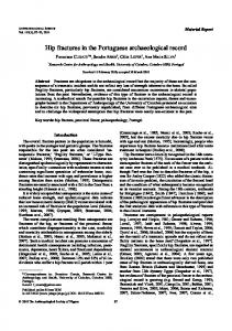
Figure 1. An extracapsular, intertrochanteric, fracture in an old male (Paradela, Barcelos). Figure 2. Increased opacity in the intertrochanteric region.
A hip fracture was observed in an old male (> 50 years old: Ferembach et al., 1980; Lovejoy et al., 1985; Wasterlain, 2000). The skeleton is fairly complete. However, the vertebra, the ribs, the upper limbs and the hip bones are fragmentary. The majority of the long bones from the lower limb are well-preserved. This skeleton shows degenerative lesions in the left shoulder, hip, and costal joints. Severe joint destruction is observed in the right hand and in the left feet. Degenerative focuses are also present in the vertebral fragments. Furthermore, a case of ankylosis is present between the 7th cervical and the 1st thoracic vertebrae, affecting both body and apophyseal joints. The macroscopic study revealed a deformity in the intertrochanteric ridge of the left femur with bone mass deposition, creating an irregular folder. As a result, there is a shortening of the femoral neck (Figure 1). Radiographically, there is an increased opacity of the affected zone (Figure 2). Additionally, a fracture line is seen below the intertrochanteric crest. The lesions described are congruent with an extracapsular fracture, of the intertrochanteric type. At a biomechanical level, this traumatic event caused a visible limb asymmetry with acute involvement of the knee joint, with a Table 2. Study reference Curate (2009) Curate et al. (2010) Mays (2006) Mensforth and Latimer (1989) Present study Present study Present study
possible consequence to the individual’s mobility. The crude prevalence of proximal femur fractures in this sample is 1.0% (1/100, Table 2). The true prevalence is 2.4% (1/42). Case 3: Convent of São Francisco, Santarém (14th–17th centuries) The convent of São Francisco of Santarém (central Portugal) was founded around 1242 and abandoned during the 17th century. During a partial excavation of the church, 132 individuals (103 adults), from both sexes and all age groups, were unearthed (Silva, 1999). An aged female (> 50 years: Ferembach et al., 1980; Lovejoy et al., 1985; Brooks and Suchey, 1990) exhibits a remodeled extracapsular, intertrochanteric, hip fracture of the left femur. The skeleton is incomplete but reasonably well preserved. Moderate osteoarthrosis was noted in the tarsal bones. Although the left femur is broken and incomplete (the distal segment is absent), it is possible to observe the fracture repair in the intertrochanteric region. Several degenerative alterations are present, namely a severe secondary osteoarthrosis in the proximal femoral articulation and the closure of the acetabulum. The true prevalence for hip
Crude prevalence of hip fractures in various skeletal samples Provenience
Identified skeletal collection, University of Coimbra, Portugal Santa Clara, Coimbra, Portugal Ancaster, UK Hamann-Todd Collection, USA São Julião, Constância, Portugal São Francisco, Santarém, Portugal Paradela, Portugal
Chronology
n
Fractured
%
19th–20th centuries 14th–17th centuries 3rd–4th centuries 19th–20th centuries 14th–19th centuries 14th–17th centuries 12th–19th centuries
98 71 16 938 106 103 100
2 1 1 23 1 1 1
2.0 1.4 6.3 2.5 0.9 1.0 1.0
90
ANTHROPOLOGICAL SCIENCE
F. CURATE ET AL.
fractures in this sample is 1.0% (1/103, Table 1). The crude prevalence is also 1.0% (1/103, Table 2). Case 4: Church of São Julião, Constância (14th–19th centuries) In the years of 2002 and 2003, during the urban renewal of the historical centre of Constância (Portugal), 151 skeletons (106 adults and 45 non adults) were unearthed from the Church of São Julião, dated from the 14th–19th centuries (Assis, 2007). Among the several pathological cases, an old female skeleton was observed (> 50 years: Ferembach et al., 1980; Lovejoy et al., 1985; Wasterlain, 2000), with an intracapsular fracture of the left femur. The skeleton is fairly well preserved but incomplete. The preserved joints display degenerative lesions, more conspicuous in the hip bone, and in the lumbar and sacrum vertebrae. The sacrum shows porosity and perforation of the upper body surface. Musculoskeletal stress lesions are present in the upper and lower limbs. In the right leg there is a severe periosteal reaction of unknown aetiology at the site of insertion of the tibia–fibular ligament. An atypical neck angle in the left femur not present in the matched pair was noted. The alterations are characterized by a decrease in the neck length and by a slight medial-posterior rotation of the femoral head. A small bony ridge crosses the femoral neck in the anterior view. Moreover, a bone folder is present in the superior border of the femoral head. These bone abnormalities are compatible with a consolidated cervical (intracapsular) hip fracture. According to the Gardner classification, the head rotation suggests a valgus impaction fracture. The crude prevalence of hip fractures in this studybase is 0.9% (1/106), while the true prevalence is 2.3% (1/ 43). The crude prevalence in older females (> 50 years) is 8.3% (1/12, Table 1). Case 5: Juncal necropolis (16th–20th centuries) Juncal (Porto de Mós) is a small village located in the centre of Portugal. Its old necropolis, closed in the early 20th century, is contiguous to the S. Miguel Church which was built in 1780. In 2006, during an emergency archaeological survey, human bone remains were exhumed from an ossuary. A left femur fragment with severe pathological alterations in the proximal joint was recovered. This femur probably belongs to an adult male (Wasterlain, 2000). The estimation of age at death was impossible due to the absence of the remaining skeleton. Macroscopically the bone lesions are characterized by a medial-posterior rotation of the femur head, levelled at the growth cartilage border, and subsequent reduction of the femoral neck angle. A slight bony callus is noted above the trochanteric fossa. The femur head shows acute degenerative lesions, or osteoarthrosis, with marginal lipping, sclerosis and eburnation. Severe bone growth is present at the site of insertion of the piriform, obturator internus and gemelli muscles. New bone deposition is present in the proximal quarter of the diaphysis. The alterations described are consistent with a subcapital neck fracture (an intracapsular fracture), with a varus rotation of the head. Alternatively, this case might represent a slipped capital femoral epiphysis (Brenkel et al., 1986; Ankarath et al., 2002). Slipped capital epiphysis is a Salter-Harris type 1 fracture
Figure 3. A comminuted fracture at the base of the neck of the femur in an old male (Church of Nossa Senhora da Conceição, Seixal).
through the proximal femoral physis and usually occurs in adolescents at about the growth spurt. There is a sheering force across the epiphysis at the hypertrophic zone that allows a slip, usually in the varus direction (Adler, 2008). Against this differential diagnostic is the fact that this fracture frequently results in osteonecrosis and chondrolysis because of the tenuous nature of the blood supply, which is clearly not the case. Case 6: Church of Nossa Senhora da Conceição, Seixal (18th–19th centuries) The church of Nossa Senhora da Conceição, Seixal (south Portugal), was built in 1500 and partially destroyed in 1755, during the Grande Terramoto de Lisboa (Great Earthquake of Lisbon). It was abandoned in the 19th century. The associated necropolis was used only for a short period, between 1755 and the end of the 19th century (Lopes, 2002). During an emergency excavation 30 adult individuals were recovered (11 females; 10 males). An elderly male (> 50 years: Lovejoy et al., 1985; Brooks and Suchey, 1990; Bruzek, 1991) exhibited a traumatic lesion in the right proximal femur (Figure 3). The total separation of the bone in three pieces is compatible with a comminuted fracture at the base of the neck of the femur (extracapsular fracture). The fracture had not healed although the abundant new bone production proves that some time had elapsed between the moment of the fracture and the death of the individual. A severe non-specific infectious disease affected the whole femur and the tibia. Osteopenia, i.e. generalized bone loss (González-Reimers et al., 2002), was not detected on plain radiological analysis. As such, this fracture is probably secondary to a harsh traumatic episode, like a violent fall or a sturdy blow to the right side.
Discussion In this study we describe six previously unpublished cases of hip fracture, from various Portuguese archaeological sites dating from the Late Neolithic to the beginning of the 20th century. The individuals observed did not show signs of other fractures.
Vol. 119, 2011
The few existing palaeopathological studies which documented hip fractures convey a true prevalence ranging from 0.6% (Ives, 2007) to 2.0% (Curate, 2009), and a crude prevalence ranging from 1.4% (Curate et al., 2010) to 6.3% (Mays, 2006). Intermediary values were reported by Curate et al. (2010), for the true prevalence (Table 1); and Mensforth and Latimer (1989) and Curate (2009), for the crude prevalence (Table 2). Therefore, the crude prevalence rates found in the different samples in the present study are usually lower than the ones reported in the available literature. The true prevalence rates observed in the study samples are similar to the rates found in the literature. Fracture patterns within a population are highly informative, because age and sex influences the occurrence and nature of the trauma (Lovell, 1997). So-called fragility fractures are typically associated with moderate trauma at sites containing significant quantities of trabecular bone; incidence rates that increase with aging; and higher frequency among females (Melton, 1995). In the described hip fracture cases, taken as a whole, males and females were equally affected (two females; two males; one possibly female, and one possibly male). Women display a significantly higher bone fragility, which is a consequence of their smaller-sized bony structures, inferior peak bone mass, rapid bone loss after menopause, thinner cortical thickness, and a greater trabecular disconnection (Orwoll, 2000). In addition, women may incur greater microarchitectural damage than men and adapt less effectively by periosteal apposition, a factor that further contributes to gender differences in bone geometry (Seeman, 2003). Therefore, hip fractures are usually most common in women (Gillespie, 2001; Brunner et al., 2003; Nolla and Rozadilla, 2004) but some modern clinical studies found a higher incidence among males (e.g. Solomon, 1968; Zhang et al., 2000). Four of the affected individuals were older adults (> 50 years), which is in agreement with the general epidemiological expectation that older individuals are more prone to this type of fracture (Anderson and Cooper, 1999; Brunner et al., 2003). With increasing age, modifications taking place in bone quantity, quality and microarchitecture influence the resistance of trabecular bone to local collapse, augmenting fracture risk (Nagaraja et al., 2007). According to anatomical location, three intracapsular and three extracapsular fractures were recorded. In modern epidemiological studies, intracapsular fractures are generally more common than extracapsular fractures (Kannus et al., 1996; Michaëlsson et al., 1999; Zhang et al., 2000), but not always (Maghraoui et al., 2005). These two types of hip fracture are usually considered together in aetiological studies (Michaëlsson et al., 1999). Nevertheless, individuals with a trochanteric fracture (i.e. extracapsular) are generally more osteoporotic than those with cervical hip fractures (i.e. intracapsular), and have higher postfracture mortality (Seeley et al., 1991; Kannus et al., 1996; Nurmi et al., 2003). The elderly individual from the Church of Nossa Senhora da Conceição (Case 6) suffered an extracapsular, comminuted, fracture, and died shortly after, possibly because a secondary infection. Infectious pathogens are responsible for a high mortality rate after hip fracture (Sharma et al.,
HIP FRACTURES IN PORTUGAL
91
2003; Fenton et al., 2008). Mortality in past populations from hip fractures would unquestionably have been much higher (Brickley, 2002), but repair was possible (Stimson, 1883). Of the six described fractures, five were remodelled, suggesting long survival of the individuals after trauma. Furthermore, three of the affected individuals showed signs of eburnation in the head of the femur, which demonstrate the preservation of limb mobility (Stimson, 1883)—although eburnation could be present before the fracture. More than half of the patients who live autonomously before sustaining a hip fracture, face enduring disability and prolonged institutionalization as hip fractures promote total or partial incapacity of the affected limb (Melton et al., 2003; Physician’s Guide, 2003). As such, the remodelling associated with the fractures and the long-term survival of some of these individuals are suggestive of a solid community support, at least during recovery. Proximal femur fractures have been acknowledged as a sign of osteoporosis for more than 150 years (Cooper, 1822). Currently, nearly all hip fractures occur as an outcome of a fall by an individual with reduced bone strength (Melton et al., 2003; Physician’s Guide, 2003; Di Monaco et al., 2006; Mukamal et al., 2007). As such, it is probable that at least some of the described hip fractures are a consequence of bone loss. However, one of the cases (Case 6) is probably the result of a high-energy trauma. Indeed, about one in six hip fractures result from severe trauma (Melton, 1995).
Conclusion Hip fractures are regarded as unusual traumatic events in the archaeological record (Ortner, 2003; Agarwal et al., 2004). Notwithstanding, there are increasing numbers of palaeopathological reports that refer to this type of fracture (e.g. Stroud and Kemp, 1993; Roberts and Manchester, 1995; Dequeker et al., 1997; Bartonícek and Vlcek, 2001; Campillo, 2001; Ibáñez, 2001; Mays, 2006; Ives, 2007; Salter-Pedersen, 2007; Curate et al., 2010). Although the proportion of the population that lived to over 70 years of age was certainly smaller in the past (Brickley, 2002), some people definitely lived into old age (Jackes, 2000), enough to experience osteoporotic fractures (Brickley, 2002). It is apparent from classic medical texts (e.g. Paré, 1575; Cooper, 1822; Malgaigne, 1842; Smith, 1847) and palaeopathological studies focused on identified skeletal collections (Mensforth and Latimer, 1989; Curate, 2009) that fragility fractures occurred with relatively high frequency in aged individuals from populations with lifestyles different from those found nowadays, who obviously did not benefit from modern medicine. This review was intended to expand the information about hip fractures in the archaeological record. While this type of fracture was probably rarer in the past as compared to modern populations, it is increasingly evident that it indeed affected individuals in past communities. A careful revision of old, forgotten, archaeological reports and the directed study of large skeletal series will surely convey additional information about fractures of the proximal femur in the past.
92
ANTHROPOLOGICAL SCIENCE
F. CURATE ET AL.
Acknowledgments The authors wish to thank the Fundação para a Ciência e Tecnologia (Research Grant SFRH/BD/22773/2005) for the financial support of this research.
References Adler B. (2008) Slipped capital femoral epiphysis. eMedicine Radiology. http://emedicine.medscape.com/article/413810-overview. Agarwal S., Dumitriu M., Tomlinson G., and Grynpas M. (2004) Medieval trabecular bone architecture: the influence of age, sex, and lifestyle. American Journal of Physical Anthropology, 124: 33–44. Alvrus A. (1999) Fracture patterns among the Nubians of Semna South, Sudanese Nubia. International Journal of Osteoarchaeology, 9: 417–429. Anderson F. and Cooper C. (1999) The influence of osteoporosis in trauma. Trauma, 1: 181–192. Ankarath S., Giannoudis P., and Scott B. (2002) Delay in diagnosis of slipped upper femoral epiphysis. Journal of the Royal Society of Medicine, 95: 356–358. Assis S. (2007) A memória dos rios no quotidiano dos homens: contributo de uma série osteológica proveniente de Constância para o conhecimento dos padrões ocupacionais. Unpublished Master’s Thesis, Department of Anthropology, University of Coimbra, Coimbra. Bartonícek J. and Vlcek E. (2001) Femoral neck fracture—the cause of death of Emperor Charles IV. Archives of Orthopaedic and Trauma Surgery, 121: 353–354. Brenkel I., Prosser A., and Pearse M. (1986) Slipped capital femoral epiphysis: continuing problem of late diagnosis. British Medical Journal, 293: 256–257. Brickley M. (2002) An investigation of historical and archaeological evidence for age-related bone loss and osteoporosis. International Journal of Osteoarchaeology, 12: 364–371. Brooks S. and Suchey J. (1990) Skeletal age determination based on the os pubis: a comparison of the Acsádi-Nemeskéri and Suchey-Brooks methods. Human Evolution, 5: 227–238. Brunner L., Eshilian-Oates L., and Kuo T. (2003) Hip fractures in adults. American Family Physician, 67: 537–542. Bruzek J. (1991) Fiabilité des procédés de détermination du sexe a partir de l’os coxal. Implications à l’étude du dimorphisme sexuel de l’homme fossile. Ph.D. thesis, Institut de Paléontologie Humaine, Muséum National d’Histoire Naturelle, Paris. Buzon M. and Richman R. (2007) Traumatic injuries and imperialism: the effect of Egyptian colonial strategies at Tombos in Upper Nubia. American Journal of Physical Anthropology, 133: 783–791. Campillo D. (2001) Introducción a la paleopatología. Bellaterra, Barcelona. Canale S. (1998) Campbell’s Operative Orthopaedics. Mosby, St Louis. Cooper A. (1822) A treatise on dislocations and fractures of the joints. Bransby B. Cooper, London. Cummings S., Kelsey J., Newitt M., and O’Dowd K. (1985) Epidemiology of osteoporotic fractures. Epidemiology Reviews, 7: 178–208. Curate F. (2009) Perda de osso cortical e fracturas osteoporóticas na colecção de esqueletos identificados do Museu Antropológico da Universidade de Coimbra. In: Cerdà, M., and Garcia-Prosper, E. (eds.), Investigaciones HistóricoMédicas sobre Salud y Enfermedades en el Pasado. Grupo Paleolab & Sociedad Española de Paleopatología, Valencia, pp. 421–434. Curate F., Lopes C., and Cunha E. (2010) A 14th–17th century osteoporotic hip fracture from the Santa Clara-a-Velha Convent in Coimbra (Portugal). International Journal of Osteoarchaeology, 20: 591–596.
Dequeker J., Ortner D., Stix A., Cheng X., Brys P., and Boonen S. (1997) Hip fracture and osteoporosis in a XIIth Dynasty female skeleton from Lisht, Upper Egypt. Journal of Bone and Mineral Research, 12: 881–888. Di Monaco M., Vallero F., Di Monaco R., Tappero R., and Cavanna A. (2006) Bone mineral density in hip-fracture patients with Parkinson’s disease: a case control study. Archives of Physical Medicine and Rehabilitation, 87: 1459–1462. Djuric M., Roberts C., Rakocevic Z., Djonic D., and Lešic A. (2006) Fractures in late medieval skeletal populations From Serbia. American Journal of Physical Anthropology, 130: 167–178. Domett K. and Tayles N. (2006) Adult fracture patterns in prehistoric Thailand: a biocultural interpretation. International Journal of Osteoarchaeology, 16: 185–199. Fenton P., Singh K., and Cooper M. (2008) Clostridium difficile infection following hip fracture. Journal of Hospital Infection, 68: 376–377. Ferembach D., Schwidetzky I., and Stloukal M. (1980) Recommendations for age and sex diagnosis of skeletons. Workshop of European Anthropologists. Journal of Human Evolution, 9: 517–549. Ferreira M. and Silva A.M. (2002) A case of osteomyelitis in the hip of a Medieval Portuguese male skeleton. Antropologia Portuguesa, 19: 65–70. Garcia S. (2007) Maleitas do corpo em tempos medievais. Unpublished Ph.D. Thesis, Department of Anthropology, University of Coimbra, Coimbra. Gillespie W. (2001) Extracts from “Clinical Evidence”: hip fracture. British Medical Journal, 321: 968–975. González-Reimers E., Velasco-Vázquez J., Arnay-de-la-Rosa M., Santolaria-Fernández F., Gómez-Rodríguez M., and Machado-Calvo M. (2002) Double-energy X-ray absorptiometry in the diagnosis of osteopenia in ancient skeletal remains. American Journal of Physical Anthropology, 118: 134–145. Grauer A. and Roberts C. (1996) Paleoepidemiology, healing and possible treatment of trauma in the medieval cemetery of St. Helen-on-the-Walls, York, England. American Journal of Physical Anthropology, 100: 531–544. Grynpas M. (2003) The role of bone quality on bone loss and bone fragility. In: Agarwal S. and Stout S. (eds.), Bone Loss and Osteoporosis—An Anthropological Perspective. Kluver Academic/Plenum Publishers, New York, pp. 33–44. Hamilton F. (1860) A Practical Treatise on Fractures and Dislocations. Henry C. Lea, Philadelphia. Ibáñez M. (2001) Aspectos antropológicos y paleopatológicos de las inhumaciones prehistóricas del Tabaya (Aspe, Alicante). In: Martín M. and Rodríguez F. (eds.), Dónde estamos? Pasado, presente y futuro de la paleopatología. Universidad Autónoma de Madrid, Madrid, pp. 263–268. Ives R. (2007) An investigation of vitamin D deficiency osteomalacia and age-related osteoporosis in six post-medieval urban collections. Ph.D. Thesis, University of Birmingham. Jackes M. (2000) Building the bases for paleodemographic analysis: adult age determination. In: Katzenberg A. and Saunders S. (eds.), Biological Anthropology of the Human Skeleton. Wiley Liss, New York, pp. 417–466. Judd M. and Roberts C. (1998) Fracture patterns at the Medieval Leper hospital in Chichester. American Journal of Physical Anthropology, 105: 43–55. Jurmain R. (1999) Stories from the Skeleton. Behavioural Reconstruction in Human Osteology. Interpreting the Remains of the Past. Gordon and Breach, Amsterdam. Kannus P., Parkkari J., Sievänen H., Heinoen A., Vuori I., and Järvinen M. (1996) Epidemiology of hip fractures. Bone, 18: 57–63. Lee M. and Ertl J. (2008) Subtrochanteric hip fractures. eMedicine Orthpidic Surgery. http://emedicine.medscape.com/article/ 1247329-overview. Lenze E., Munin M., Skidmore E., Amanda D., Rogers J., and Whyte E. (2007) Onset of depression in elderly persons after
Vol. 119, 2011
hip fracture: implications for prevention and early intervention of latelife depression. Journal of the American Geriatrics Society, 55: 81–86. Lopes C. (2002) Material osteológico exumado do n.°2 da Rua 1.° de Dezembro (Seixal): Estudo laboratorial do material de inumações primárias. Unpublished report. Bioanthropos, Coimbra. Lovejoy C. and Heiple K. (1981) The analysis of fractures in skeletal populations with an example from the Libben Site, Ottowa County Ohio. American Journal of Physical Anthropology 55: 529–541. Lovejoy C.O., Meindl R.S., Pryzbeck T.R., and Mensforth R.P. (1985) Chronological metamorphosis of the auricular surface of the ilium: a new method for the determination of adult skeletal age at death. American Journal of Physical Anthropology, 68: 15–28. Lovell N. (1997) Trauma analysis in paleopathology. Yearbook of Physical Anthropology, 40: 139–170. Magaziner J., Hawkes W., Hebel J., Zimmerman S., Fox K., and Dolan M. (2000) Recovery from hip fracture in eight areas of function. Journal of Gerontology, 55: M498–507. Maghraoui A., Koumba B., Jroundi I., Achemlal L., Bezza A., and Tazi M. (2005) Epidemiology of hip fractures in 2002 in Rabat, Morocco. Osteoporosis International, 16: 597–602. Malgaigne J. (1842) Traité des fractures et des luxations. Baillière, Paris. Marcus R. (1996) The nature of osteoporosis. In: Marcus R., Feldman D., and Kelsey J. (eds.), Osteoporosis. Academic Press, San Diego, pp. 647–659. Mays S. (2006) Age-related cortical bone loss in women from a 3rd–4th century AD population from England. American Journal of Physical Anthropology, 129: 518–528. Melton L., III (1995) Epidemiology of fractures. In: Riggs B. and MeltonL., III (eds.), Osteoporosis—Etiology, Diagnosis, and Management. Lippincott-Raven Publishers, Philadelphia, pp. 225–247. Melton L., III, Gabriel S., Crowson C., Tosteson A., Johnell O., and Kanis J. (2003) Cost-equivalence of different osteoporotic fractures. Osteoporosis International, 14: 383–388. Mensforth R. and Latimer B. (1989) Hamann-Todd Collection Aging Studies: osteoporosis fracture syndrome. American Journal of Physical Anthropology, 80: 461–479. Michaëlsson K., Weiderpass E., Farahmand B., Baron J., Persson P.-G., Zidén L., Zetterberg C., and Ljunghall S. (1999) Differences in risk factor patterns between cervical and trochanteric hip fractures. Osteoporosis International, 10: 487–494. Mitchell P. (2006) Trauma in the Crusader period city of Caesarea: a major port in the medieval eastern Mediterranean. International Journal of Osteoarchaeology, 16: 493–505. Mukamal K., Robbins J., Cauley J., Kern L., and Siscovick D. (2007) Alcohol consumption, bone density, and hip fracture among older adults: the cardiovascular health study. Osteoporosis International, 18: 593–602. Nagaraja S., Lin A., and Guldberg R. (2007) Age-related changes in trabecular bone microdamage initiation. Bone, 40: 973–980. Neves W., Barros A., and Costa M. (1999) Incidence and distribution of postcranial fractures in the prehistoric population of San Pedro de Atacama, North Chile. American Journal of Physical Anthropology, 109: 253–258. Nolla J. and Rozadilla A. (2004) Atlas de osteoporose. Revisfarma, Lisboa. Nurmi I., Narinem A., Luthje P., and Tanninen S. (2003) Cost analysis of hip fracture treatment among the elderly for the public health services: a 1-year prospective study in 106 consecutive patients. Archives of Orthopaedic and Trauma Surgery, 123: 551–554. Ortner D. (2003) Identification of Pathological Conditions in Human Skeletal Remains. Academic Press, San Diego. Orwoll E. (2000) Perspective: assessing bone density in men. Journal of Bone and Mineral Research, 15: 1867–1870. Paré A. (1575) Ouevres. Gabriel Buon, Paris.
HIP FRACTURES IN PORTUGAL
93
Pervez H., Parker M., Pryor G., Lutchman L., and Chirodian N. (2002) Classification of trochanteric fracture of the proximal femur: a study of the reliability of current systems. Injury, 33: 713–715. Physician’s Guide (2003) Physician’s Guide to Prevention and Treatment of Osteoporosis Developed by The National Osteoporosis Foundation, Washington, DC. Radley D., Gottlieb D., Fisher E., and Tosteson A. (2008) Comorbidity risk-adjustment strategies are comparable among persons with hip fracture. Journal of Clinical Epidemiology, 61: 81–87. Riggs B. and Melton L., III (1986) Involutional osteoporosis. New England Journal of Medicine, 314: 1676–1684. Roberts C. and Manchester K. (1995) The Archaeology of Disease. Cornell University Press, Ithaca, NY. Roche J., Wenn R., Sahota O., and Moran C. (2005) Effect of comorbidities and postoperative complications on mortality after hip fracture in elderly people: prospective observational cohort study. British Medical Journal, 331: 1374–1378. Salter-Pedersen E. (2007) A 15th century osteoporotic hip fracture with complications. Poster presented at the 34th Paleopathology Association Annual Meeting (North America), Philadelphia. Seeley D., Browner W., Nevitt M., Genant H., Scott J., and Cummings S. (1991) Which fractures are associated with low appendicular bone mass in elderly women? The Study of Osteoporotic Fractures Research Group. Annals of Internal Medicine, 115: 837–842. Seeman E. (2003) Periosteal bone formation—a neglected determinant of bone strength. New England Journal of Medicine, 349: 320–323. Sharma P., Bomireddy R., and Phillips S. (2003) Clostridium difficile associated diarrhoea after internal fixation of intertrochanteric femoral fractures. European Journal of Clinical Microbiology & Infectious Diseases, 22: 615–618. Sievänen H., Kannus P., and Järvinen T. (2007) Bone quality: an empty term. PLoS Medicine, 4: e27. Silva A.M. (1999) Estudo Paleobiológico dos esqueletos exumados do Convento de São Francisco de Santarém na Campanha de 1996. Unpublished report, University of Coimbra, Coimbra. Silva A.M. (2002) Antropologia funerária e Paleobiologia das populações portuguesas (litorais) do Neolítico final/Calcolítico. Coimbra, Departamento de Antropologia da FCTUC. Ph.D. thesis, Department of Anthropology, University of Coimbra, Coimbra. Silva A.M. (2003) Portuguese populations of the Late Neolithic and Chalcolithic periods exhumed from collective burials: an overview. Anthropologie, XLI/1–2: 55–64. Smith R. (1847) A treatise on fractures in the vicinity of joints, and on certain forms of accidental and congenital dislocations. Hodges and Smith, Dublin. Solomon L. (1968) Osteoporosis and fracture of the femoral neck in the South African Bantu. Journal of Bone and Joint Surgery (Br), 50B: 2–13. Stimson L. (1883) A practical treatise on fractures and dislocations. Lea & Febiger, New York and Philadelphia. Strømsøe K. (2004) Fracture fixation problems in osteoporosis. Injury, 35: 107–113. Stroud G. and Kemp R. (1993) Cemeteries of the church and priory of St Andrew’s, Fishergate. The archaeology of York. The Medieval cemeteries 12/2. Council for British Archaeology, York Archaeological Trust, York. Wasterlain R.S. (2000) Morphé: análise das proporções entre os membros, dimorfismo sexual e estatura de uma amostra da colecção de esqueletos identificados do Museu de Antropologia da Universidade de Coimbra. Master thesis in Anthropology, Department of Anthropology, Coimbra University, Coimbra. Zhang L., Cheng A., Bai Z., Lu Y., Endo N., Dohmae Y., and Takahashi H. (2000) Epidemiology of cervical and trochanteric fractures of the proximal femur in 1994 in Tangshan, China. Journal of Bone Mineraliztion and Metabolism, 18: 84–88.