Scientific Research and Essay Vol. 3 (11), pp. 524-530, November, 2008 Available online at http://www.academicjournals.org/SRE ISSN 1992-2248 © 2007 Academic Journals
Full Length research Paper
Comparative studies on alkaline phosphatase (ALP), alkaline phosphatase amplification (AMP) horseradish peroxidase (HRP), penicillinase (PNC) and avidin – biotin penicillinase amplification ELISA in detection of maize streak virus (MSV) in maize plants and Cicadulina mbila (leafhoppers) insect vector Abolade S. Afolabi1* and George Thottappilly1,2 1
Biotechnology Advanced Laboratory, Sheda Science and Technology Complex, PMB 186, Abuja, FCT, Nigeria. Biotechnology, Sahrdaya College of Engineering, Kodakara. Residence: Teekay Villa, P. O. Karoor, Via. Thazhekkad680 697, Trichur Dist., Kerala State, India.
2
Accepted 13 November, 2008
Comparative study of dilution end point on various detecting enzymes in ELISA techniques to determine sensitivity level for detection of Maize Steak Virus (MSV), both in the maize plant and the insect vector Cicadulina mbila (leafhopper), was carried out using Alkaline phosphatase (ALP), Horseradish peroxidase (HRP), Alkaline phosphatase amplification (AMP) and Penicillinase (PNC) and Avidin- Biotin Penicillinase amplification based ELISA. Assays with the PNC-based ELISA were as sensitive as analogous assays performed with ALP and HRP for the virus in infected maize plant. The HRP and the Penicillinase- based ELISA were able to detect virus in three and crushed leafhoppers respectively. Only the Avidin- Biotin Penicillinase amplification technique was able to detect viruses in one single leafhopper. As expected, the ALP and the alkaline phosphatase enzymatic amplification systems were not useful in virus detection in the insect vector due to the high background of the healthy leafhoppers. This could be due to insects containing their own phosphatase stem. The protein A coat Penicillinase technique was not useful due to nonspecific reaction. Key words: Enzyme Linked Immunosorbent Assay, maize streak virus, Cicadulina mbila, alkaline phosphatase, alkaline phosphatase amplification, horseradish peroxidase, penicillinase based ELISA. INTRODUCTION Enzyme linked Immunosorbent Assay (ELISA), developed as alternatives to radio- immunoassay, is a simple and versatile serological assay based on antigen – antibody reaction where antibody or antigen is adsorbed on a solid phase. ELISA makes use of enzymes attached to one of the reactants (conjugate) to allow quantification,
*Corresponding author: E mail:
[email protected];
[email protected]. Tel: +234 (0) 803 488 1092 or +234 (0)1 470 5247
through color development after addition of a suitable substrate/chromogen. It can detect the presence of antigens that are recognized by an antibody or test for antibodies that recognize an antigen. Pioneered independently in 1971 by Van Weemen et al. The microtitre form of the technique was extended to plant viruses diagnosis by Clark and Adams (1977) using the technique of Voller et al., (1974). The search for a rapid, sensitive, reproducible and inexpensive method of virus detection in plants especially in the developing countries necessitated a cheaper reporter' enzyme (and its substrate) and a simplified, cost
Afolabi and Thottappilly
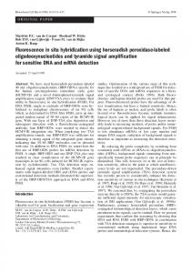
effective and sensitive ELISA procedure. Two enzymes that have been conventionally used in ELISA for the detection of plant viruses are alkaline phosphatase (ALP) (Voller et al., 1974, Joshi et al., 1978, Voller et al., 1978) and horseradish peroxidase (HRP) (Rennard et al., 1980, Tsang et al., 1995, Polák and Caristek, 2008) both of which have problems associated with their use. These problems include high cost, limited availability, the potential carcinogenic activity of some of the substrates and the hazardous nature of the hydrolyzed products of the substrates. Penicillinase based ELISA has also been demonstrated to be useful in identification of viruses (Yolken et al., 1984; Sudarshana and Reddy, 1989; Singh and Barker, 1992). The use of Biotin - Avidin / Streptavidin complex has also been used to amplify ELISA to detect low concentration of viral particles (Zrien et al., 1986; Nielson et al., 1987). Avidin is a basic glycoprotein (MW 68 Kd) which has a high affinity (dissociation constant of avidin is measured to be KD 10-15 M - Wilchek and Bayer 1989) for the small (MW 24Kd) water- soluble vitamin biotin (vitamin H). Biotin, which has four binding sited for Avidin and can be conjugated (biotinylation) to a variety of biological molecules, including antibodies and enzymes. The biotinylated protein can thus bind to more than one molecule of avidin. Maximum of three enzyme- avidin complex can then bind to biotinylated molecules resulting in amplification. This property is responsible for amplification when avidin-biotin complex is used for immunological detection. However, avidin has two distinct disadvantages when used in immuno-cytochemical detection systems. It has a high iso-electric point of approximately 10, and therefore, is positively charged at neutral pH. Consequently, it may bind non-specifically to negatively charged structures. The second disadvantage is that avidin is a glycoprotein and reacts with molecules such as lectins via the carbohydrate moiety. These two problems are overcome with the substitution of Streptavidin; a protein (MW 60 Kd) isolated from the bacterium Streptomyces avidinii, for avidin. Streptavidin has an iso-electric point close to neutral pH and therefore possess few strongly charged groups at the near neutral pH used in immuno-cytochemical detection systems. Furthermore, Streptavidin is not a glycoprotein and therefore does not bind to lectins. The physical properties of Streptavidin therefore make this protein much more desirable for use in immuno-cytochemical detection systems than Avidin. In the ELISA Amplification System, bound alkaline phosphatase label catalyses the dephosphorylation of nicotin+ amide adenine dinucleotide phosphate (NADP ), (instead + of p-nitro-phenyl phosphate substrate); the NAD so + formed then catalytically activates an NAD -specific redox cycle, yielding an intensely colored formazan dye (Figure 1). Each molecule of product from the first reaction takes part in many cycles of the second reaction thus amplification (Stanley et al., 1985). ß lactamase, generally known as penicillinase (PNC) has
525
several features that make it a very desirable enzyme label for ELISA tests. These include a relatively small molecular weight (24,000 Dalton), a high turnover rate, not too expensive and is more readily available (together with its substrate Penicillin G) in developing countries than other enzymes. In addition, its hydrolyzed products are not known to be hazardous. Consequently, the Penicillinaseenzyme based ELISA was compared with alkaline phosphatase, alkaline phosphatase amplification (based on the affinity of Avidin/Streptavidin for biotinylated molecule for amplification), and horseradish peroxidase for sensitivity and suitability in the detection of maize steak virus both in plant and in Cicadulina insect vector. MATERIALS AND METHODS Sources of maize plants and Cicadulina mbila (insect vector) infected with maize streak virus (MSV) Maize streak virus was obtained from the test plants routinely kept in the virology screen-house of the International Institute of Tropical Agriculture (IITA), Ibadan. The insects (vector) Cicadulina mbila (leafhoppers) used in this work were obtained from the facility of the IITA Entomology Unit. C. mbila (leafhoppers) were made viruliferous by feeding them on maize streak virus infected maize plants. Healthy leafhoppers were raised on millet and prior to use (5 to 7 days), the millet pots were removed and replaced by pots of healthy maize plants in their cage for feeding for 2 days. The pots of maize plants were then removed (replaced with millet again) and transferred into an insect-proof cage where the plants were checked for maize streak virus (MSV) symptoms. These non- viruliferous leaf hoppers were used as negative controls in the experiment conducted alongside with viruliferous leafhoppers. Antisera and ELISA conjugates All antisera used were polyclonal antibodies produced in rabbits immunized with preparation of purified virus particles at the Virology laboratory of the International Institute of Tropical Agriculture (IITA), Ibadan. The protein A-Horseradish peroxidase conjugate, the anti rabbit gamma globulin-Alkaline phosphate conjugate, enzyme Penicillinase, Avidin, Biotin and goat anti-rabbit Fc specific antibody were purchased from Sigma Chemical Company, Sigma-Aldrich Company Ltd., The Old Brickyard, New Road, Gillingham, Dorset, SP8 4XT (UK). There were three separate and different conjugations (1) Penicillinase to the goat anti-rabbit Fc specific antibody (2) Avidin to goat anti-rabbit Fc specific antibody and (3) Biotin to enzyme Penicillinase. All performed according to the protocol of Singh and Barker (1992). Briefly, a ratio of 1:0.05 (gamma globulin: enzyme, Avidin and Biotin) was dissolved in distilled water and put in a dialysis tube and tied. Dialysis bag was then suspended in 500 ml half strength Phosphate saline buffer (PBS) with 1 ml of 25% Glutaraldehyde (final concentration of 1%). Dialysis was carried on until straw color began to appear. The tube was then taken out and dialyzed against fresh 500 ml of 1/2 strength PBS, three times to remove the remaining and unreacted Glutaraldehyde. The conjugate was diluted with distilled water (to 10 ml) and bovine serum albumin at 5 mg/ml was added and stored in the refrigerator. Before use, the conjugate and antibody were titrated as in checkerboard to determine the appropriate com- bination to be used in ELISA tests. Enzyme linked immunosorbent assay (ELISA) procedure All tests were carried out using polystyrene microtitre plates
526
Sci. Res. Essays
NADH
P iodonitrotetrazolium violet Diaphorase
Red color
Acetaldehyde
Alcohol dehydrogenase
NAD
Ethanol
AP
NADP Figure 1. Enzyme Amplification system: Bound Alkaline phosphatase acts directly on the substrate NADPH whose product initiates a secondary cyclic enzyme reaction resulting in a colored product. The process is repeated several times to give amplification.
(Dynateck®). To determine the optimum condition for both the Horseradish peroxide and Penicillinase conjugate system, a checkerboard titration was performed (not shown). The optimum titre of 1:1000 of coating antibody and 1:500 conjugate for the Horseradish peroxide while that of the two Penicillinase- based ELISA was 1:1000 antibody and 1:1000 conjugate. All dilutions were made with phosphate buffered saline (PBS) containing one or two drops of Tween 20. For the Horseradish peroxidase, the micro-titre plates were coated with protein A (1:1000 dilution) using carbonate coating buffer with 2 drops of Tween 20 and incubated at 37oC for 2.5 hours. This was followed by washing 3 times using PBS-Tween. The plates were then tapped dried. The virus infected plant samples and controls were crushed in phosphate buffered saline (PBS) extraction buffer containing (PVP) (PBS-Tween + 2% PVP) at 1:5 dilution (w/v) for the healthy and diseased plants at two dilutions (1:5 and 1:25). Healthy leafhoppers were at two dilutions of 1:10 and 1:25 while the diseased hoppers were in three dilutions (1:10, 1:25 and 1:50). The samples of the 'plate Ag trapped ELISA were extracted in coating buffer with 2% PVP. The plates were incubated at 4oC overnight, washed three times with PBS-Tween and blocked with 5% Carnation® non-fat milk incubated at 37oC for 30 min. After washing, the detecting antibody was added at dilution of 1:1000 and incubated at 37oC for 2.5 h. After washing 3 times with PBS-Tween, the conjugates were added at appropriate concentration 1:500, 1:1000 and 1:1000 Horseradish peroxidase, alkaline phosphatase and Penicillinase- based respectively. All Plates were incubated for 2.5 h at 37oC. Substrate preparation Penicillinase 20 mg Bromthymol blue (BTB) was dissolved in 50 ml 0.2 m NaOH and neutralized to pH 7.2 starting initially with concentrated HCl, and using it more and more dilute. The volume was made up to 100 ml with distilled water. This can then be stored at 4oC. It is stable for approximately 90 days. Before use, 2 mg/ml Penicillin-G was added and the pH readjusted to 7.2. 200 uL was added to each well.
Peroxidase 10 mg/ml of 3, 31 5, 51 Tetramethyl-benzidine was dissolved in Dimethyl sulfoxide and stored at 4oC. 1 ml of Na Acetate/Citric acid buffer pH 5.8, 100 uL of Tetramethyl-benzidine and 10 uL of 6% H2O2 were added and made up to 10 ml with distilled water. This was used fresh. The blue color produced after reaction was not stopped, but read spectro-photometrically. Alkaline phosphate 1 mg/ml Para nitro-phenyl phosphate was dissolved in 9.7% Diethanolamine pH 9.8 buffer. 100 uL was used for each well. Readings were taken visually and spectrophotometrically at 30 min, one hour and after 4oC overnight incubation intervals. Alkaline phosphate was read at 405 nm, Horseradish peroxidase (nonstopped reaction) at 625 or 655 nm and Penicillinase at 625 (or 655 nm). All experiments were repeated three times. Alkaline phosphate amplification Instead of using p-nitro-phenyl phosphate as a substrate, Amplifier solution and made as follows was used: 1.100 mg iodonitrotetrazolium violet (INT) (mol wt 505.7), Sigma grade 1, and 1.2 ml ethyl alcohol (200 proof) was dissolved using a stirring bar at 20oC for 10 - 15 min in a total volume of 40 ml 0.025 M NaPO4 buffer, pH 7.0, with 0.02% NaN3. 2. After the material is completely dissolved, 1.2 mg diaphorase (EC 1.6.4.3) (from microorganisms), Boehringer (Indianapolis, Md.) and 7.5 mg alcohol dehydrogenase (EC 1.1.1.1), Boehringer was added and gently swirled to dissolve. 3. This was enough for eight 96- well plates, using 50 ul/well, and used within several hours. 4. The reaction was allowed to proceed for 35 – 45 min at room temperature. The intensely colored formazan dye product from reaction was read at 492 nm. (The longer the incubation, the more NAD that was generated and the greater was the signal produced by the amplification step).
Afolabi and Thottappilly
527
Table 1. Absorbance values of alkaline phosphatase ELISA (405 nm). Hydrolysis of p-nitro-phenyl phosphate to pnitro phenol resulted in color change from colorless to yellow. Since yellow absorbs maximally at 405 nm, there is, therefore, an increase in absorbance from negative (colorless) to positive (yellow).
Maize plant Dilution of plant leaf extract (w/v) Healthy Infected Threshold = 2 x O. D. of Negative reading 1 : 10 0.001 1.025 = , > 0.002 1 : 20 0.001 0.709 = , > 0.002 1 : 100 0.000 0.055 = , > 0.002 0.1 g of Viruleferous Cicadulina mbila (leafhopper) crushed in buffer Dilution of insect extracts (w/v) Healthy Infected Threshold = 2 x O. D. of Negative reading 1 : 10 0.137 0.253 =, > 0.274 1 : 20 0.125 0.194 =, > 0.250 1 : 100 0.098 0.125 =, > 0.196 Unit of Viruleferous Cicadulina mbila (leafhopper) Dilution of insect extracts (w/v) Healthy Infected Threshold = 2 x O. D. of Negative reading Insect crushed in 105 µl of buffer 3 leafhoppers 0.065 0.098 =, > 0.13 2 leafhoppers 0.048 0.057 =, > 0.096 1 leafhopper 0.008 0.009 =, > 0.16
RESULTS Using dilution end-point for comparison, Tables 1 and 5 clearly showed that alkaline phosphatase and alkaline phosphatase enzyme amplification system, while able to detect the virus in the maize plants, were not able to detect the maize streak virus in the leafhopper insect vector. The background signal was high. This could be because insects do have alkaline phosphatase, which, consequently interfere with this procedure. It is also worth mentioning that the reaction in the enzyme amplification system was rapid. The reaction was readable visually within ten minutes of addition of the substrate complex as compared to minimum of 45 min with regular alkaline phosphatase ELISA. The Horseradish peroxidase system was not only sensitive in the detection of the virus in the maize plant at a dilution of 1:100 (like the alkaline phosphatase), it was also able to detect the virus in all crushed and diluted virus samples of 0.1 g crushed leafhoppers. However, it was unable able to detect virus in two and single leafhopper, but able to do so in three leafhoppers crushed together (Table 2). The Penicillinase ELISA (Table 3) was equally able to detect virus in maize plant, 1:100 dilution of 0.1 gm crushed leafhoppers and like the HRP three leafhoppers crushed together. However, the color formation was pronounced and readable within one hour. However, for positive samples, there is a decrease in absorbance value, rather than increase as seen in the Alkaline and Horseradish based reaction. This is because, blue color, which is negative absorbs maximally at 630 nm and as it turns yellow (positive), there is corresponding decrease in absorbance.
However, the Avidin-Biotin- penicillinase amplification ELISA was more sensitive in the detection of the virus in a single C. mbila (leafhoppers) insect vector Table 4. The protein A coat Penicillinase technique was not useful due to non-specific reaction (data not shown). DISCUSSION Enzyme-linked Immunosorbent Assays (ELISAs) combine the specificity of antibodies with the sensitivity of simple enzyme assays, by using antibodies or antigens coupled to an easily assayed enzyme thus providing a useful measurement of antigen or antibody concentration. There are two main variations on this method: (a) Direct detection of the presence of antigens that are recognized by an antibody /antibodies (b) Indirect detection of antigen by detecting the antibodies that has reacted with it using anti gamma globulin. Substrates are critical for the detection and visualization steps of an ELISA. The choice of substrate depends on a number of factors, including the degree of sensitivity required, different instrumentation and filter requirements, safety issues, and choice of enzyme. Most commonly used enzymes are Horseradish peroxidase (HRP) and alkaline phosphatase (AP). HRP has been shown historically to be more sensitive than AP primarily due to its faster catalytic rate. However, HRP reactions may be self-limiting due to substrate inhibition of enzyme. In contrast, AP exhibits a slower catalytic rate, but is not selflimiting. In addition, reaction rates remain linear over long periods; therefore, sensitivity can be improved by allowing the reaction to proceed for a longer time. AP substrates also tend to be less toxic and thus easier to handle. Peroxidases catalyze the oxidation of a wide variety
528
Sci. Res. Essays
Table 2. Absorbance values of Horseradish peroxidase ELISA (655 nm). Enzymatic product of horseradish peroxidase activity resulted in color change from colorless to blue. Blue absorbs maximally at 655 nm, hence there is also increase in absorbance from negative (colorless) to positive (blue)
Maize plant Dilution of plant leaf extract (w/v) Healthy Infected Threshold = 2 x O. D. of Negative reading 1 : 10 0.020 1.240 =, > 0.040 1 : 20 0.010 0.802 =, > 0.020 1 : 100 0.010 0.080 =, > 0 .020 0.1 gm of Viruleferous Cicadulina mbila (leafhopper) crushed in buffer Dilution of insect extracts (w/v) Healthy Infected Threshold = 2 x O. D. of Negative reading 1 : 10 0.019 0.950 =, > 0.038 1 : 20 0.013 0.930 =, > 0.026 1 : 100 0.010 0.630 =, > 0.020 Unit of Viruleferous Cicadulina mbila (leafhopper) Dilution of insect extracts (w/v) Healthy Infected Insect crushed in 105 µl of buffer Threshold = 2 x O. D. of Negative reading 3 leafhoppers 0.025 0.500 =, > 0.050 2 leafhoppers 0.018 0.320 =, > 0.036 1 leafhopper 0.075 0.093 =, > 0.150
Table 3. Absorbance values of Penicillinase based ELISA (655 nm). Enzymatic hydrolysis of penicillin to penicilloic acid resulted in lowering of pH. There was a color change of the Bromthymol blue, pH indicator (neutral) to yellow (acidic). Blue color absorbs maximally at 630 / 655, hence there is a decrease in absorbance from blue (negative) to yellow (positive)
Maize plant Dilution of plant leaf extract (w/v) Healthy Infected Threshold = 2/3 of Negative reading 1 : 10 1.902 0.283 =,, < 1.27 1 : 20 1.930 0.531 =, < 1.29 1 : 100 1.903 0.689 =, < 1.27 0.1 gm of Viruleferous Cicadulina mbila (leafhopper) crushed in buffer Dilution of insect extracts (w/v) Healthy Infected Threshold = 2/3 x O. D. of Negative reading 1 : 10 1.877 0.379 =, < 1.25 1 : 20 1.823 0.432 =, < 1.22 1 : 100 1.933 0.533 =, < 1.29 Unit of Viruleferous Cicadulina mbila (leafhopper) Dilution of insect extracts (w/v) Healthy Infected Insect crushed in 105 µl of buffer Threshold = 2/3 x O. D. of Negative reading 3 leafhoppers 1.155 0.530 =, < 0.77 2 leafhoppers 1.153 0.997 =, < 0.77 1 leafhopper 1.162 1.022 =, < 0.77
of substrates, including benzidine and other aromatic amines. However, benzidine is carcinogenic in animal and man (Haley, 1975) and mutagenic to bacteria following activation by hepatic microsomes in the Ames test (Ames et al., 1972). Originally, o-phenylene diamine (OPD) was the best chromogen for horseradish peroxidase but today, the use of 3, 3’, 5, 5’- tetramethylbenzidine as non-mutagenic chromogen for the end point determination has been a major improvement. However,
it is expensive especially for the developing world. In the Penicillinase based ELISA, the Penicillinsubstrate is hydrolyzed to Penicilloic acid. Change in the pH is observed when the Bromthymol blue indicator changes from blue to greenish tinge to yellow from neutral to acidic pH. This hydrolyzed product is not considered to be toxic compared to Para nitro phenol produced in the alkaline phosphatase based ELISA. Moreover, the Penicillinase based ELISA reagents are considerably cheaper
Afolabi and Thottappilly
529
Table 4. Absorbance values of Avidin-Biotin Penicillinase based ELISA (630 nm). Enzymatic hydrolysis of penicillin to penicilloic acid resulted in lowering of pH. There was a color change of the Bromthymol blue, pH indicator (neutral) to yellow (acidic). Blue color absorbs maximally at 630 / 655, hence there is a decrease in absorbance from blue (negative) to yellow (positive).
Maize plant Dilution of plant leaf extract (w/v) Healthy Infected Threshold = 2/3 x O. D. of Negative reading 1 : 10 1.860 0.111 =, < 1.24 1 : 20 1.860 0.172 =, < 1.24 1 : 100 1.829 0.244 =, < 1.22 0.1gm of Viruleferous Cicadulina mbila (leafhopper) crushed in buffer Dilution of insect extracts (w/v) Healthy Infected Threshold = 2/3 x O. D. of Negative reading 1 : 10 1.886 0.201 =, < 1.26 1 : 20 1.866 0.213 =, < 1.24 1 : 100 1.593 0.359 =, < 1.06 Unit of Viruleferous Cicadulina mbila (leafhopper) Dilution of insect extracts (w/v) Healthy Infected Threshold = 2/3 x O. D. of Negative reading Insect crushed in 105 µl of buffer 3 leafhoppers 1.312 0.388 =, < 0.87 2 leafhoppers 1.259 0.439 =, < 0.84 1 leafhopper 1.517 0.497 =, < 1.01
Table 5. Absorbance values of Alkaline Phosphatase -Enzymatic Amplification System based ELISA (492 nm) (AMP). Reduced form of nicotinamide adenine dinucleotide phosphate NADPH), is dephosphorylated to reduced nicotinamide adenine dinucleoide (NADH). NADH activates a secondary enzyme system, which reduces a tetrazolium salt (iodonitrotetrazolium violet or INT-violet) to form an intensely colored formazan dye.
Maize plant Dilution of plant leaf extract (w/v) Healthy Infected Threshold = 2 x O. D. of Negative reading 1 : 10 0.092 1.172 =, > 0.184 1 : 20 0.078 1.076 =, > 0.156 1 : 100 0.073 0.652 =, > 0.146 0.1gm of Viruleferous Cicadulina mbila (leafhopper) crushed in buffer Dilution of insect extracts (w/v) Healthy Infected Threshold = 2 x O. D. of Negative reading 1 : 10 0.369 0.746 =, > 0.738 1 : 20 0.259 0.511 =, > 0.518 1 : 100 0.300 0.300 =, > 0.600 Unit of Viruleferous Cicadulina mbila (leafhopper) Dilution of insect extracts (w/v) Healthy Infected Threshold = 2 x O. D. of Negative reading Insect crushed in 105 µl of buffer 3 leafhoppers 0.389 0.532 =, > 0.778 2 leafhoppers 0.307 0.499 =, > 0.614 1 leafhopper 0.300 0.399 =, > 0.600
and has the added advantage that the color change from blue (negative) to yellow (positive) is more pronounced and can be easily visualized without the need for a spectrophotometer plate reader. All these make Penicillinase particularly useful in developing world where such instruments are, although not so expensive, are not readily available in every laboratory. In this work, the Penicillinase-based ELISA assays were found not only to be equally sensitive as those assays with ALP and HRP in
the detection the maize steak virus. in detecting virus in plants but also, better in the detection of viruses in Cicadulina mbila insect vector. REFERENCES Ames BN, Sims P, Grover PL (1972). Epoxides of Carcinogenic Polycyclic Hydrocarbons Are Frameshift Mutagens. Sci. 176: 47-49. Clark MF, Adams AN (1977). Characteristics of the Microplate Method of Enzyme-Linked Immunosorbent Assay for the Detection of Plant
530
Sci. Res. Essays
Viruses. J. Gen. Virol. 34: 475-483. Haley TJ (1975). Benzidine revisited: a review of the literature and problems associated with the use of benzidine and its congeners. Clin. Toxicol. 8: 13-42. Joshi UM, Raghocan V, Zemro G, Sheth AR, Borkar PS, Ramechandran S (1978). In: S.B. Pal (Ed) Enzyme labeled immunoassay of hormones and drugs. pp. 233-245. Nielson CM, Hanson K, Anderson HMK, Gerstoft J, Vestergaard BF (1987). A biotin avidin amplified inhibition enzyme immunoassay for detection of CMV antibodies in human serum. J. Virol.l Methods 16: 195 208. Polák J, Caristek R (2008). Use of Horseradish-Peroxidase Labelled Antibodies in ELISA for Plant Virus Detection. J. Phytopathol. 122 (3): 200-207. Rennard SI, Berg R, Martin GR, Foidart JM, Robey PG (1980). Enzyme-linked immunoassay (ELISA) for connective tissue components. Anal. Biochem.1; 104(1): 205-214. Singh S, Barker H (1992). Comparison of penicillinase-based and alkaline phosphatase-based enzyme-linked immunosorbent assay for the detection of six potato viruses. Potato Res. 34: 451–457. Stanley CJ, Johannsson A, Self CH (1985). Enzyme amplification can enhance both the speed and the sensitivity of immunoassays. J. Immunol. Methods, 83: 89-95. Sudarshana MR, Reddy DVR (1989). Penicillinase based enzyme linked Immunosorbent assay for detection of plant viruses. J. Virol. methods 26: 45-52.
Tsang VCW, Greene RM, Pilcher JB (1995). Optimization of the Covalent Conjugating Procedure (NaIO4) of Horseradish Peroxidase to Antibodies for Use in Enzyme-Linked Immunosorbent Assay. Journal of Immunoassay and Immunochemistry, 16 (4): 395 – 418.van Weemen BK, Schurs AH (1971). Immunoassay using antigen-enzyme conjugates. FEBS Lett 15: 232-236. Voller A, Bartlett A, Bidwell DE (1978). Enzyme immunoassays with special reference to ELISA techniques. J. Clin. Pathol. 31(6): 507– 520. Voller A, Bidwell D, Huldt G, Engvall E (1974). A microplate method of enzyme-linked immunosorbent assay and its application to malaria. Bull World Health Organ. 51(2): 209–211. Yolken RH, Wee SB, Regenmottel MV (1984). The use of beta lactamase in enzyme immunoassay for detection of microbial antigens. J. Immunol. Methods 73: 109-123. Zrien M, Burckand J, van Regenmortel MV (1986). Use of the Biotin Avidin system for detecting a broad range of serologically related viruses by ELISA. J. Virol. Method 13: 121-128.