Cell Host & Microbe
Review Host Immune Response to Infection and Cancer: Unexpected Commonalities Romina S. Goldszmid,1 Amiran Dzutsev,1,2 and Giorgio Trinchieri1,* 1Cancer
and Inflammation Program, National Cancer Institute, Frederick, MD 21702, USA Biomedical Research, Inc., Frederick, MD 21701, USA *Correspondence:
[email protected] http://dx.doi.org/10.1016/j.chom.2014.02.003 2Leidos
Both microbes and tumors activate innate resistance, tissue repair, and adaptive immunity. Unlike acute infection, tumor growth is initially unapparent; however, inflammation and immunity affect all phases of tumor growth from initiation to progression and dissemination. Here, we discuss the shared features involved in the immune response to infection and cancer including modulation by commensal microbiota, reactive hematopoiesis, chronic immune responses and regulatory mechanisms to prevent collateral tissue damage. This comparative analysis of immunity to infection and cancer furthers our understanding of the basic mechanisms underlying innate resistance and adaptive immunity and their translational application to the design of new therapeutic approaches. Introduction Organisms resist infections by establishing barriers and activating different classes of innate resistance and adaptive immunity. The tissue in which the infection occurs regulates the strength, quality, and type of the immune response for efficient pathogen eradication and tissue repair while limiting collateral tissue damage (Matzinger and Kamala, 2011). However, some microbes have evolved evasion mechanisms that thwart these host responses, resulting in uncontrolled or chronic infection associated with pathologic damage. Unlike the traditional view of immunity as a response to foreign microbes and molecules, the primary trigger for innate resistance, which is followed by adaptive immunity in higher organisms, may be the sensing of altered self, such as changes in tissue homeostasis or integrity (Matzinger, 2002; Medzhitov, 2008). Innate receptors respond to microbe-associated molecular patterns (MAMPs) and also endogenous mediators of inflammation released by damaged tissues (damage-associated molecular patterns [DAMPs]). These tissue-intrinsic inflammatory responses are amplified by the recruitment of hematopoietic cells that express innate receptors for exogenous and endogenous ligands present in the inflamed tissue and set the stage, via antigen presentation and activation of T and B cells, for adaptive immunity. Tumors may originate at sites of chronic inflammation that are either caused by infectious pathogens or often aseptic. Nevertheless, tumors eventually establish an almost symbiotic relationship with their host that mimics chronic infections or cohabitation with commensal microorganisms and maintain their presence by suppressing excessive inflammation and antitumor immune responses. The molecular alterations in the transformed cells or the surrounding epithelial and stromal cells are associated with a proinflammatory wound-healing microenvironment that, in addition to providing tumor growth and angiogenic factors, affects tissue remodeling and decreases the expression of molecules involved in cell-to-cell adhesion, initiating the epithelial-mesenchimal progression, which can lead to tumor cell invasion and progression (Edme et al., 2002; Sal-
cedo et al., 2013). Nucleic acids and other products released upon cell stress or death in more advanced tumors or, following cytotoxic therapy, are recognized by innate sensors, resulting in the inflammation and activation of antigen-presenting cells (APCs) (Ma et al., 2013b). Thus, like infection, tumors influence and are affected by inflammation and immunity from their early initiation to progression, dissemination, and eventually death of the host. Cancer-associated inflammation not only affects local tumor growth and dissemination but is also responsible for severe cancer-associated comorbidities, such as anorexia, cachexia, and immunosuppression, making cancer a systemic, rather than localized, disease (Trinchieri, 2012). Malignant growth originates within differentiated tissues and maintains some of the functional, morphologic, and immune traits of the tissue of origin, representing a ‘‘caricature’’ of the original tissue (Pierce and Speers, 1988). The immune response elicited by the tumor will be, in most cases, unable to eradicate it and will establish a Darwinian environment that selects the genetically fittest cancer cells (tumor editing and escape) that evolve into aggressive malignant tumors or, in some cases, remain temporarily in equilibrium with nonmalignant host cells (Schreiber et al., 2011). Given the parallels between immunity to infection and cancer, this review focuses on the similarities and differences in these processes and how these two fields can help each other in advancing our understanding of innate resistance and immunity. Inflammation and Cancer Inflammation and immunity are inherent characteristics of cancer, and avoiding immune destruction and tumor-promoting inflammation are now among the hallmarks of cancer (Hanahan and Weinberg, 2011). Up to a quarter of human cancers are related to infection or infection-associated chronic inflammation. Helicobacter pylori, human papillomaviruses (HPVs), EpsteinBarr virus (EBV), and hepatitis B and C viruses are the most common etiopathogenic factors (Parkin, 2006). Some oncogenic pathogens directly transform the tumor-forming cells (e.g., EBV or HPV), whereas others (e.g., H. pylori) establish an Cell Host & Microbe 15, March 12, 2014 ª2014 Elsevier Inc. 295
Cell Host & Microbe
Review inflammatory milieu favoring tumor generation. Other cancers initiate in tissues chronically inflamed for causes other than infection; e.g., tissue injury by physical or chemical insults or genetic diseases. Altered composition (dysbiosis) of the intestinal microbiota or its physical interaction with hematopoietic cells also regulate inflammation and has been shown to be a cause of cancer (Goldszmid and Trinchieri, 2012; Jobin, 2012; Rao et al., 2006). The class of inflammatory and immune response observed in tumors is determined by the characteristics of the originating tissue, the proinflammatory mediators released by the tumor cells or their stroma, and the nature of tissue damage, pathogens, or commensals associated with carcinogenesis. Although most proinflammatory cytokines may have pleiotropic pro- or antitumor effects, the classes of immune response with a predominant role in tissue repair and angiogenesis (e.g., the Th-2 response or the alternatively activated M2 macrophages) are most likely tumor promoting, whereas tissue and tumor damaging responses (e.g., Th1 or classically activated M1 macrophages) are generally associated with antitumor effects. Chronic inflammation promotes cancer through multiple mechanisms. Genomic instability and DNA damage, mediated in part by reactive oxygen species (ROS), may cause genetic and epigenetic mutations that initiate cell transformation and cancer (Schetter et al., 2010). Inflammation also promotes tumor progression by inducing tissue remodeling, supporting angiogenesis, and providing growth factors (Schetter et al., 2010). In all tumors, regardless of their etiopathogenesis, cancer-associated inflammation is present and maintains a tumor-promoting milieu as well as an immunosuppressive environment, allowing the tumor to escape immunity (Grivennikov et al., 2010). Hematopoietic Response in Infection and Cancer Hematopoietic stem cell (HSC) proliferation, the formation of monocytes and granulocytes, extramedullary hematopoiesis, and reactive increase of circulating neutrophils and inflammatory monocytes are well-known responses to acute and chronic infections. The regulation of bone marrow (BM) HSCs is an integral part of the first response to inflammation or stress. HSCs express innate receptors as well as chemokine and cytokine receptors, and their mobilization, proliferation, and renewal is regulated by cytokines released during infection (King and Goodell, 2011). Leukocytosis, or an increase in white blood cells, has also been frequently associated with cancer and attributed to tumor cell production of colony-stimulating factors, which induce HSC activation and differentiation (Ascensao et al., 1987). Additionally, the contribution of myeloid cells to tumor pathogenesis by promoting angiogenesis, tissue invasion, and metastasis formation has been recently established (Shojaei et al., 2007; Yan et al., 2010). Also, both the tumor microenvironment and inflammation associated with infections endow some mobilized myeloid cells with immunosuppressive activity (Gabrilovich et al., 2012). HSCs are present in BM-specialized endosteal arteriolar niches as quiescent or self-renewing cells (Kunisaki et al., 2013). The large majority of HSCs are dormant and divide a couple of times per year, whereas 5%–10% of HSCs are active, divide once a month, and participate in their self-renewal while generating a continuous output of all the hematopoietic lineages in order to maintain homeostasis (Pietras et al., 2011). In 296 Cell Host & Microbe 15, March 12, 2014 ª2014 Elsevier Inc.
response to injury observed in both infections and cancer, quiescent HSCs are detached from their endosteal niche and induced into cell cycle and differentiation in vascular niches (Figure 1). The factors regulating reactive hematopoiesis include earlyacting cytokines such as interleukin 3 (IL-3), IL-6, and Fms-like tyrosine kinase 3 ligand as well as myeloid differentiating factors such as granulocyte colony-stimulating factor (G-CSF), macrophage (M) CSF, and granulocyte (G)-M CSF (King and Goodell, 2011). Acute exposure to either type I interferons (IFNs) or IFN-g triggers activation and cycling of HSCs and, together with tumor necrosis factor (TNF), induces and skews the differentiation of monocytic lineages (Baldridge et al., 2011; de Bruin et al., 2012; Essers et al., 2009). However, chronic exposure to these factors induces a loss of HSC repopulation ability, possibly contributing to the hematopoietic defects observed in chronic infections or cancer (Baldridge et al., 2011). Other factors include neutrophil proteases that mobilize the HSCs from the endosteal niches and ATP, which acts on purinergic receptors on HSCs to induce cell cycling (Rossi et al., 2012). Ligands for the innate immune Toll-like receptors (TLRs), such as endotoxin, DNA, and endogenous ligands, directly activate HSCs or act indirectly on mesenchimal and inflammatory cells (Boettcher et al., 2012; Boiko and Borghesi, 2012). Tumor cells can affect hematopoiesis by producing some of these factors, such as IL-6, G-CSF, or GM-CSF, or activating stromal cells to produce these cytokines. GM-CSF produced by pancreatic cancer cells regulates myeloid inflammation and is controlled by signaling through oncogenic Kras (Bayne et al., 2012). Similarly, GM-CSF is induced by oncogenic Hras in skin carcinogenesis in an IL-1-dependent manner (Salcedo et al., 2013). Low levels of type I IFN present in tumors are important for antitumor immunity and also centrally affect hematopoiesis (Fuertes et al., 2013). Additionally, damaged tumor cells release DAMPs involved in HSC mobilization. For example, mitochondrial damage releases DNA and formylpeptides, ligands for TLR9, and the chemoattractant formyl peptide receptors, respectively, and ATP is released by tumor cells damaged by anthracyclines, a class of anticancer chemotherapeutics (Ma et al., 2013a; Zhang et al., 2010). The small number of HSCs normally present in circulation is dramatically increased in response to inflammation, and their homing to peripheral organs such as the liver, lung, and, in experimental animals, spleen establishes foci of extra medullary hematopoiesis. BM emargination of neutrophils depends on the expression of ligands for the chemokine receptor CXCR2 (Ko¨hler et al., 2011), whereas signaling through TLRs induces the secretion of the chemokine ligand CCL2, which stimulates the egression of monocytes expressing the cognate chemokine receptor CCR2 into the peripheral blood (Serbina and Pamer, 2006). Then, CCR2+ inflammatory monocytes migrate in a mostly CCR2-independent way to inflamed tissues or tumors, where they can differentiate into distinct subsets of activated myeloid cells sharing characteristics with macrophages and dendritic cells (DCs) (Serbina and Pamer, 2006). Myeloid Cells in Infection and Cancer Myeloid-derived cells in cancer, infection, or other stress conditions have been identified with different names, including reactive neutrophils, inflammatory monocytes, macrophages,
Cell Host & Microbe
Review Figure 1. Reactive Hematopoiesis in Infection and Cancer Factors produced by infected tissues or tumors and their stroma induce the mobilization of HSCs from endosteal arteriolar niches to vascular niches and promote their proliferation and differentiation. In response to chemotactic stimuli, HSCs, monocytes, and neutrophils marginalize in the blood circulation. HSCs reach organs such as liver and spleen, where extramedullary hematopoiesis occurs. Neutrophils and inflammatory monocytes enter infected tissues and tumor stroma, where monocytes differentiate into macrophage- or DClike cells. In infections, these inflammatory myeloid cell types, partly in response to MAMPs and IFN-g secreted by NK cells, produce cytokines that favor the generation of effector Th cells, often Th1, and CTL. In tumors, various mediators present in the microenvironment lead to the appearance of myeloid cells with immunosuppressive activity and favoring tumor growth and angiogenesis. In both infections and cancer, the differentiation of the myeloid cells in the inflamed tissue is modulated by the presence of the gut microbiota. Myeloid cells with immunosuppressive activities are also observed in infections where they may contribute to the resolution of inflammation and immunity, whereas myeloid cells in the tumor can become immunostimulating if properly stimulated and if the activity of suppressive factors such as IL-10 is inhibited.
tumor-associated macrophages, DCs, immature myeloid cells, myeloid suppressor cells, and others, often referring to cell subsets with identical or indistinguishable surface phenotypes (Table 1). The unified terminology of myeloid-derived suppressor cells (MDSCs) has been proposed for myeloid cells present in the BM, spleen, or tumor microenvironment and able to suppress T cell responses (Gabrilovich et al., 2007). Reactivity with RB68C5 antibody (anti-GR1) was initially used for the identification of these cells. RB6-8C5 antibody recognizes the neutrophilspecific Ly6G antigen but also cross-reacts with Ly6C expressed on inflammatory monocytes. On the basis of Ly6C and Ly6G expression, two major subsets of monocytic MDSC-M and granulocytic MDSC-G, respectively, have been characterized (Gabrilovich et al., 2012). Cells with the phenotypic characteristics of the so-called MDSCs in the BM represent a differentiation stage of the granulocytic-monocytic lineage, and their increase in inflammation probably reflects the reactive amplification of myelopoiesis over erythropoiesis and lymphopoiesis. The augmented frequency of these cells in the spleen could be due to the homing of reactive myeloid cells or reflect extramedullary hematopoiesis in this organ. The immunosuppressive activity of reactive myeloid cells in spleen and tumors is not a property of subsets of normal mature or immature myeloid cells (Gabrilovich and Nagaraj, 2009) but instead appears to be endowed by the in-
flamed microenvironment. Their immunosuppressive mechanisms have been studied in both cancer and infection. The major mechanism for MDSC-G is ROS production with high expression of the NOX2 NADPH oxidase that generates ROS, whereas MDSC-M expresses nitric oxide (NO) synthase 2 that produces NO (Gabrilovich et al., 2012). Other immunosuppressive mechanisms of tumor-associated myeloid cells are expression of arginases, surface galectin 9, and disintegrin and metalloproteinase domain-containing protein 17, depletion of L-cysteine, and production of transforming growth factor b, indoleamine-pyrrole 2,3-dioxygenase (IDO), and IL-10 (Gabrilovich et al., 2012). The immunosuppression mediated by reactive myeloid cells in infection is critical for the resolution of acute inflammation and exhaustion of immunity. In chronic lymphocyte choriomeningitis virus infection, they accumulate and may play a role in its maintenance (Norris et al., 2013). Also, reactive myeloid cells may contribute to immunodepression in HIV-infected patients (Qin et al., 2013). During Toxoplasma gondii infection in mice, monocyte-derived cells infiltrating the infected tissues are required for the induction of a prototypic Th1 response by producing IL-12, which induces IFN-g production and Th1 differentiation (Goldszmid et al., 2012). However, T. gondii acute infection also induces IL-6 in BM stromal cells, reactive hematopoiesis, and the expansion of regulatory myeloid cells that suppress the lung-associated immune system through NO production (Chou et al., 2012; Voisin et al., 2004). The cytokines regulating the protective Th1 response to T. gondii, IL-12, and IFN-g endow myeloid cells with NO-dependent immunosuppressive activity (Voisin et al., 2004). Similarly, the use of IL-12 to elicit antitumor Cell Host & Microbe 15, March 12, 2014 ª2014 Elsevier Inc. 297
Cell Host & Microbe
Review Table 1. Myeloid Cells Associated with Infection and Cancer Tumor Microenvironment
Infection Site
Ly6Chi inflammatory PMN MDSC-Mo monocytes
MDSC-G
CD45
+
+
+
+
CD11b
++
++
++
++
Gr-1
++
+++
++
+++
Ly6C
+++
++
+++
++
Ly6G
N.E.
+++
N.E.
+++
F4/80
+
N.E.
+
N.E.
CD68
+
N.E.
+
N.E.
CD115
++
+/
++
+/
CCR2
++
+
++
+
Cx3CR1
+
N.E.
+
N.E.
CXCR2
+
++
+
++
CXCR4
+
+
+
+
CD124
+
+
+
+
Ly6B (7/4) +++
++
+++
++
CD117
N.E.
N.E.
N.E.
N.E.
CD11c
+/
N.E.
+/
N.E.
MHC II
+/
+/
+/
N.E.
MHC I
+
+
+
+
CD49d
++
+
++
+
CD80
+
+
+
+
RNS
++
+
++
+
ROS
+
++
+
++
Induce strong Th1 pathogen killing
Immunosuppressive promote tumor growth
The table shows the surface phenotype and ability to produce reactive nitrogen and oxygen species (RNS and ROS) commonly reported for inflammatory monocytes and polymorphonuclear cells (PMNs) at infection sites in comparison to tumor-associated MDSC-M and MDSC-G. In infection, these myeloid cells are usually reported to induce a strong Th1 response and pathogen killing, although, in some circumstances, they may be immunosuppressive. Tumor-associated myeloid cells are usually found to be immunosuppressive. N.E., not expressed.
immune responses in experimental cancer therapy resulted in a transient immunosuppression mediated by NO production from reactive myeloid cells (Koblish et al., 1998). However, in polymicrobial sepsis induced in mice by cecal ligation and puncture after an initial Th1-type inflammation, expansion of reactive myeloid cells producing IL-10 induces the suppression of T cell responses and Th2 polarization, possibly reflecting the lifethreatening immunosuppression following clinical sepsis (Delano et al., 2007). IL-10 production can be associated with Th2 response, but IL-10 induction in IFN-g-producing Th1 cells can mediate the downregulation of the Th1 response in parasitic and viral infections (Jankovic and Trinchieri, 2007). Thus, the immunosuppressive mechanisms of tumor-associated myeloid cells are characteristics of both classical (M1) and alternative (M2) macrophage activation. During tumor progression, reactive myeloid cells may mediate immunosuppression by either the self-limiting mechanism of Th1 inflammation resolution (e.g., NO, ROS, and IL-10 production) or switching to a wound repair 298 Cell Host & Microbe 15, March 12, 2014 ª2014 Elsevier Inc.
and angiogenic protumor Th2 inflammation with expression of arginase, TGF-b, and IL-10. Repolarization of the tumorinfiltrating myeloid cells by blocking IL-10 removes the immunosuppressive brake and allows TLR ligands to induce a tumor-eradicating Th1 inflammation and cytotoxic T cell response (Guiducci et al., 2005). Myeloid cell populations associated with infection and tumors are heterogeneous and often described as immature cells. However, the immunosuppressive activity in these cells is not due to their immaturity or the expansion of specialized subsets. Their functions are very specialized and typical of differentiated cells rather than immature cells. Phenotypic and gene expression analysis of the reactive myeloid cells have shown differences from their unactivated counterparts (monocytes, macrophages, and neutrophils) but did not identify them as immature myeloid cells. In infection models (e.g., T. gondii or Listeria monocytogenes infections), tissue-infiltrating monocytes differentiate into macrophages and DC subsets, important for sustaining the protective Th1 response, that are morphologically and phenotypically equivalent to the immunosuppressive myeloid cells (Goldszmid et al., 2012). Differentiation of monocytes into Th1 promoting cells in T. gondii infection is controlled by natural killer (NK)-cell-produced IFN-g (Goldszmid et al., 2012). IFN-g and TNF not only induce reactive myelopoiesis but also shift the myeloid differentiation toward monocytes and macrophages and act even on differentiated neutrophils to induce morphological and functional changes, including the appearance of immature nuclear morphology (de Bruin et al., 2012). Subsets of neutrophils exist with different morphological appearance and functions. Even terminally differentiated neutrophils can be induced to proliferate changing their morphological appearance and increasing their half-life (Beyrau et al., 2012). Thus, additional studies are needed for a full understanding of the nature and mechanism of mobilization, differentiation, and activation of immunosuppressive reactive myeloid cells. Licensing of Immunity by the Commensal Microbiota At epithelial surfaces, the microbiota modulates local inflammation and immunity. However, the commensal microbiota, particularly in the gut, also regulates systemic innate resistance and adaptive immunity. The development of the immune system after birth depends on exposure to the commensal microbiota. In addition, the microbiota modulates an inflammatory and immune tone necessary for protection against infections. Some translocation of microbes and their products through the skin and mucosal barriers takes place in physiological conditions, and this permeability increases during infections or inflammation. In HIV patients, microbial translocation increases blood endotoxin and microbial DNA and is considered to be a cause of the systemic immune activation important for T cell depletion and AIDS immunodeficiency (Qin et al., 2013). The gut microbiota is required for the initiation of mouse lung immunity against infection with respiratory viruses, increasing the constitutive expression of the pro-IL-1b and pro-IL-18 genes involved in proinflammatory responses as well as the ability to produce and respond to IFN (Abt et al., 2012; Ichinohe et al., 2011) (Figure 2). The commensal microbiota also poises inflammatory genes such as Ifnb1, Il6, and Tnf for transcription by inducing epigenetic chromatin modifications (Ganal et al.,
Cell Host & Microbe
Review Figure 2. Contribution of the Commensal Microbiota to the Resistance to Infection and Anticancer Therapy Effectiveness The presence of the gut microbiota is essential for resistance in the lungs to respiratory virus infection and for the success of anticancer therapies in subcutaneous tumors. The microbiota controls the efficacy of CpG-oligonucleotide (ODN) therapy combined with anti-IL-10R treatment by enabling myeloid cells to produce TNF and the early genotoxic effect of oxaliplatin by enabling them to release ROS. Gut transmucosal translocation of bacteria induced by total-body irradiation (TBI) or cyclophosphamide (CTX) treatment is required for the anticancer effectiveness of adoptive T cell transfer and for the induction of adaptive immunity following CTX-induced immunogenic cell death. The skin microbiota control skin immunity in a compartmentalized way and is required for an efficient immune response against L. major infection.
2012). The importance of commensal-mediated innate immune signaling is illustrated by the fact that TLR ligands, administered locally in the lung or systemically, rescue anti-influenza immunity in antibiotic-treated mice (Ichinohe et al., 2011). Although the gut microbiota has systemic effects beyond intestinal mucosal immunity, the control of skin immunity is compartmentalized, and resistance to cutaneous infection with Leishmania major requires the presence of skin microbiota (Naik et al., 2012). The commensal microbiota also affects inflammation-dependent carcinogenesis, tumor immunity, and response to therapy (for a more detailed discussion about the role of microbiota in colon cancer, see the review by Sears and Garrett [2014] in this issue of Cell Host & Microbe). Mice unable to produce, process, or respond to IL-18 display dysbiosis and have an increased susceptibility to chemical colon and liver carcinogenesis that can be transferred to WT mice by cohousing (Elinav et al., 2013; Salcedo et al., 2010). Colonic infection with Helicobacter hepaticus, a bacterium in the intestinal microbiota, influences distant carcinogenesis by complex opposing mechanisms: it enhances the number of small intestine and colon polyps in the APCmin/+ mouse model of colon cancer and mammary carcinoma in APCmin/+/Rag2 / mice and increases chemical and viral transgenic liver carcinogenesis (Fox et al., 2010), but it also induces IL-10-producing T regulatory (T-reg) cells that protect against intestinal and mammary carcinogenesis (Rao et al., 1996). Furthermore, in the Atm / mouse model of ataxia-telangiectasia, natural or induced variation in intestinal microbiota modifies lymphoma incidence (Yamamoto et al., 2013). These profound effects of the gut microbiota on carcinogenesis in distant organs are mediated by the modulation of the TNF-dependent systemic inflammatory tone, oxidative stress, and leukocyte or epithelial cell genotoxicity (Fox et al., 2010; Westbrook et al., 2012; Yamamoto et al., 2013).
In murine studies, the gut microbiota has been shown to modulate myeloidderived cell functions in the tumor microenvironment required for response to immune and chemotherapy. Subcutaneous tumors of germ-free (GF) or antibiotic-treated mice are impaired in their ability to respond to CpG oligonucleotide immunotherapy and platinum chemotherapy (Iida et al., 2013). In these mice, tumor-infiltrating, myeloid-derived cells have impaired production of various cytokines, including TNF, following CpG oligonucleotide treatment and show deficient ROS production and cytotoxicity following chemotherapy. The presence of several Gram+ and Gram bacterial spp correlates with the response of tumor myeloid cells to CpG oligonucleotides, whereas certain commensal Lactobacillus spp. were found to decrease the response (Iida et al., 2013). In addition to direct tumor cell cytotoxicity, some antineoplastic agents (e.g., cyclophosphamide [CTX]) induce an adaptive antitumor immune response. In mice, CTX alters the composition of the intestinal microbiota and induces mucositis, or inflammation of the mucous membranes, associated with translocation of Gram+ bacteria into draining lymph nodes that enhance the generation of antitumor Th17 and memory Th1 immune responses (Viaud et al., 2013). Accordingly, GF or antibiotic-treated mice show relative resistance to CTX chemotherapy (Viaud et al., 2013). Thus, the activation of APCs and induction of an antitumor immune response by immunogenic death induced by chemotherapy is not only dependent on the release of endogenous DAMPs by damaged tissue but is also primed and/or enhanced by the contribution of MAMPs from commensal bacteria. With a somewhat analogous mechanism, total-body irradiation by inducing mucosal damage and microbial translocation increase the efficacy of adoptively transferred tumor-specific CD8+ T cells (Paulos et al., 2007). Type I IFNs: More than Antiviral Cytokines Type I IFNs are secreted polypeptides produced by virally infected cells that induce a cell-intrinsic antiviral status in neighboring cells that prevents the spread of the infection (Trinchieri, 2010). However, type I IFNs are induced by not only viral Cell Host & Microbe 15, March 12, 2014 ª2014 Elsevier Inc. 299
Cell Host & Microbe
Review infections but also MAMPs and DAMPs (Trinchieri, 2010). Many innate receptors activated by these ligands can trigger the production of type I IFNs that play important roles in modulating the differentiation and activated gene expression profile in inflammatory cells. Notably, TLR3, TLR7, TLR8, and TLR9 sense endosomal nucleic acid, whereas, in the cytosol, the RIG-I-like helicase receptors recognize RNA, whereas DNA is sensed by stimulator of interferon gene directly or through a recently defined family of DNA sensors (Bhat and Fitzgerald, 2013). In addition to microbial nucleic acids, in tissues damaged by infection, tumor growth, or cytotoxic therapy, both nuclear and mitochondrial DNA may be released and sensed by inflammatory and other cells resulting in the production of IFN. Type I IFNs also mediate several so-called ‘‘anticellular’’ activities, including the inhibition of cell proliferation, mobilization of HSCs, inhibition, and skewing of hematopoietic cell differentiation, antiangiogenesis, and immunomodulatory effects such as activation of NK cell functions, modulation of DC activation, migration, and antigen presentation functions, promotion of T cell response and memory, and differentiation of plasma cells (Trinchieri, 2010). Unlike their role in the innate response to acute viral infections, type I IFN have contrasting and often deleterious effects in the resistance to bacteria and play a role in the susceptibility to bacterial superinfection complicating many viral infections (Trinchieri, 2010). Type I IFN immunosuppressive effects also dominate and contribute to limiting the antiviral effect of the IFN-g-producing CD4+ T cells and through NK cell activation of the CD8+ cytotoxic T cells, thus contributing to chronic viral infections (Odorizzi and Wherry, 2013). The immunosuppression mediated by type I IFN in infection has been shown to be mediated in part by the induction of IL-10 and the programmed death 1 (PD1) ligand PDL1, which initiates immune inhibitory signals (Odorizzi and Wherry, 2013). Type I IFN induced during infection, if maintained for an extended time, may result in immunosuppression or autoimmune and hematologic pathology (Trinchieri, 2010). However, constitutive physiological low-level IFN-b transcriptionally controlled by c-Jun and NF-kB rather than interferon regulatory factors is maintained by the commensal microbiota (Abt et al., 2012; Ganal et al., 2012; Gough et al., 2012). Constitutive IFN-b is required for maintaining hematopoietic homeostasis, optimal proinflammatory cytokine production by myeloid cells, and signaling in response to not only IFN but also unrelated cytokines (Abt et al., 2012; Gough et al., 2012). In the absence of tonic IFN-b signaling, animals are more susceptible to infection and cancer, have increased bone resorption and HSC mobilization, and an IFN-g-dependent inflammatory pathology (Gough et al., 2012). The functions of type I IFN, including its immunomodulating, antiangiogenic, antiproliferative, and proapoptotic effects, have made it an obvious candidate for therapy in hematopoietic and solid tumors, although its use has now been superseded by targeted therapies (Trinchieri, 2010) due to severe toxicity that includes autoimmune, inflammatory, hematological, and neurological side effects. High levels of type I IFNs, such as those used in therapy, have a direct antitumor effect by regulating cell proliferation, apoptosis, and autophagy. However, low levels of type I IFNs are also present in tumors because of either basal IFN-b production or active induction by DAMPs (Fuertes et al., 300 Cell Host & Microbe 15, March 12, 2014 ª2014 Elsevier Inc.
2013). Type I IFNs in the tumor microenvironment induce IL-10 from T-reg cells that control the protumor and proangiogenic IL-17 (Stewart et al., 2013). It has been recently shown that endogenous type I IFN acting on hematopoietic cells retards the growth of mouse transplantable and carcinogen-induced tumors, in particular through the activation of NK cells and CD8a+ DCs as well as the inhibition of Th17 cells (Diamond et al., 2011; Fuertes et al., 2013; Stewart et al., 2013). Low levels of type I IFN are also required for the initial production of IL-12 by CD11b+ DCs, whereas higher levels inhibit it (Gautier et al., 2005). Although radiotherapy directly mediates tumor cell DNA damage and cytotoxicity, ablative radiation of tumors induces type I IFN production that acts on immune cells and is required for treatment efficacy (Burnette et al., 2011). Lymphoid Structures in Infected Tissues and Tumors Although progressing tumors fail to induce protective immunity, an ongoing chronic immune response to the tumor is often present, and the extent and characteristics of tumor lymphocytic infiltration has prognostic significance (Bindea et al., 2013). Chronically inflamed tissues in infections, autoimmunity, and cancer develop ectopic lymphoid follicles named tertiary lymphoid organs (TLOs) (Neyt et al., 2012) (Figure 3). TLOs are similar to secondary lymphoid organs but lack the capsule surrounding the lymph nodes and resemble the organized lymphoid tissue of the gut. They are composed of B and T cells in continuous but separated anatomical structures, DCs, and macrophages; they also have stromal elements characteristic of secondary lymphoid tissues (Link et al., 2011). As in secondary lymphoid organs, lymphoid tissue inducer (LTi) cells, a subset of innate lymphoid cells, are important for TLO formation. LTi constitutively produce the cytokines lymphotoxin a (LTa) and LTb that, along with LIGHT—another TNF family cytokine—are sufficient to transform mesenchimal stromal cells into lymphoid tissue organizer cells expressing CXCL13 or CCL19 and CCL21 that recruit other cellular elements that form the TLOs (van de Pavert et al., 2009). CD11b+ DCs are essential for TLO maintenance via LTb production (GeurtsvanKessel et al., 2009). However, IL-17- and IL-5-producing T cells can also act on mesenchimal stromal cells, leading to TLO formation in the absence of LTi cells (Rangel-Moreno et al., 2011). TLOs function as an additional line of immune defense, allowing local priming of naive T cells, and are necessary for effective immune responses (Yu et al., 2004). By examining CD19 expression as a marker for B cells in different cancer samples in public data sets (https://proteinatlas.org), we observed ectopic lymphoid tissues in almost all cancer types except gliomas. Other evidence has shown that the number of TLOs and presence of activated Th1-polarizing DCs in solid tumors positively correlates with patient survival (Dieu-Nosjean et al., 2008; Martinet et al., 2011). In mice, the induction of TLOs with LIGHT resulted in the rejection of established tumors (Yu et al., 2004). However, TLOs may also suppress antitumor immune response and accelerate tumor growth (Shields et al., 2010). It has been suggested that, following immunogenic cell death induced by certain chemotherapy agents, antigen presentation may occur in the tumor bed independently of draining lymph nodes or TLOs (Ma et al., 2013a). Additional in vivo imaging studies are required to directly identify the relationship between cellular
Cell Host & Microbe
Review
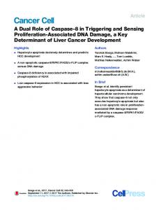
Figure 3. Tertiary Lymphoid Organs in Infection and Cancer (A) In chronic infections, such as in the lung of influenza-virus-infected mice shown here, TLOs are present and characterized by anatomically contiguous but separated T (CD4+ or CD8+) and B cell areas (B220+) and the presence of conventional (CD11C+) or follicular DCs (FDCs), suggesting localized sites of T and B cell responses (reproduced with permission from GeurtsvanKessel et al., 2009). (B–D) TLOs are often present in tumors as visualized by the B cell marker CD19 (from http://www.proteinatlas.org).
components of TLOs and tumors. Overall, TLO generation could be considered a compensatory mechanism for chronically inflamed tissues that have decreased lymphatic drainage and APC migration (Thaunat et al., 2006). Negative Regulation of Immunity in Infection and Cancer The immune response is selectively controlled in different tissues to achieve a balance between an effective response and minimal collateral damage until immune safe checkpoints are initiated (Matzinger and Kamala, 2011). The level of protection is particularly robust in organs with limited spontaneous regeneration (e.g., the central nervous system) (Iliff et al., 2012), whereas, in other tissues, a more regulatory type of control is established. Many vital organs express high levels of TGF-b (and its activating enzyme, MMP9) that directly suppress T and NK cell responses and induce T-reg cells. In the intestine, the cohabitation with commensal bacteria is facilitated by the presence of IL-10producing T-reg cells that are induced by a subset of CD103 expressing DCs primed by the mucus-forming protein mucin 2 (MUC2) (Shan et al., 2013) and activated by environmental cues, such as bacterial short-chain fatty acids, which act via free fatty acid receptor 2 or the TLR2 agonist polysaccharide A found on the bacterial surface (Round et al., 2011; Smith et al., 2013). Many tumors hijack the mechanisms of tolerance induction, for instance, by expressing high levels of TGF-b (Meulmeester and Ten Dijke, 2011) and IDO (Wainwright et al., 2012), or mimic the intestinal tolerant microenvironment by the presence of activated IL-10-producing T-reg cells (Stewart
et al., 2013) (Figure 4). Tumor-associated T-reg cells have the opposite effects on tumor progression by regulating proangiogenic Th17 cells but also the antitumor T cell response; the transient depletion of T-reg cells slow tumor progression and improve the efficacy of radiation therapy (Bos et al., 2013; Stewart et al., 2013). During acute infection, the number of antigen-specific T cells rapidly increases, peaking around days 7–10. Once the pathogen is cleared, the majority of T cells undergo apoptosis, leaving a small number of memory cells able to rapidly respond to secondary challenge. In chronic infection, the immune response is suppressed by various mechanisms to prevent tissue damage, and T cells acquire an exhausted phenotype characterized by the loss of effector molecules (IFN-g, TNF, granzymes, and perforin) and the upregulation of inhibitory molecules, such as cytotoxic T lymphocyte-associated antigen 4 (CTLA4 or CD152), PD-1 (or CD279), lymphocyte-activation gene 3 (LAG3 or CD223), T cell immunoglobulin and mucin protein 3 (TIM-3), adenosine receptor, killer-cell immunoglobulin-like receptors, and several others. Although tumors rarely induce an acute immune response, T cells in the tumor environment have an exhausted phenotype and are restrained by both tissue-intrinsic tolerogenic mechanisms as well as mechanisms responsible for the contraction of the immune response to infections (Crespo et al., 2013). T cell exhaustion is reversible by the removal of inhibitory signals. CTLA4 competes with CD28 for binding to CD80/CD86 on DCs and is an important negative regulator in infection and cancer (Kaufmann and Walker, 2009; Leach Cell Host & Microbe 15, March 12, 2014 ª2014 Elsevier Inc. 301
Cell Host & Microbe
Review Figure 4. Main Immune Checkpoints Negatively Regulating the Immune Response in Infection and Cancer T cells in the tumor environment have an exhausted phenotype, and their proliferation and activation are restrained by both tissue-intrinsic tolerogenic mechanisms that limit excessive inflammatory and immune response to infection as well as by the mechanisms responsible for the contraction of the immune response to infection.
et al., 1996). CTLA4 also scavenges CD80 from the surface of DCs and induces IDO production, mechanisms that may explain how CTLA4-expressing T-reg cells inhibit T cell responses (Fallarino et al., 2005; Wing et al., 2008). PD-1 has T-cell-inhibitory properties in chronic infections and cancer (Barber et al., 2006; Zha et al., 2004). LAG-3 and TIM-3 are also highly expressed on the surface of exhausted T cells (Anderson, 2012; Blackburn et al., 2009; Norde et al., 2012). T cells also directly sense the environment and modify the strength of TCR signaling; e.g., by assessing nutrient availability and oxygen levels. Hypoxia-inducible factor I activated in the hypoxic conditions observed in chronically inflamed tissues and tumors determines the activity of cytotoxic CD8 T cells by inducing increased production of effector molecules (e.g., perforin and granzymes) as well as increasing the levels of inhibitory molecules, such as CTLA4 and LAG3 (Doedens et al., 2013). Nutrient availability, due to low blood supply or altered cell metabolism, mostly sensed by mammalian target of rapamycin (mTOR) and general control nonrepressed 2, also affects T cell responses (McGaha et al., 2012). Myeloid, stromal, and tumor cells similarly modulate mTOR activity and strength of TCR signaling in T cells by producing enzymes that deplete essential amino acids such as IDO, which regulates tryptophan, in immediate T cell environment. Conclusions Unlike vaccines against pathogens, cancer vaccines have so far obtained only limited clinical success, which is most likely due to the persistency of an immunosuppressive tumor microenvironment. As discussed above, the immune environment created by the tumor recapitulates that present in chronic infections in 302 Cell Host & Microbe 15, March 12, 2014 ª2014 Elsevier Inc.
which homeostatic mechanisms aimed at preventing excessive inflammation and tissue damage establish immunological checkpoints that prevent an effective immune response able to completely eliminate the pathogens or destroy the tumor. Thus, immunoinhibitory mechanisms responsible for chronic infection, such as the local production of IL-10, type I IFN, IDO, and TGF-b, and ligands for inhibitory coreceptors are also present in the tumor microenvironment (Diamond et al., 2011; Guiducci et al., 2005; Odorizzi and Wherry, 2013; Pardoll, 2012; Stewart et al., 2013). Therefore, it is not surprising that the effectiveness of procedures simply directed to induce a new response to tumor antigens or enhance the natural response is prevented by the same mechanisms that limit the ability of immunosurveillance to prevent the growth of the tumors. Many of the mechanisms regulating the immune responses to infections and cancer discussed in this review are derived from studies in experimental animals. Although it is important to avoid an automatic extrapolation from mouse studies to humans due to the differences in the immune systems between the two species and the limitations of the experimental models of cancer, the recent success in cancer immunotherapy indicates that the information from mouse studies can be useful for the design of clinical approaches. Thus, in both chronic infectious disease and tumor immunotherapy, much effort has been devoted to developing strategies for reversing immunosuppression; e.g., better adjuvants or blockers of immunosuppressive factors such as TFG-b, IDO, or IL-10. The most promising advances have been obtained with antibodies that block coinhibitory molecules. The monoclonal antibody to CTLA4 (ipilimumab), approved for use in patients with metastatic melanoma, increases patients’ survival most likely by amplifying the endogenous antitumor T cell response. Antibodies against PD1 receptor or its ligand PD-L1 induce significant clinical responses in patients with melanoma or other solid tumors, such as lung cancer (Pardoll, 2012). The effectiveness of these new drugs suggests that, in a proportion of patients, the endogenous immune response to cancer is sufficient to induce tumor regression. Thus, there is an optimistic expectation that the combination of immunocheckpoint inhibitors with therapies such as cancer vaccines or certain chemotherapies that amplify the antitumor immune response might offer better chances for therapeutic success. Recent studies have also identified the existence of
Cell Host & Microbe
Review memory T cells with stem-cell-like properties in viral infections and tumors that maintain the ability to develop in potent effector T cells; the expansion of these cells or reprogramming terminally differentiated tumor-infiltrating lymphocytes to confer stem-celllike properties might be used to augment immunotherapies against cancer (Gattinoni et al., 2012). The promise of these new therapeutic approaches proves the usefulness of comparative studies in infectious and cancer immunity for a better understanding of their basic mechanisms and translational application for the development of new therapies. REFERENCES Abt, M.C., Osborne, L.C., Monticelli, L.A., Doering, T.A., Alenghat, T., Sonnenberg, G.F., Paley, M.A., Antenus, M., Williams, K.L., Erikson, J., et al. (2012). Commensal bacteria calibrate the activation threshold of innate antiviral immunity. Immunity 37, 158–170. Anderson, A.C. (2012). Tim-3, a negative regulator of anti-tumor immunity. Curr. Opin. Immunol. 24, 213–216. Ascensao, J.L., Oken, M.M., Ewing, S.L., Goldberg, R.J., and Kaplan, M.E. (1987). Leukocytosis and large cell lung cancer. A frequent association. Cancer 60, 903–905. Baldridge, M.T., King, K.Y., and Goodell, M.A. (2011). Inflammatory signals regulate hematopoietic stem cells. Trends Immunol. 32, 57–65. Barber, D.L., Wherry, E.J., Masopust, D., Zhu, B., Allison, J.P., Sharpe, A.H., Freeman, G.J., and Ahmed, R. (2006). Restoring function in exhausted CD8 T cells during chronic viral infection. Nature 439, 682–687. Bayne, L.J., Beatty, G.L., Jhala, N., Clark, C.E., Rhim, A.D., Stanger, B.Z., and Vonderheide, R.H. (2012). Tumor-derived granulocyte-macrophage colonystimulating factor regulates myeloid inflammation and T cell immunity in pancreatic cancer. Cancer Cell 21, 822–835. Beyrau, M., Bodkin, J.V., and Nourshargh, S. (2012). Neutrophil heterogeneity in health and disease: a revitalized avenue in inflammation and immunity. Open Biol 2, 120134. Bhat, N., and Fitzgerald, K.A. (2013). Recognition of Cytosolic DNA by cGAS and other STING-dependent sensors. Eur. J. Immunol. Bindea, G., Mlecnik, B., Tosolini, M., Kirilovsky, A., Waldner, M., Obenauf, A.C., Angell, H., Fredriksen, T., Lafontaine, L., Berger, A., et al. (2013). Spatiotemporal dynamics of intratumoral immune cells reveal the immune landscape in human cancer. Immunity 39, 782–795. Blackburn, S.D., Shin, H., Haining, W.N., Zou, T., Workman, C.J., Polley, A., Betts, M.R., Freeman, G.J., Vignali, D.A., and Wherry, E.J. (2009). Coregulation of CD8+ T cell exhaustion by multiple inhibitory receptors during chronic viral infection. Nat. Immunol. 10, 29–37. Boettcher, S., Ziegler, P., Schmid, M.A., Takizawa, H., van Rooijen, N., Kopf, M., Heikenwalder, M., and Manz, M.G. (2012). Cutting edge: LPS-induced emergency myelopoiesis depends on TLR4-expressing nonhematopoietic cells. J. Immunol. 188, 5824–5828. Boiko, J.R., and Borghesi, L. (2012). Hematopoiesis sculpted by pathogens: Toll-like receptors and inflammatory mediators directly activate stem cells. Cytokine 57, 1–8. Bos, P.D., Plitas, G., Rudra, D., Lee, S.Y., and Rudensky, A.Y. (2013). Transient regulatory T cell ablation deters oncogene-driven breast cancer and enhances radiotherapy. J. Exp. Med. 210, 2435–2466. Burnette, B.C., Liang, H., Lee, Y., Chlewicki, L., Khodarev, N.N., Weichselbaum, R.R., Fu, Y.X., and Auh, S.L. (2011). The efficacy of radiotherapy relies upon induction of type i interferon-dependent innate and adaptive immunity. Cancer Res. 71, 2488–2496. Chou, D.B., Sworder, B., Bouladoux, N., Roy, C.N., Uchida, A.M., Grigg, M., Robey, P.G., and Belkaid, Y. (2012). Stromal-derived IL-6 alters the balance of myeloerythroid progenitors during Toxoplasma gondii infection. J. Leukoc. Biol. 92, 123–131.
Crespo, J., Sun, H., Welling, T.H., Tian, Z., and Zou, W. (2013). T cell anergy, exhaustion, senescence, and stemness in the tumor microenvironment. Curr. Opin. Immunol. 25, 214–221. de Bruin, A.M., Libregts, S.F., Valkhof, M., Boon, L., Touw, I.P., and Nolte, M.A. (2012). IFNg induces monopoiesis and inhibits neutrophil development during inflammation. Blood 119, 1543–1554. Delano, M.J., Scumpia, P.O., Weinstein, J.S., Coco, D., Nagaraj, S., KellyScumpia, K.M., O’Malley, K.A., Wynn, J.L., Antonenko, S., Al-Quran, S.Z., et al. (2007). MyD88-dependent expansion of an immature GR-1(+)CD11b(+) population induces T cell suppression and Th2 polarization in sepsis. J. Exp. Med. 204, 1463–1474. Diamond, M.S., Kinder, M., Matsushita, H., Mashayekhi, M., Dunn, G.P., Archambault, J.M., Lee, H., Arthur, C.D., White, J.M., Kalinke, U., et al. (2011). Type I interferon is selectively required by dendritic cells for immune rejection of tumors. J. Exp. Med. 208, 1989–2003. Dieu-Nosjean, M.C., Antoine, M., Danel, C., Heudes, D., Wislez, M., Poulot, V., Rabbe, N., Laurans, L., Tartour, E., de Chaisemartin, L., et al. (2008). Longterm survival for patients with non-small-cell lung cancer with intratumoral lymphoid structures. J. Clin. Oncol. 26, 4410–4417. Doedens, A.L., Phan, A.T., Stradner, M.H., Fujimoto, J.K., Nguyen, J.V., Yang, E., Johnson, R.S., and Goldrath, A.W. (2013). Hypoxia-inducible factors enhance the effector responses of CD8(+) T cells to persistent antigen. Nat. Immunol. 14, 1173–1182. Edme, N., Downward, J., Thiery, J.P., and Boyer, B. (2002). Ras induces NBT-II epithelial cell scattering through the coordinate activities of Rac and MAPK pathways. J. Cell Sci. 115, 2591–2601. Elinav, E., Nowarski, R., Thaiss, C.A., Hu, B., Jin, C., and Flavell, R.A. (2013). Inflammation-induced cancer: crosstalk between tumours, immune cells and microorganisms. Nat. Rev. Cancer 13, 759–771. Essers, M.A., Offner, S., Blanco-Bose, W.E., Waibler, Z., Kalinke, U., Duchosal, M.A., and Trumpp, A. (2009). IFNalpha activates dormant haematopoietic stem cells in vivo. Nature 458, 904–908. Fallarino, F., Orabona, C., Vacca, C., Bianchi, R., Gizzi, S., Asselin-Paturel, C., Fioretti, M.C., Trinchieri, G., Grohmann, U., and Puccetti, P. (2005). Ligand and cytokine dependence of the immunosuppressive pathway of tryptophan catabolism in plasmacytoid dendritic cells. Int. Immunol. 17, 1429–1438. Fox, J.G., Feng, Y., Theve, E.J., Raczynski, A.R., Fiala, J.L., Doernte, A.L., Williams, M., McFaline, J.L., Essigmann, J.M., Schauer, D.B., et al. (2010). Gut microbes define liver cancer risk in mice exposed to chemical and viral transgenic hepatocarcinogens. Gut 59, 88–97. Fuertes, M.B., Woo, S.R., Burnett, B., Fu, Y.X., and Gajewski, T.F. (2013). Type I interferon response and innate immune sensing of cancer. Trends Immunol. 34, 67–73. Gabrilovich, D.I., and Nagaraj, S. (2009). Myeloid-derived suppressor cells as regulators of the immune system. Nat. Rev. Immunol. 9, 162–174. Gabrilovich, D.I., Bronte, V., Chen, S.H., Colombo, M.P., Ochoa, A., OstrandRosenberg, S., and Schreiber, H. (2007). The terminology issue for myeloidderived suppressor cells. Cancer Res. 67, 425, author reply 426. Gabrilovich, D.I., Ostrand-Rosenberg, S., and Bronte, V. (2012). Coordinated regulation of myeloid cells by tumours. Nat. Rev. Immunol. 12, 253–268. Ganal, S.C., Sanos, S.L., Kallfass, C., Oberle, K., Johner, C., Kirschning, C., Lienenklaus, S., Weiss, S., Staeheli, P., Aichele, P., and Diefenbach, A. (2012). Priming of natural killer cells by nonmucosal mononuclear phagocytes requires instructive signals from commensal microbiota. Immunity 37, 171–186. Gattinoni, L., Klebanoff, C.A., and Restifo, N.P. (2012). Paths to stemness: building the ultimate antitumour T cell. Nat. Rev. Cancer 12, 671–684. Gautier, G., Humbert, M., Deauvieau, F., Scuiller, M., Hiscott, J., Bates, E.E., Trinchieri, G., Caux, C., and Garrone, P. (2005). A type I interferon autocrineparacrine loop is involved in Toll-like receptor-induced interleukin-12p70 secretion by dendritic cells. J. Exp. Med. 201, 1435–1446. GeurtsvanKessel, C.H., Willart, M.A., Bergen, I.M., van Rijt, L.S., Muskens, F., Elewaut, D., Osterhaus, A.D., Hendriks, R., Rimmelzwaan, G.F., and Lambrecht, B.N. (2009). Dendritic cells are crucial for maintenance of tertiary
Cell Host & Microbe 15, March 12, 2014 ª2014 Elsevier Inc. 303
Cell Host & Microbe
Review lymphoid structures in the lung of influenza virus-infected mice. J. Exp. Med. 206, 2339–2349.
Ma, Y., Galluzzi, L., Zitvogel, L., and Kroemer, G. (2013b). Autophagy and cellular immune responses. Immunity 39, 211–227.
Goldszmid, R.S., and Trinchieri, G. (2012). The price of immunity. Nat. Immunol. 13, 932–938.
Martinet, L., Garrido, I., Filleron, T., Le Guellec, S., Bellard, E., Fournie, J.J., Rochaix, P., and Girard, J.P. (2011). Human solid tumors contain high endothelial venules: association with T- and B-lymphocyte infiltration and favorable prognosis in breast cancer. Cancer Res. 71, 5678–5687.
Goldszmid, R.S., Caspar, P., Rivollier, A., White, S., Dzutsev, A., Hieny, S., Kelsall, B., Trinchieri, G., and Sher, A. (2012). NK cell-derived interferon-g orchestrates cellular dynamics and the differentiation of monocytes into dendritic cells at the site of infection. Immunity 36, 1047–1059. Gough, D.J., Messina, N.L., Clarke, C.J., Johnstone, R.W., and Levy, D.E. (2012). Constitutive type I interferon modulates homeostatic balance through tonic signaling. Immunity 36, 166–174. Grivennikov, S.I., Greten, F.R., and Karin, M. (2010). Immunity, inflammation, and cancer. Cell 140, 883–899. Guiducci, C., Vicari, A.P., Sangaletti, S., Trinchieri, G., and Colombo, M.P. (2005). Redirecting in vivo elicited tumor infiltrating macrophages and dendritic cells towards tumor rejection. Cancer Res. 65, 3437–3446. Hanahan, D., and Weinberg, R.A. (2011). Hallmarks of cancer: the next generation. Cell 144, 646–674. Ichinohe, T., Pang, I.K., Kumamoto, Y., Peaper, D.R., Ho, J.H., Murray, T.S., and Iwasaki, A. (2011). Microbiota regulates immune defense against respiratory tract influenza A virus infection. Proc. Natl. Acad. Sci. USA 108, 5354– 5359. Iida, N., Dzutsev, A., Stewart, C.A., Smith, L., Bouladoux, N., Weingarten, R.A., Molina, D.A., Salcedo, R., Back, T., Cramer, S., et al. (2013). Commensal bacteria control cancer response to therapy by modulating the tumor microenvironment. Science 342, 967–970. Iliff, J.J., Wang, M., Liao, Y., Plogg, B.A., Peng, W., Gundersen, G.A., Benveniste, H., Vates, G.E., Deane, R., Goldman, S.A., et al. (2012). A paravascular pathway facilitates CSF flow through the brain parenchyma and the clearance of interstitial solutes, including amyloid b. Sci. Transl. Med. 4, ra111. Jankovic, D., and Trinchieri, G. (2007). IL-10 or not IL-10: that is the question. Nat. Immunol. 8, 1281–1283. Jobin, C. (2012). Colorectal cancer: CRC—all about microbial products and barrier function? Nat Rev Gastroenterol Hepatol 9, 694–696. Kaufmann, D.E., and Walker, B.D. (2009). PD-1 and CTLA-4 inhibitory cosignaling pathways in HIV infection and the potential for therapeutic intervention. J. Immunol. 182, 5891–5897. King, K.Y., and Goodell, M.A. (2011). Inflammatory modulation of HSCs: viewing the HSC as a foundation for the immune response. Nat. Rev. Immunol. 11, 685–692. Koblish, H.K., Hunter, C.A., Wysocka, M., Trinchieri, G., and Lee, W.M. (1998). Immune suppression by recombinant interleukin (rIL)-12 involves interferon gamma induction of nitric oxide synthase 2 (iNOS) activity: inhibitors of NO generation reveal the extent of rIL-12 vaccine adjuvant effect. J. Exp. Med. 188, 1603–1610. Ko¨hler, A., De Filippo, K., Hasenberg, M., van den Brandt, C., Nye, E., Hosking, M.P., Lane, T.E., Ma¨nn, L., Ransohoff, R.M., Hauser, A.E., et al. (2011). G-CSFmediated thrombopoietin release triggers neutrophil motility and mobilization from bone marrow via induction of Cxcr2 ligands. Blood 117, 4349–4357. Kunisaki, Y., Bruns, I., Scheiermann, C., Ahmed, J., Pinho, S., Zhang, D., Mizoguchi, T., Wei, Q., Lucas, D., Ito, K., et al. (2013). Arteriolar niches maintain haematopoietic stem cell quiescence. Nature 502, 637–643.
Matzinger, P. (2002). The danger model: a renewed sense of self. Science 296, 301–305. Matzinger, P., and Kamala, T. (2011). Tissue-based class control: the other side of tolerance. Nat. Rev. Immunol. 11, 221–230. McGaha, T.L., Huang, L., Lemos, H., Metz, R., Mautino, M., Prendergast, G.C., and Mellor, A.L. (2012). Amino acid catabolism: a pivotal regulator of innate and adaptive immunity. Immunol. Rev. 249, 135–157. Medzhitov, R. (2008). Origin and physiological roles of inflammation. Nature 454, 428–435. Meulmeester, E., and Ten Dijke, P. (2011). The dynamic roles of TGF-b in cancer. J. Pathol. 223, 205–218. Naik, S., Bouladoux, N., Wilhelm, C., Molloy, M.J., Salcedo, R., Kastenmuller, W., Deming, C., Quinones, M., Koo, L., Conlan, S., et al. (2012). Compartmentalized control of skin immunity by resident commensals. Science 337, 1115– 1119. Neyt, K., Perros, F., GeurtsvanKessel, C.H., Hammad, H., and Lambrecht, B.N. (2012). Tertiary lymphoid organs in infection and autoimmunity. Trends Immunol. 33, 297–305. Norde, W.J., Hobo, W., van der Voort, R., and Dolstra, H. (2012). Coinhibitory molecules in hematologic malignancies: targets for therapeutic intervention. Blood 120, 728–736. Norris, B.A., Uebelhoer, L.S., Nakaya, H.I., Price, A.A., Grakoui, A., and Pulendran, B. (2013). Chronic but not acute virus infection induces sustained expansion of myeloid suppressor cell numbers that inhibit viral-specific T cell immunity. Immunity 38, 309–321. Odorizzi, P.M., and Wherry, E.J. (2013). Immunology. An interferon paradox. Science 340, 155–156. Pardoll, D.M. (2012). Immunology beats cancer: a blueprint for successful translation. Nat. Immunol. 13, 1129–1132. Parkin, D.M. (2006). The global health burden of infection-associated cancers in the year 2002. Int. J. Cancer 118, 3030–3044. Paulos, C.M., Wrzesinski, C., Kaiser, A., Hinrichs, C.S., Chieppa, M., Cassard, L., Palmer, D.C., Boni, A., Muranski, P., Yu, Z., et al. (2007). Microbial translocation augments the function of adoptively transferred self/tumor-specific CD8+ T cells via TLR4 signaling. J. Clin. Invest. 117, 2197–2204. Pierce, G.B., and Speers, W.C. (1988). Tumors as caricatures of the process of tissue renewal: prospects for therapy by directing differentiation. Cancer Res. 48, 1996–2004. Pietras, E.M., Warr, M.R., and Passegue´, E. (2011). Cell cycle regulation in hematopoietic stem cells. J. Cell Biol. 195, 709–720. Qin, A., Cai, W., Pan, T., Wu, K., Yang, Q., Wang, N., Liu, Y., Yan, D., Hu, F., Guo, P., et al. (2013). Expansion of monocytic myeloid-derived suppressor cells dampens T cell function in HIV-1-seropositive individuals. J. Virol. 87, 1477–1490.
Leach, D.R., Krummel, M.F., and Allison, J.P. (1996). Enhancement of antitumor immunity by CTLA-4 blockade. Science 271, 1734–1736.
Rangel-Moreno, J., Carragher, D.M., de la Luz Garcia-Hernandez, M., Hwang, J.Y., Kusser, K., Hartson, L., Kolls, J.K., Khader, S.A., and Randall, T.D. (2011). The development of inducible bronchus-associated lymphoid tissue depends on IL-17. Nat. Immunol. 12, 639–646.
Link, A., Hardie, D.L., Favre, S., Britschgi, M.R., Adams, D.H., Sixt, M., Cyster, J.G., Buckley, C.D., and Luther, S.A. (2011). Association of T-zone reticular networks and conduits with ectopic lymphoid tissues in mice and humans. Am. J. Pathol. 178, 1662–1675.
Rao, J.B., Chamberlain, R.S., Bronte, V., Carroll, M.W., Irvine, K.R., Moss, B., Rosenberg, S.A., and Restifo, N.P. (1996). IL-12 is an effective adjuvant to recombinant vaccinia virus-based tumor vaccines: enhancement by simultaneous B7-1 expression. J. Immunol. 156, 3357–3365.
Ma, Y., Adjemian, S., Mattarollo, S.R., Yamazaki, T., Aymeric, L., Yang, H., Portela Catani, J.P., Hannani, D., Duret, H., Steegh, K., et al. (2013a). Anticancer chemotherapy-induced intratumoral recruitment and differentiation of antigen-presenting cells. Immunity 38, 729–741.
Rao, V.P., Poutahidis, T., Ge, Z., Nambiar, P.R., Boussahmain, C., Wang, Y.Y., Horwitz, B.H., Fox, J.G., and Erdman, S.E. (2006). Innate immune inflammatory response against enteric bacteria Helicobacter hepaticus induces mammary adenocarcinoma in mice. Cancer Res. 66, 7395–7400.
304 Cell Host & Microbe 15, March 12, 2014 ª2014 Elsevier Inc.
Cell Host & Microbe
Review Rossi, L., Salvestrini, V., Ferrari, D., Di Virgilio, F., and Lemoli, R.M. (2012). The sixth sense: hematopoietic stem cells detect danger through purinergic signaling. Blood 120, 2365–2375. Round, J.L., Lee, S.M., Li, J., Tran, G., Jabri, B., Chatila, T.A., and Mazmanian, S.K. (2011). The Toll-like receptor 2 pathway establishes colonization by a commensal of the human microbiota. Science 332, 974–977. Salcedo, R., Worschech, A., Cardone, M., Jones, Y., Gyulai, Z., Dai, R.M., Wang, E., Ma, W., Haines, D., O’hUigin, C., et al. (2010). MyD88-mediated signaling prevents development of adenocarcinomas of the colon: role of interleukin 18. J. Exp. Med. 207, 1625–1636. Salcedo, R., Cataisson, C., Hasan, U., Yuspa, S.H., and Trinchieri, G. (2013). MyD88 and its divergent toll in carcinogenesis. Trends Immunol. 34, 379–389. Schetter, A.J., Heegaard, N.H., and Harris, C.C. (2010). Inflammation and cancer: interweaving microRNA, free radical, cytokine and p53 pathways. Carcinogenesis 31, 37–49. Schreiber, R.D., Old, L.J., and Smyth, M.J. (2011). Cancer immunoediting: integrating immunity’s roles in cancer suppression and promotion. Science 331, 1565–1570. Sears, C.L., and Garrett, W.S. (2014). Microbes, Microbiota, and Colon Cancer. Cell Host Microbe 15, this issue, 317–328.
Trinchieri, G. (2010). Type I interferon: friend or foe? J. Exp. Med. 207, 2053– 2063. Trinchieri, G. (2012). Cancer and inflammation: an old intuition with rapidly evolving new concepts. Annu. Rev. Immunol. 30, 677–706. van de Pavert, S.A., Olivier, B.J., Goverse, G., Vondenhoff, M.F., Greuter, M., Beke, P., Kusser, K., Ho¨pken, U.E., Lipp, M., Niederreither, K., et al. (2009). Chemokine CXCL13 is essential for lymph node initiation and is induced by retinoic acid and neuronal stimulation. Nat. Immunol. 10, 1193–1199. Viaud, S., Saccheri, F., Mignot, G., Yamazaki, T., Daille`re, R., Hannani, D., Enot, D.P., Pfirschke, C., Engblom, C., Pittet, M.J., et al. (2013). The intestinal microbiota modulates the anticancer immune effects of cyclophosphamide. Science 342, 971–976. Voisin, M.B., Buzoni-Gatel, D., Bout, D., and Velge-Roussel, F. (2004). Both expansion of regulatory GR1+ CD11b+ myeloid cells and anergy of T lymphocytes participate in hyporesponsiveness of the lung-associated immune system during acute toxoplasmosis. Infect. Immun. 72, 5487–5492. Wainwright, D.A., Balyasnikova, I.V., Chang, A.L., Ahmed, A.U., Moon, K.S., Auffinger, B., Tobias, A.L., Han, Y., and Lesniak, M.S. (2012). IDO expression in brain tumors increases the recruitment of regulatory T cells and negatively impacts survival. Clin. Cancer Res. 18, 6110–6121.
Serbina, N.V., and Pamer, E.G. (2006). Monocyte emigration from bone marrow during bacterial infection requires signals mediated by chemokine receptor CCR2. Nat. Immunol. 7, 311–317.
Westbrook, A.M., Wei, B., Hacke, K., Xia, M., Braun, J., and Schiestl, R.H. (2012). The role of tumour necrosis factor-a and tumour necrosis factor receptor signalling in inflammation-associated systemic genotoxicity. Mutagenesis 27, 77–86.
Shan, M., Gentile, M., Yeiser, J.R., Walland, A.C., Bornstein, V.U., Chen, K., He, B., Cassis, L., Bigas, A., Cols, M., et al. (2013). Mucus enhances gut homeostasis and oral tolerance by delivering immunoregulatory signals. Science 342, 447–453.
Wing, K., Onishi, Y., Prieto-Martin, P., Yamaguchi, T., Miyara, M., Fehervari, Z., Nomura, T., and Sakaguchi, S. (2008). CTLA-4 control over Foxp3+ regulatory T cell function. Science 322, 271–275.
Shields, J.D., Kourtis, I.C., Tomei, A.A., Roberts, J.M., and Swartz, M.A. (2010). Induction of lymphoidlike stroma and immune escape by tumors that express the chemokine CCL21. Science 328, 749–752.
Yamamoto, M.L., Maier, I., Dang, A.T., Berry, D., Liu, J., Ruegger, P.M., Yang, J.I., Soto, P.A., Presley, L.L., Reliene, R., et al. (2013). Intestinal bacteria modify lymphoma incidence and latency by affecting systemic inflammatory state, oxidative stress, and leukocyte genotoxicity. Cancer Res. 73, 4222–4232.
Shojaei, F., Wu, X., Zhong, C., Yu, L., Liang, X.H., Yao, J., Blanchard, D., Bais, C., Peale, F.V., van Bruggen, N., et al. (2007). Bv8 regulates myeloid-celldependent tumour angiogenesis. Nature 450, 825–831. Smith, P.M., Howitt, M.R., Panikov, N., Michaud, M., Gallini, C.A., Bohlooly-Y, M., Glickman, J.N., and Garrett, W.S. (2013). The microbial metabolites, shortchain fatty acids, regulate colonic Treg cell homeostasis. Science 341, 569–573. Stewart, C.A., Metheny, H., Iida, N., Smith, L., Hanson, M., Steinhagen, F., Leighty, R.M., Roers, A., Karp, C.L., Mu¨ller, W., and Trinchieri, G. (2013). Interferon-dependent IL-10 production by Tregs limits tumor Th17 inflammation. J. Clin. Invest. 123, 4859–4874. Thaunat, O., Kerjaschki, D., and Nicoletti, A. (2006). Is defective lymphatic drainage a trigger for lymphoid neogenesis? Trends Immunol. 27, 441–445.
Yan, H.H., Pickup, M., Pang, Y., Gorska, A.E., Li, Z., Chytil, A., Geng, Y., Gray, J.W., Moses, H.L., and Yang, L. (2010). Gr-1+CD11b+ myeloid cells tip the balance of immune protection to tumor promotion in the premetastatic lung. Cancer Res. 70, 6139–6149. Yu, P., Lee, Y., Liu, W., Chin, R.K., Wang, J., Wang, Y., Schietinger, A., Philip, M., Schreiber, H., and Fu, Y.X. (2004). Priming of naive T cells inside tumors leads to eradication of established tumors. Nat. Immunol. 5, 141–149. Zha, Yy., Blank, C., and Gajewski, T.F. (2004). Negative regulation of T-cell function by PD-1. Crit. Rev. Immunol. 24, 229–237. Zhang, Q., Raoof, M., Chen, Y., Sumi, Y., Sursal, T., Junger, W., Brohi, K., Itagaki, K., and Hauser, C.J. (2010). Circulating mitochondrial DAMPs cause inflammatory responses to injury. Nature 464, 104–107.
Cell Host & Microbe 15, March 12, 2014 ª2014 Elsevier Inc. 305