CaMKI was initially recognized as an entity distinct from the first CaM kinases identified, phosphorylase kinase and isoforms of MLCK, by its ability to ...
The EMBO Journal vol.14 no.15 pp.3679-3686, 1995
Human calcium-calmodulin dependent protein kinase 1: cDNA cloning, domain structure and activation by phosphorylation at threonine-177 by calcium-calmodulin dependent protein kinase I kinase Bodduluri Haribabul, Sara S.Hook, Michele A.Selbert3, Elaine G.Goldstein3, Eric D.Tomhave, Arthur M.Edelman3, Ralph Snyderman1l2 and Anthony R.Means4 Department of Pharmacology, 'Department of Medicine and
2Department of Immunology, Duke University Medical Center, Durham, NC 27710 and 3Department of Pharmacology and Toxicology, State University of New York at Buffalo, Buffalo, NY 14214, USA 4Corresponding author
Human Ca2+-calmodulin (CaM) dependent protein kinase I (CaMKI) encodes a 370 amino acid protein with a calculated Mr of 41 337. The 1.5 kb CaMKI mRNA is expressed in many different human tissues and is the product of a single gene located on human chromosome 3. CaMKI 1-306, was unable to bind Ca2+-CaM and was completely inactive thereby defining an essential component of the CaM-binding domain to residues C-terminal to 306. CaMKI 1-294 did not *bind CaM but was fully active in the absence of Ca2+CaM, indicating that residues 295-306 are sufficient to maintain CaMKI in an auto-inhibited state. CaMKI was phosphorylated on Thrl77 and its activity enhanced -25-fold by CaMKI kinase in a Ca2+-CaM dependent manner. Replacement of Thrl77 with Ala or Asp prevented both phosphorylation and activation by CaMKI kinase and the latter replacement also led to partial activation in the absence of CaMKI kinase. Whereas CaMKI 1-306 was unresponsive to CaMKI kinase, the 1-294 mutant was phosphorylated and activated by CaMKI kinase in both the presence and absence of Ca2+-CaM although at a faster rate in its presence. These results indicate that the auto-inhibitory domain in CaMKI gates, in a Ca2+-CaM dependent fashion, accessibility of both substrates to the substrate binding cleft and CaMKI kinase to Thrl77. Additionally, CaMKI kinase responds directly to Ca2+-CaM with increased activity. Keywords: auto-inhibition/calciumlcalmodulin/phosphorylation/protein kinase
Introduction Ca2+-calmodulin dependent protein kinases (CaM kinases) are a structurally related group a subfamily within the larger
of enzymes that constitute family of protein serine/
threonine kinases (Hanks and Quinn, 1991). Currently recognized members of' the CaM kinase family are: phosphorylase kinase, myosin light chain kinase (MLCK) and CaM kinases (CaMKs) I, II, III and IV (reviewed in Edelman et al., 1987; Hanson and Schulman, 1992). Oxford University Press
CaMKI was initially recognized as an entity distinct from the first CaM kinases identified, phosphorylase kinase and isoforms of MLCK, by its ability to phosphorylate the synaptic vesicle-associated protein, synapsin I, and distinct from CaMKII by its preference for site 1 (Ser9) over sites 2 and 3 of synapsin I (Kennedy and Greengard, 1981). CaMKI has since been purified and characterized from bovine and rat brain (Naim and Greengard, 1987; DeRemer et al., 1992a,b; Mochizuki et al., 1993a; Lee et al., 1994). It is a monomeric enzyme of -40 kDa, however, analysis of the purified brain preparations by SDS-PAGE has revealed multiple polypeptides of 37-43 kDa (Naim and Greengard, 1987; DeRemer et al., 1992a,b; Ito et al., 1994). The precise relationships between these various protein products have yet to be definitively established. Clones encoding CaMKI have been isolated from rat brain (Picciotto et al., 1993) and fetal lung (Cho et al., 1994) cDNA libraries. Proteins of 332 and 374 amino acids, respectively, were reported to be encoded by these cDNAs although the shortened length of the former appears to be due to a sequencing artifact. A number of recent observations are consistent with an important role for CaMKI in the transduction of Ca2+ signals in a variety of cellular contexts. First, multiple in vitro protein substrates for CaMKI have been identified, including: synapsins I and II (Nairn and Greengard, 1987; DeRemer et al., 1992a,b); cyclic AMP-response element binding protein (CREB) (Sheng etal., 1991); and the cystic fibrosis transmembrane conductance regulator (CFTR) (Picciotto et al., 1992). Second, CaMKI has been shown to phosphorylate synthetic peptides based on several proteins including synapsin I (DeRemer et al., 1992b) and glycogen synthase (DeRemer et al., 1992b; Mochizuki et al., 1993a). Based on a study of synthetic peptides modeled on Ser9 of synapsin I, Lee et al. (1994) concluded that CaMKI recognizes mainly, if not entirely, primary sequence, rather than higher-order structural determinants in its substrates. Moreover, its minimal primary sequence substrate recognition motif: Hyd-Xaa-Arg-Xaa-Xaa-(Ser/ Thr)*-Xaa-Xaa-Xaa-Hyd is only somewhat more elaborate than that of the broad specificity CaM kinase, CaMKII (Hyd-Xaa-Arg-Xaa-Xaa-[Ser/Thr]*; Pearson et al., 1985; Stokoe et al., 1993). Thus, based on in vitro data, CaMKI appears to have the potential for recognizing a multiplicity of substrates in vivo. Finally, as reported here for human and previously for rat (Picciotto et al., 1992; Cho et al., 1994), CaMKI has a widespread tissue distribution, implying the existence of physiological targets beyond the small number identified to date. It has recently been appreciated that in addition to its Ca2+-CaM dependence, CaMKI must be in the phosphorylated state to be maximally active. This phenomenon was first demonstrated for purified rat brain CaMKI (an
3679
B.Haribabu et al. Hcamk Rcamk Rcamk Rcamk Dcamk Ycamk
I I II IV II I
Hcamk Rcamk Rcamk Rcamk Dcamk Ycamk
I I II
Hcamk Rcamk Rcamk Rcamk Dcamk Ycamk
I I II IV
Hcamk Rcamk Rcamk Rcamk Dcamk Ycamk
I I II IV II I
Hcamk Rcamk Rcamk Rcamk Dcamk Ycamk
I I II IV II I
Hcamk Rcamk Rcamk Rcamk Dcamk Ycamk
I I II IV II I
IV II I
II I
MLGA--VEGPRWKQAEDIRD---------------IYDFRDVLGTGAFS 32
.P.............. .-............. ..... ... MATITCTRFTEE.QLFEE..K.... 25 MLKVTVPSCPSSPCSSVTSSTENLVPDYWID.SKR---DPLS.---------------FFEVESE..R..T. 54 MAAPAACTRFSDN..IKEE..K.... 26
.DDKVSEKESSP..T.EDSEGKMAHVQPASYVNKKK.V.GKT..A.T.G 49 EVIILAEDKRTQKLVAIKtCIAKEALEGKEGSME --- NEIAVLHKIKHPNIVALDDI YESGGHLYLIMQLVSGGELFDRIVEKGFYTERDASRLI FQVDAV 12 9 ..................... K ....................... K..... 129 . FD. .T.. ED. .AREY.S.A ... HC.Q.I.E.. 123 I.YRCKQ.G...PY.L.-VL.KTVDK.--IVR--- T..G..LRLS .. IK.KE.F.TPTEIS.VLE.T..Y.S ... ADAVK.I.E.. 148 I.KRCVQ.S.GFEF.A.I.NTKK.TARD--FQKLER.ARICR.LH ..... R.H.SIQEENYH ..VFD.T.. ED. .ARE ..S.A . HC.Q.I.ES. 124 V.RQ.KNTE.GED ..V.ILI.K ..K.NKVQL.ALYD.LDI.QRLH ...... FK.WF ..KDKF.I.T. .AK ....... LK. .KF .E. .V.ILVEI.S.. 149
V.RRCVKVLAGQEY.A.I.NTKK.SARD--HQKLER.ARICRLL ...... R.H.SISEE ..H
22 7 KYLHDLGIVHRDLKPENLLYYSLDEDSKIMISDFGLSK-MEDPGSVLSTACGTPGYVAPEVI&AQKPYSKAVDCWSIGVIAYILLCGYPPFYDENDAK-LF 227 .-..
LHC.QM.V...LAKKAVIA........... ..AIEV.GEQQAWFGFA .L... .R..G.P........ ..... LRKG.PV.L.WC ..LVW-.QHR.Y A .. EN.ATPAP.APLK.A .....-IVEHQVLMK.V . C... I.RGCA.GPE ..M..V.I.T .... FE ..... RGDQFM. LA.KAKGAAVKLA ... W. .-.HDR.Y NHC.QN.V ....... .. . V. AIEVQGDHQAWFGFA ..... .KKE..G.S.I....... ..M.SQN .IDKSDE.PLVVA ... IA.RLKSDEEL.YKPA.SL ........ T.DGHG.PC.I ...... T.T . SA.RA.RVQD-FL ....
EQILK1EY--EFDSPYWDDISDSAKDFIRHLMEKDPEKRFTCEQALQHPWIAGDTALDKNIH-QSVSZQ
~~~~~~~~~~~~~~ 222 247 223 248
AKSEVKLGTS 324
.............. --......... .. L.NKMLTIN.S..I.AAE..K ... SHRSTVASCM.R.ETVDCL..FN.RR.L.G.ILT.MLATRNFSGGKSGG RR ..NC. .--Y.I.-.W ..EV.LN ... LVKK.IVL ..K..L.TF .. ------------...
324
QEGQGQTASHGELLTPVAGGPAAGCCCRDCCVEPGTELSPTLPHQL* ...T.. ..P.AP.PSSRAMD
370
NPHIHLMGDESACIAYIRITQYLDAGGIPRTAQSEETRVWHRRDGKWQIVHFHRSGAPSVLPH* ETDDQEMKRNSEETLKSVEEEMDPKAEEEAAAVGLGVPPQQDAILPEY*
370 332 478 474 490 446
Q..K.iGA.--D.P..E..TVTPE.
320
VT--GKAANFV.MDTAQKKLQEFN.RR.L.A.VKAVW 320 S..KAGA.--DYP..E..TVTPE..NL.NQMLTVN.N..I.AAE..KC CQRERVASVV.R.ETVDCL..FN.RR.L.G.ILT.MLATRNFS-SRSMI 320 DECTTG..PVK.HR....SV.NK..Q..LKALNL..S..P.AAEL.ED... .I---CTELKT.-NLLPGLKEGLD.RQ.FRNSVERVRLNMKIQ..RDLYL 344
374 NKKIDG--VKESSESTNTT-- IEDEDTK-VRKQEI IKVTEQ. IEAISNGDFESYTKMCDPGMTAFEPEALGNLVEGLDFHRFYFENLWSRNSKPVHTTIL 415 --- -ASSRLGSASSSHTNIQESNKASSEAQPAQDGKDKTDP. ENXMQAGDHEAAlIAADETMKLQSEEVEEEEGVKEEEEEEEEEEETSRMVPQEPEDRL 426 TKKGEGSQVKESTDSSSTT--LEDDDIIAARRQET IKITEQ. IEAINSGDFDGYTKICDPHLTAFEPEALGNLVEGIDFHKFYFENVLGKNCKKINTTIL 418 EQTESDSDFDEGSQANGSVP . LK--- -ATDTSQLSKK. . EEEQSKLKSELTSKAFAQLVNTVLAEKEKFLNINRVCSSDSDLPGSDIKSLDEAKEKPEGK 440
NPHVHLLGEEAACIAYVRLTQYIDKQGHAHTHQSEETRVWHKRDNKWQNVHFHRSASAKISGATTFDFIPQK* DTKTEE*
Fig. 1. Alignment of the amino acid sequence of human CaMKI with other multifunctional CaM kinases. The pile-up program of the GCG software was used to align and compare the amino acid sequences of human CaMKI (Hcamk I), rat CaMKI (Rcamk 1), rat CaMKII (Rcamk II), rat CaMKIV (Rcamk IV), Drosophila CaMKII (Dcamk) and the yeast CaM kinase (Ycamk). Residues identical to the human CaMKI are indicated with a '.' and gaps in the alignment are indicated as a dash (-). The amino acid sequence is numbered on the right. Lys49 and Thrl77 which are the targets of site directed mutagenesis in human CaMKI are shown in bold and with an asterisk on top. The last residues in the two C-terminal truncation mutants (at amino acids 294 and 306, respectively) of human CaMKI are indicated with '1.'. A tentatively assigned auto-inhibitory and CaM-binding domain is shown in bold italics.
isoform, 43 kDa) by DeRemer et al. (I 992b) and confirmed by Mochizuki et al. (1 993b) for an isoform termed CaMKV which may be identical to CaMKIa. The activating phosphorylation was initially interpreted to be autocatalytic based on its Ca2+-CaM dependence (DeRemer et al., 1992b). However, it was simultaneously reported that a similar magnitude of activation was produced by an activator separated from CaMKIa during the latter's purification (DeRemer et al., 1992a). Purification from porcine brain of the activator, a monomeric, -52 kDa protein, yielded the following insights into CaMKI regulation (Lee and Edelman, 1994, 1995): (i) based on experiments with the ATP-affinity analogue fluorosulfonylbenzoyl adenosine (FSBA), the activator itself appears to be a protein kinase and thus may be designated CaMKI kinase; (ii) activating autophosphorylation occurs at a negligibly slow rate in comparison with phosphorylation by CaMKI kinase; (iii) activation by CaMKI kinase also requires Ca2+-CaM; (iv) CaMKI has an almost absolute dependence for its activity upon phosphorylation by CaMKI kinase; and (v) in the activated state, CaMKI retains the dependence of its activity upon Ca2+-CaM. These findings indicate that CaMKI is a component of a kinase cascade which is
3680
Ca2+-CaM regulated but theoretically may be subject to regulation by other intra- or extracellular signals. In view of its potential physiological importance and complex regulatory controls, we undertook, and report here, the cDNA cloning of CaMKI from a human HL-60 cell cDNA library. CaMKI is a 370 amino acid protein, expressed in many different human tissues and likely encoded by a single gene located on human chromosome 3. Human CaMKI is regulated by two important mechanisms: classic relief of intrasteric inhibition mediated by identified CaM-binding and auto-inhibitory domains; and phosphorylation by CaMKI kinase at Thrl77, a residue equivalent to the activating phosphorylation sites of a number of other important signal transducing protein kinases (Marshall, 1994).
Results Human CaMKI cDNA cloning A novel protein kinase-like clone was previously identified in attempts to isolate G-protein coupled receptor kinases in neutrophils (Haribabu and Snyderman, 1993). This fragment was used to isolate a full-length cDNA from a
Human CaM kinase I
Specific Activity
(jmol
Iys49
1
WT
OST
CaMK I
ala49
1 -
KIN K49A *19 I..
.
..-
IGST
IGSTl
I
Iys49
1
T1 77S
[GST | Iys49
1
T1 77D
Iys49 I
I
1r01G TIt 1
GS| 1
1-294;T1 77A
Iys49
-1
Iys49
1
Iys49
0.32 + 0.06
0
0
II
I
serI77 CaMK I
370
asp177
370
0
0.19 + 0.11
0
0.08 + 0.02
0
0.73
thri77
thril7
30 0
ala177 a
asp177
CaMK I
0
294
0.19 + 0.02
0.19 + 0.02
0.21 + 0.05
0.22 + 0.07
2.31 + 1.17
1.19 + 0.24
294 I
rS fCaMK I
1-294;T1 77D I GST
Ca2+'CaM
370
ala177
CaMK I I
+
370
CaMK I
CaMK I
-%J%F%f
1-294
thril7
|OGST ICOaMK I lys49
-Ca2+'CaM 0
CaMK I 1
Ti 77A
370
thri77
min" mg-')
294
l
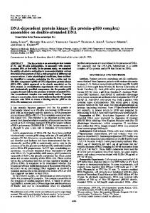
Fig. 2. Schematic representation of CaMKI wild type and mutant enzymes and their specific activities. CaMKI wild type and mutant enzymes expressed in Ecoli as GST fusion proteins are represented by boxes with amino acid coordinates shown on top of each box and relevant substitutions and deletions as indicated. For measurement of specific activities CaMKI wild type and mutant enzymes were pre-incubated (in the absence of CaMKI kinase) and assayed as described in Table I with the exception that pre-incubation and assay were performed in both the absence and presence of Ca2+ (I mM excess of EGTA) and CaM (1 ,uM). Each point represents the mean (± SE) of two to five determinations (except for T177D which was a single determination) with each determination the average of two time points.
differentiated HL-60 cell library. The complete sequence contained a single open-reading frame encoding a protein of 370 amino acids and a calculated Mr of 41 337 that was 97% identical to rat CaM kinase I (Picciotto et al., 1993; Cho et al., 1994). This sequence has been deposited in the EMBL database under accession number L41816. Northern analysis showed the presence of CaM kinase I mRNA in a wide range of tissues and Southern analysis was consistent with the presence of a single gene. Hybridization to a panel of human-hamster somatic cell hybrids was also consistent with a single gene that mapped to human chromosome 3 (data not shown).
CaMKI domain structure Based on the alignment of the primary sequence of human CaMKI with those of other multifunctional CaM kinases, it was possible to identify amino acid residues potentially important for CaMKI function (Figure 1). To evaluate these hypotheses, the wild type and a number of substitution and truncation mutants of CaMKI were expressed in Escherichia coli as GST fusion proteins and purified.
Figure 2 is a schematic representation of their structures and their specific peptide kinase activities determined in the presence and absence of Ca2+-CaM. Wild type CaMKI displayed a specific peptide kinase activity in the presence of Ca2+-CaM of 0.32 ,umol/min/mg, a value comparable with that of purified rat brain CaM kinase Ia without readdition of its activator protein (DeRemer et al., 1992a). As expected from analyses of other protein kinase mutants (Taylor and Rodzio-Andzelm, 1994), modification of the essential lysine at the ATP binding site to an alanine (K49A) resulted in an inactive enzyme. Replacement of Thrl77 with other uncharged amino acids (Ala or Ser), slightly lowered the specific activity while substitution with Asp enhanced activity suggesting that the introduction of negative charge at this position has an activating effect on the enzyme. To localize its auto-inhibitory and CaMbinding domains, C-terminal truncations of CaMKI at residues 294 and 306 were made (Figure 2). Although the full-length enzymes were all inactive in the absence of Ca2+-CaM, both truncations removed the ability of CaMKI to bind CaM (data not shown). The truncation at 294
3681
B.Haribabu et al. Mr(X 1 0
.:;
9766-
*
w
_
43-
c
c 4000 E
29-
c;
-
_. _ 3000
+
4 + +
6 +
+
8 +
9 10 + +
--
+
-
+
--
+
-
2 2.
":a' -ClM + + .l!lMK t Kindso - ±
i 2000 Q
+
Fig. 4. Phosphorylation of CaMKI wild type and T 177A, T 177S and Kin- mutant enzymes by CaMKI kinase. CaMKI (wild type, T177A, T177S and Kin-) enzymes, (all at final concentrations of 15 ,g/ml) were 0
5
10
15
20
25
30
Preincubation Time (min)
Fig. 3. Time course and requirements for CaMKI wild type activation. CaMKI wild type, (0.4 ,tg/ml), was pre-incubated in the presence (closed symbols) or absence (open symbols) of CaMKI kinase (0.1 tg/ml) for the indicated time periods of pre-incubation, at 30°C, with the following additions (as final concentrations; CaC12 and MgCl2 are given as concentrations in excess of EGTA and EDTA concentrations, respectively): 10 mM MgCl2, 0.2 mM ATP, 1.5 mM EGTA (squares); or 10 mM MgCl2, 0.2 mM ATP,1 mM CaCl2, I tM CaM (circles). Peptide-kinase activity was then measured using 100 tM synapsin site-l peptide as substrate in the presence of Ca2+ (1 mM in excess of EGTA) and CaM (1 tM) as described in Materials and methods for I and 2 min. Background subtractions: for each condition, activity in the absence of pre-incubation (zero time of preincubation) has been subtracted to control for activation occurring during the assay itself and where appropriate, the activity of CaMKI kinase alone has also been subtracted. Each point represents the mean of the I and 2 min time points.
produced an enzyme that was equally active in the presence and absence of Ca2+-CaM whereas truncation at 306 produced an enzyme that was inactive in both the presence and absence of Ca2+-CaM. These results are consistent with the auto-inhibitory and CaM-binding domains, or essential portions of them, encompassing residues 295306 and residues C-terminal to 306, respectively. As with the wild type enzyme, substitution of Thrl77 with an uncharged residue (Ala) produced no change in activity of the 1-294 mutant enzyme while substitution of a negatively charged residue (Asp) produced marked enzyme activation.
Mechanism of activation of CaMKI by CaMKI kinase Bacterially expressed wild type CaMKI and mutant enzymes were examined for their abilities to be phosphorylated and activated by purified porcine brain CaMKI kinase. As shown in Figure 3, the full length or wild type CaMKI was rapidly activated by pre-incubation with CaMKI kinase and MgATP. Complete activation also required Ca2+-CaM, although as previously observed (Lee and Edelman, 1994), a small increase in CaMKI activity was also seen in the presence of CaMKI kinase in the absence of Ca2+-CaM. This CaMKI was also phosphorylated by CaMKI kinase (Figure 4). The lack of significant autophosphorylation (lanes 1 and 7) and the equivalent phosphorylation of the kinase-negative mutant and wild type enzymes (lane 8 versus lane 10), confirm that the activation of CaMKI is due to its direct phosphorylation by the activator and that the latter is appropriately designated
3682
incubated in the presence of I mM CaCb, 1 tM CaM and in the presence and absence of CaMKI kinase (O. I ,g/ml) as indicated, for 10 min, at 30°C. All incubations also contained: 50 mM Tris, pH 7.6, 0.5 mM DTT, 10 mM MgCl2 and 48 tM [7y-32P]ATP (- 1.6x 10⁴ c.p.m./pmol). Reactions were terminated by boiling in SDS-3-mercaptoethanol dissociation solution and were electrophoresed in a 10% polyacrylamide-SDS gel. Lanes: I and 2, wild type; 3 and 4, T177A; 5 and 6, T177S; 7 and 8, wild type; 9 and 10, Kin-. Each lane represents the electrophoresis of 0.3 ,g of CaMKI. The figure is a composite of the resultant autoradiograms of two separate experiments (lanes 1-6 and 7-10, respectively). CaMKI migrates with an apparent Mr of -66 kDa due to fusion with GST. The band at -30 kDa (lanes 2 and 8) appears to represent a small proportion of the CaMKI in this preparation that was proteolyzed during purification from the bacterial lysate. Table I. Activation of CaMKI wild type and mutant enzymes by CaMKI kinase CaMKI
WT KINT177A T177S T177D 1-306 1-294 I-294T177A 1-294T177D
Specific activity (,umol/min/mg) -CaMKI kinase
+-CaMKI kinase
0.32±0.06 0 (.19+0.11 0.08±+ 0.02 0.73 0 0.15±+0.03 0.22+0.07 1.19±0.24
7.89-+1.62 0.08+0.03 0.12+0.09 1.37-+0.2() 0.69 0.01 1.24+0.53 0.09±0.03 1.36±0.83
CaMKI (wild type and mutant) enzymes, were preincubated in the presence or absence of CaMKI kinase (0.1 ,ug/ml) and in the presence of (as final concentrations; CaC12 and MgCl2 are given as concentrations in excess of EGTA and EDTA concentrations respectively): 10 mM MgCI2, 0.2 mM ATP, 1 mM CaCl2, 1 .tM CaM for 20 min, at 30°C. Peptide kinase activity was then measured using 100 .tM Synapsin site-l peptide as substrate in the presence of Ca2+ (1 mM in excess of EGTA) and CaM (I ,uM) as described in Materials and methods for 1-20 min. Background subtraction: the activity of CaM kinase I kinase alone has been subtracted. Each point represents the mean (+S.E.) of 2-5 determinations (except for T177D which was a single determination) with each determination the average of two time points.
CaMKI kinase (Lee and Edelman, 1995). Replacement of Thrl77 with non-phosphorylatable Ala or Asp residues prevented phosphorylation (lanes 3 and 4 and data not shown, respectively) thereby identifying Thrl77 as the site of phosphorylation by CaMKI kinase. The T177S mutant was phosphorylated but at a reduced level relative to the wild type enzyme (lanes 5 and 6) indicating a preference of CaMKI kinase for threonine as the phosphoacceptor amino acid. As shown in Table I, there was a strong correlation between Thrl77 phosphorylation and
Human CaM kinase I 9000 8000
Mr(X 10-3) -
9766-
7000
9
6000 E
5000
43-
4000 0
29-
-
3000
2000 1 000H
Ca2--CaM CaMKI Kinase
0
0
5
10
15
20
Preincubation Time (min)
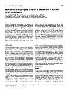
Fig. 5. Time course and requirements for CaMKI (1-294) activation. CaMKI (1-294) (5 jg/ml) was pre-incubated in the presence (closed symbols) or absence (open symbols) of CaMKI kinase (0.02 jg/ml) for the indicated time periods of pre-incubation, at 300C, with the following additions (as final concentrations; CaCI2 and MgCI2 are given as concentrations in excess of EGTA and EDTA concentrations, respectively): 10 mM MgCI2, 0.2 mM ATP, 2 mM EGTA (squares); or 10 mM MgCI2, 0.2 mM ATP, 1 mM CaCI2, 1 jM CaM (circles). Peptide-kinase activity was then measured using 100 ,uM synapsin site-1 peptide as substrate in the presence of Ca2+ (1 mM in excess of EGTA) and CaM (1 gM) (circles) or 1.3 mM EGTA (squares) as described in Materials and methods for 1 and 2 min. Background subtractions: for each condition, activity in the absence of preincubation (zero time of pre-incubation) has been subtracted to control for activation occurring during the assay itself and where appropriate, the activity of CaMKI kinase alone has also been subtracted. Each point represents the mean of two determinations with each determination the mean of the I and 2 min time points.
Ca2+-CaM dependent peptide kinase activity of CaMKI. The specific activity of wild type CaMKI increased by -25-fold from 0.32 ,umol/min/mg to 7.89 ,umol/min/mg upon phosphorylation by CaMKI kinase. In contrast, the specific activity of the T177A mutant was unchanged following incubation with CaMKI kinase. The T177S mutant showed an 18-fold increase in activity upon phosphorylation but its maximal specific activity was -20% of that of the wild type enzyme consistent with its lower level of phosphorylation. The increased basal activity of the Ti 77D mutant suggests that the introduction of the negatively charged carboxylate moiety mimicked Thr phosphorylation although the latter was considerably more efficient in activating the enzyme. The kinasenegative mutant, although phosphorylated to the same extent as wild type CaMKI, did not exhibit appreciable activity. Thus, whatever conformational change accompanies activation cannot override the requirement for Lys49 to participate in ATP binding. The role of Ca2+-CaM in the phosphorylation and activation of CaMKI by CaMKI kinase was investigated using the C-terminal truncation mutants. Figure 5 shows a time course of activation of CaMKI 1-294 by CaMKI kinase. In contrast to the full-length enzyme, the 1-294 mutant enzyme was activated to the same extent in the absence of Ca2+-CaM as in its presence, although Ca2+CaM accelerated the rate of activation. In parallel fashion to its activation (Figures 3 and 5), full-length CaMKI was not detectably phosphorylated in the absence of Ca2+CaM whereas the 1-294 mutant was significantly phos-
1
2
3 4 5 6 7 8
+
+
-
-
-
+
-
+-_
+
+-_++
9 10 11 12 +
-
_+-_ +
+
Fig. 6. Phosphorylation of CaMKI wild type and 1-294 and 1-306 mutant enzymes by CaMKI kinase in the presence and absence of Ca2+-CaM. CaMKI (wild type, 1-294 and 1-306) enzymes, (at final concentrations of 15 jg/ml), were phosphorylated as indicated, in the presence of I mM CaCI2, 1 jM CaM or 2 mM EGTA and the presence and absence of CaMKI kinase (0.1 ,ug/ml) as described in the legend to Figure 4. Lanes: 1-4, wild type; 5-8, 1-294; 9-12, 1-306. Each lane represents the electrophoresis of 0.3 gg of CaMKI. CaMKI migrates with an apparent Mr of -66 kDa due to fusion with GST. The band at -30 kDa (lane 1) appears to represent a small proportion of the CaMKI in this preparation that was proteolyzed during purification from the bacterial lysate.
phorylated under these conditions (Figure 6, lane 4 versus lane 8). The 1-306 truncation mutant was not phosphorylated by CaMKI kinase in either the presence or absence of Ca2+-CaM (lanes 9-12). A comparison of the kinase activities of these mutants in the absence and presence of CaMKI kinase is given in Table I. CaMKI 1-306 was inactive and unresponsive to CaMKI kinase whereas the activity of the 1-294 truncation mutant was stimulated 8fold by CaMKI kinase. Collectively, these data indicate that the auto-inhibitory domain of CaMKI gates, in a Ca2+-CaM dependent fashion, accessibility of Thrl77 to CaMKI kinase and that CaMKI kinase itself appears to be directly regulated by Ca2+-CaM. As with the fulllength enzyme, replacement of Thri77 of the 1-294 mutant with non-phosphorylatable Ala and Asp residues prevented activation and the latter substitution led to a CaMKI kinase independent partial activation.
Discussion Although CaMKI was one of the first biochemically identified Ca2+-CaM dependent multifunctional enzymes (Kennedy and Greengard, 1981), lack of a cloned sequence until recently has limited our understanding of the structure and function of this enzyme. The first cDNA of rat CaMKI was predicted to encode a protein of 332 amino acids (Picciotto et al., 1993). However, the extended identity of the human CaMKI described here to the recently identified 374 amino acid rat CaMKI cDNA (Cho et al., 1994) suggests that the earlier deduction of a shorter protein was probably a sequencing artifact. While the rat and human sequences exhibit very little difference in the first 362 amino acids, the C-terminal end of the protein is more divergent. This may represent species variation of the non-essential part of the protein. CaMKI purified from bovine or rat brain has been reported to consist of multiple polypeptides with Mrs by SDS-PAGE of: 42, 39 and
3683
B.Haribabu et al.
37 kDa (Nairn and Greengard, 1987); 43, 39 and 37 kDa (DeRemer et al., 1992a,b); and 41 and 40 kDa (Ito et al., 1994). Identification of a single CaMKI gene localized to chromosome 3 suggests that these variants are likely the result of post-transcriptional and/or post-translational modification of the products of a single gene. The generation of a Ca2+-CaM independent enzyme by truncation at amino acid 294 and a completely inactive enzyme by truncation at 306 shows that the mechanism of activation of CaMKI by Ca2+-CaM has a similar structural basis to that of other Ca2+-CaM dependent protein kinases. Ile294 in CaMKI is at an equivalent position in its primary sequence to Leu313 of CaMKIV or Leu290 of CaMKII (Figure 1). Truncation at this site led to Ca2`-CaM independent enzymes in the latter two cases (Cruzalegui et al., 1992; Cruzalegui and Means, 1993). Collectively, these data illustrate that all three classes of Ca2+-CaM dependent multifunctional enzymes (I, II and IV) are regulated by intrasteric auto-inhibition. The sequence between amino acids 294 and 306 of CaMKI (IKKNFAKSKWKQA) when aligned with the same regions of CaMKII and CaMKIV shows several shared and divergent features. In CaMKIIa the first five amino acids of this region (LKKFN) are sufficient for complete inhibition of the enzyme activity in the context of a truncated protein. However, deletion or substitution mutation of this region in the context of the full-length protein showed that the LKKFN residues are not essential for auto-inhibition (Cruzalegui and Means, 1992). Brickey et al. (1994) reported that His282 and Arg283 contribute to the auto-inhibition of CaMKIIa, at least with respect to ATP. Relative to the peptide substrate, mutation of Thr286 and the residues surrounding this autophosphorylation site to acidic amino acids will also at least partially relieve auto-inhibition (Fong et al., 1989; Hanson et al., 1989; Fong and Soderling, 1990; Waldman et al., 1990; Waxham et al., 1990; Hagiwara et al., 1991; Cruzalegui et al., 1992; Brickey et al., 1994). However, residues 290309 must also play a role since synthetic peptide analogs of this region competitively inhibit CaMKIIa activity with respect to substrate. A recent study of the properties of CaMKIV also suggested that the residues surrounding Thr3O8 (equivalent to Thr286 of CaMKIIa) were involved in auto-inhibition (Tokumitsu et al., 1994). The results described here for CaMKI suggest that although sequences N-terminal to Ile294 could play a role in auto-inhibition they are not sufficient, and that sequences between amino acids 294-306 must constitute at least part of a functional auto-inhibitory domain. The inability of CaMKI 1-306 to bind Ca2+-CaM localizes the CaM-binding domain or an essential part of it to residues C-terminal to 306. The CaM-binding domain of CaMKIIa was shown to extend from Phe293 to Thr3 10 based on the crystal structure of Ca2+-CaM complexed to a synthetic peptide analog of the auto-inhibitory region of the enzyme (Meador et al., 1993). If CaMKI is analogous to CaMKIIa then the CaM-binding domain would be from Asn297 to Arg314. However, if the enzyme binds CaM more similarly to smooth muscle myosin light-chain kinase (smMLCK) the binding domain could extend from Ala299 to Arg317. Regardless of which model is correct, Trp303 is predicted to occupy the C-terminal hydrophobic pocket of CaM. CaMKI truncated at 306 contains this Trp but
3684
does not bind CaM. These results are consistent with those obtained with a truncation mutant of smMLCK that also contained the homologous Trp as the C-terminal residue (Ito et al., 1991). In summary, we have shown that residues C-terminal to 306 are required for CaM binding, that residues 295-306 are sufficient to inhibit the enzyme, and that CaM binding is required to relieve autoinhibition. Further analysis is required to map more precisely the auto-inhibitory and CaM-binding domains of CaMKI . Previous studies have shown that purified brain CaMKI must be phosphorylated to exhibit maximal kinase activity (DeRemer et al., 1992b; Mochizuki et al., 1993b) and that the activating phosphorylation is catalyzed by a CaMKI kinase (DeRemer et al., 1992a; Edelman and Lee, 1994; Lee and Edelman, 1994). It was initially observed that bacterially expressed rat CaMKI autophosphorylated on Thrl77 but that this was without apparent effect on activity (Picciotto et al., 1993). Recently, Sugita et al. (1994), using a preparation designated CaM kinase kinase, identified two phosphorylation sites on rat CaMKI, Thrl77 and Thr310. Our studies reveal that the single residue of CaMKI phosphorylated by purified CaMKI kinase is Thrl77 and that phosphorylation of this residue is absolutely required for activation to occur. CaMKI kinase appears to be an enzyme of very restricted specificity in that it either did not phosphorylate or phosphorylated relatively poorly, a number of exogenous potential protein and peptide substrates we tested, including syntide-2, synapsin site I peptide, kemptide, myelin basic protein, histones, protamine, casein, phosvitin and myosin light chains (data not shown). One substrate that was phosphorylated by CaMKI kinase in addition to CaMKI was the related kinase, CaMKIV. Moreover, the site phosphorylated (Thrl96), is located at an equivalent position to Thrl77 of CaMKI (Selbert et al., 1995) and for both CaMKI and CaMKIV, phosphorylation causes a >20-fold increase in enzyme activity. Recently, two additional reports described an activator of CaMKIV and suggested that it was a Ca2+-CaM dependent CaMKIV kinase (Okuno et al., 1994; Tokumitsu et al., 1994). These proteins were purified from rat brain and exhibited different Mrs (66-68 kDa) from the porcine brain 52 kDa CaMKI kinase used here. The differences in Mr may reflect species variation, or there could exist multiple enzymes capable of activating CaMKI and CaMKIV by the same mechanism. It is also possible that activation could be produced by different mechanisms. It will therefore be important to determine whether the 68 kDa rat brain CaMKIV activator phosphorylates Thrl 96 of CaMKIV or is specific for other sites. ThrI 77 of CaMKI is at an equivalent position to the activating phosphorylation sites in cAMP dependent protein kinase, MAP kinases such as ERK1, MAP kinase kinase and cyclin dependent kinases CDC2, CDK2 and CDK4 (Taylor and Radzio-Andzelm, 1994). Recent solution of the crystal structures of PKA, ERK2 and CDK2, suggests that this threonine residue is part of a 'lip' structure at the entrance to the catalytic site (Knighton et al., 1991; DeBondt et al., 1993; Zhang et al., 1994). Phosphorylation in this 'activation loop' is central to the regulation of the catalytic activity of these three kinases (Taylor and Radzio-Andzelm, 1994). Mutational analysis
Human CaM kinase I
of PKC BII also confirms the importance of a phosphorylatable residue at this position for the catalytic activity (Orr and Newton, 1994). It has been proposed that a negative charge on this loop is important for the correct alignment of the residues participating in catalysis (Taylor and Radzio-Andzelm, 1994). Ca2+-CaM dependent protein kinases I and IV may now be added to the list of kinases regulated by phosphorylation on this 'activation loop'. Relief of intrasteric auto-inhibition and activation-loop phosphorylation represent two distinct, and fundamentally independent mechanisms, both of which must be operative for CaMKI to be maximally active. For example, whereas truncation of CaMKI at the end of the kinase domain (1294) removes its ability to bind Ca2+-CaM, the mutant still requires phosphorylation of Thrl 77 for maximal activity. And, as shown previously, the activity of phosphorylated brain CaMKI can be reduced to low levels by either dephosphorylation with protein phosphatase 2A or by removal of Ca2+-CaM from the assay (DeRemer et al., 1992b; Lee and Edelman, 1994). Yet in one important respect these two processes are mechanistically linked, namely that, as shown previously for brain CaMKI (DeRemer et al., 1992b; Lee and Edelman, 1994) and here for the human enzyme, phosphorylation of CaMKI by CaMKI kinase is itself a Ca2+-CaM dependent process. The studies described here with various mutant forms of human CaMKI provide a structural explanation for this requirement. CaMKI kinase phosphorylates full-length CaMKI only in the presence of Ca2+-CaM, and the 1294 form in either the presence or absence of Ca2+-CaM, but will not phosphorylate the inactive 1-306 form in either the presence or absence of Ca2+-CaM. Thus, Thrl 77 must be unavailable for phosphorylation in the autoinhibited conformation of CaMKI, becoming accessible through removal of the auto-inhibitory domain by either truncation in the case of the 1-294 enzyme or CaM binding to the full-length enzyme. These observations suggest a dual role for Ca2+-CaM. The binding of Ca2+_ CaM must expose Thrl77 to CaMKI kinase and also expose the active site so that substrate can bind. Phosphorylation of Thrl77 then presumably causes a further conformational change that maximally activates the enzyme. The fact that activation can occur in the presence of an overwhelming molar excess of substrate (DeRemer et al., 1992b) implies that peptide-substrate binding does not block CaMKI kinase accessibility to Thrl77. In addition to this dual role, our studies suggest that there is potentially at least a third way in which Ca2+CaM promotes CaMKI activation, that is by directly enhancing the activity of CaMKI kinase. It was previously demonstrated that CaMKI kinase binds in a Ca2+ dependent fashion to a CaM-affinity column suggesting that CaMKI kinase may be a Ca2+-CaM regulated enzyme (Lee and Edelman, 1994). The 1-294 mutant CaMKI, which has lost the ability to bind CaM, was phosphorylated and activated by CaMKI kinase at a faster rate in the presence of Ca2+-CaM than in its absence (Figures 5 and 6 and Table I). Thus, CaMKI kinase is Ca2+-CaM regulated. On the other hand, the fact that phosphorylation and maximal activation of the 1-294 mutant enzyme occurs in both the absence and presence of Ca2+-CaM suggests that CaMKI kinase must not be absolutely dependent on Ca2+-CaM for activity. We have also repurified on a small scale an
aliquot of the purified CaMKI kinase over CaMSepharose. The repurified enzyme was also capable of Ca2+-CaM independent phosphorylation of CaMKI (data not shown) suggesting that CaMKI kinase did not acquire its Ca2+-CaM independent activity by phosphorylation or proteolysis subsequent to purification. One possibility is that CaMKI kinase represents the first example of an 'atypical' CaM kinase. Such an enzyme would, hypothetically, possess a functional CaM-binding domain but no auto-inhibitory domain. In this model the active site would be accessible to substrates in the absence of Ca2+-CaM. The conformational change induced by CaM binding would then either increase substrate affinity or correctly position residues critical for catalysis. Further biochemical analysis and molecular cloning of CaMKI kinase will be required to resolve this issue.
Materials and methods Bacterial expression of CaMKI A 1.5 kbp cDNA fragment contained the entire amino acid coding region and was subcloned into pBluescript. The 5' untranslated sequence of CaMKI cDNA was removed and a BamHI site was inserted upstream of the initiating methionine by PCR. Two oligonucleotides (5'AGGGGGATTCGCCATGCTGGGGGCAGTG-3' and 5'-AATGTTGGGGTGCTT-3') were synthesized and the N-terminal -240 bp of the CaMKI coding region was amplified. PCR was carried out using Hottub DNA polymerase (Amersham), and the CaMKI cDNA clone as template for 10 cycles for 1 min at 94°C, 1 min at 45°C and 3 min at 72°C followed by a 15 min extension step at 72°C. The amplified DNA was digested with BamHI and NsiI, the 150 bp fragment was gel purified and ligated into the pBluescript CaMKI cDNA clone digested with the same enzymes. The sequence of the PCR amplified region was verified by sequencing. The 1.39 kb BamHI-EcoRI fragment from this clone was excised and subcloned into the BamHI-EcoR I sites of the bacterial expression vector pGEX-2T (Smith and Johnson, 1988). Several independent clones were analyzed for the expression of GST fusion proteins in Ecoli DH5a and BL21 (DE3). Briefly, 5.0 ml of LB medium was inoculated with a single colony and grown overnight at 37°C. The fully grown culture was diluted 5-fold in fresh LB medium and grown for an additional 1 h. Isopropyl ,B-D-thiogalactopyranoside (IPTG) was then added to a final concentration of 0.4 mM and the cultures were allowed to grow for an additional 4 h. Cell samples were analyzed by SDSPAGE for the expression of a 67 kDa GST-CaMKI fusion protein.
Preparation and expression of CaMKI mutants Substitution and deletion mutants of CaMKI were prepared using PCR. To create a Lys49 to Ala (K49A) substitution two oligonucleotides were used. The forward primer was the same as the one used above to remove the 5' untranslated sequence and the reverse primer (5'CTTGGCAATGCATGCGATGGCCACCA-3') contained the codon of Lys49 changed to an alanine and an NsiI site immediately following this codon. PCR was carried out as described above and the products were digested with BamHI-NsiI, purified and used to replace the same fragment from the wild type CaMKI cDNA clone in pBluescript. The PCR-amplified region of the K49A mutant was sequenced to confirm the presence of the intended mutation and the absence of any PCRgenerated errors. The 1.39 kb BamHI-EcoRI fragment from this clone was transferred into pGEX-2T to generate GST-K49A CaMKI (a kinasenegative CaMKI). PCR primers containing a unique SmaI site at nucleotides 657-662 of CaMKI cDNA were used in conjunction with the appropriate C-terminal primers to create the Thrl77 substitution and C-terminal deletion mutants. For generating the T 177A mutant the forward primer (5'-GAGGACCCGGGCAGTGTGCTCTCCGCCGCC3') corresponding to nucleotides 650-681 of CaMKI was made. For making T177S and T177D mutants the same forward primers but with the appropriate changes of codon 177 (nucleotides 676-678) were synthesized. These were used to amplify by PCR the C-terminal portion of the CaMKI utilizing a primer from the 3' end of the pBluescript CaMKI cDNA clone (M-13 universal primer). The amplified fragments were digested with Smal-EcoRI, gel purified and used to replace the Cterminal SmaI-EcoRI fragment in the pGEX-CaMKI clone expressing
3685
B.Haribabu et al. the wild type protein. The C-terminal truncations at 294 and 306 were created by following the same strategy described above but using reverse primers that terminate at these positions due to insertion of a stop codon and an EcoRI site. DNA from all the mutant clones was completely sequenced in the PCR amplified regions to confirm the intended mutations and lack of secondary mutations.
Purification of GST fusion proteins DHSa and BL21(DE3) cells containing the expression plasmids were grown at 37°C in 10 ml cultures overnight and used to inoculate 1.0 1 cultures. These were grown to an OD of 0.6 and IPTG was added to a concentration of 0.4 mM. After a 4 h induction at 37°C the cells were harvested by centrifugation. Soluble protein extracts were prepared by mild sonication of cells in PBS containing protease inhibitors (0.5 mM PMSF, 20 mg/ml soybean trypsin inhibitor, 10 mg/ml aprotinin, 5 mg/ml leupeptin) and 0.5 mM dithiothreitol. Cell debris was removed by centrifugation at 10 000 g for 25 min. The supematant was loaded onto a glutathione-Sepharose column, washed with PBS and eluted in 50 mM Tris-HCI pH 8.0 containing 10 mM glutathione. The purity of the enzyme preparations was verified by SDS-PAGE. A minor proteolysis product was eliminated by gel filtration using FPLC over a HiLoad 16/60 Superdex 200 column (Pharmacia-LKB Biotechnology Inc.) using 50 mM Tris-HCl (pH 8.0) as the column buffer. Purified enzymes were stored in 50% glycerol at -80°C in aliquots for several months without loss of activity.
Peptide kinase assay The activities of CaMKI wild type and mutant enzymes were assayed at 30°C in either the presence or absence of Ca2+-CaM as indicated in the figure legends, by incubation in a mixture which also contained: 50 mM Tris, pH 7.6, 0.5 mM DTT, 0.5 mg/ml bovine serum albumin (BSA), 10 mM MgCl2, 200 ,uM [y-32P]ATP (New England Nuclear/ DuPont) (-0.1 X 10³ c.p.m./pmol) and 100 IM synapsin site-l peptide. Synapsin site-1 peptide (NYLRRRLSDSNF) was custom synthesized and purified by HPLC at the Biomedical Research Core Facilities of the University of Michigan. Porcine calmodulin was obtained from Boehringer Mannheim. Quantitation of 32p incorporation into synapsin site-l peptide was determined by a P81 phosphocellulose filter-paper procedure described previously (DeRemer et al., 1 992a).
Analysis of protein phosphorylation Conditions for phosphorylation of recombinant wild type and mutant CaMKI proteins by CaMKI kinase are described in the figure legends. SDS-PAGE was performed as previously described (Lee and Edelman, 1994). CaMKI kinase was purified from pig brain as previously described in detail (Lee and Edelman, 1994), with the exceptions that the hydroxylapatite step was omitted and the order of the CaM-Sepharose and phenyl-Sepharose chromatographic steps was reversed.
Protein determinations The concentrations of CaMKI wild type and all mutant enzymes were determined by the procedure of Lowry et al. (1951) with some modifications (Peterson, 1977) using BSA as standard. CaMKI kinase concentration was determined by a colloidal gold staining method (Lee and Edelman, 1994). The concentration of CaM was determined spectrophotometrically (EM,277nm = 3300).
Acknowledgements We wish to thank Dr J.Aletta for helpful discussions, Richard Alston of the Duke Comprehensive Cancer Center for advice on multiple sequence alignment and Elizabeth Fletcher for help with the manuscript. This work was supported by grants from the National Institutes of Health (A.R.M., A.M.E. and R.S.)
References Brickey,D.A., Bann,J.G., Fong,Y.L., Perrino,L., Brennan,R.G. and Soderling,T.R. (1994) J. Biol Chem., 269, 29047-29054. Cho,F.S., Phillips,K.S., Bogucki,B. and Weaver,T.E. (1994) Biochim. Biophys. Acta, 12243, 156-160. Colbran,R.J., Fong,Y.L., Schworer,C.M. and Soderling,T.R. (1988) J. Biol. Chem., 263, 18145-18151. Cruzalegui,F.H. and Means,A.R. (1993) J. Biol. Chem., 268, 2617126178.
3686
Cruzalegui,F.H., Kapiloff,M.S., Morfin,J.P., Kemp,B.E., Rosenfeld,M.G. and Means,A.R. (1992) Proc. NatlAcad. Sci. USA, 89, 12127-1213 1. De Bondt,H.L., Rosenblatt,J., Jancarik,J., Jones,H.D., Morgan,D.O. and Kim,S.H. (1993) Nature, 363, 595-602. DeRemer,M.F., Saeli,R.J., Brautigan,D.L. and Edelman,A.M. (1992a) J. Biol. Chem., 267, 13466-13471. DeRemer,M.F., Saeli,R.J. and Edelman,A.M. (1992b) J. Biol. Chem., 267, 13460-13465. Didsbury,J.R. and Snyderman,R. (1987) FEBS Lett., 219, 259-263. Edelman,A.M., Blumenthal,D.K. and Krebs,E.G. (1987) Annu. Rev. Biochem., 56, 567-613. Fong,Y.L. and Soderling,T.R. (1990) J. Biol. Chem., 265, 11091-11097. Hagiwara,T., Ohsako,S. and Yamauchi,T. (1991) J. Biol. Chem., 266, 16401-16408. Hanks,S.K. and Quinn,A.M. (1991) Methods Enzymol., 200, 38-62. Hanson,P.I. and Schulman,H. (I 992) Annu. Rev. Biochem., 61, 559-60 1. Hanson,P.I., Kapiloff,M.S., Lou,L.L., Rosenfeld,M.G. and Schulman,H. (1989) Neuron, 3, 59-70. Haribabu,B. and Snyderman,R. (1993) Proc. Natl Acad. Sci. USA, 90, 9398-9402. Ito,M. Guerriero Jr.,V., Chen,X. and Hartshorne,D.J. (1991) Biochemistry, 30, 3498-3503. ItoT., Yokokura,H., Nairn,A.C., Nimura,Y. and Hidaka,H. (1994) Biochem. Biophys. Res. Commun., 201, 1561-1566. Kameshita,I. and Fujisawa,H. (1993) J. Biochem., 113, 583-590. Kennedy,M.B. and Greengard,P. (1981) Proc. Natl Acad. Sci. USA, 78, 1293-1297. Knighton,D.R., Wheng,J., Ten Eyck,L.F., Ashford,V.A., Xuong,N.H., Taylor,S.S. and Sowadski,J.M. (1991) Science, 253, 407-413. Lee,J.C. and Edelman,A.M. (1994) J. Biol. Chem., 269, 2158-2164. Lee,J.C. and Edelman,A.M. (1995) Biochem. Biophys. Res. Commun., in press. Lee,J.C., Kwon,Y.G., Lawrence,D.S. and Edelman,A.M. (1994) Proc. Natl Acad. Sci. USA, 91, 6413-6417. Lowry,O.H., Rosebrough,N.J., Farr,A.L. and Randall,R.J. (1951) J. Biol. Chem., 193, 265-275. Marshall,C.J. (1994) Nature, 367, 686. Meador,W.E., Means,A.R. and Quiocho,F.A. (1993) Science, 262, 1718-1721. Mochizuki,H., Ito,T. and Hidaka,H. (1993a) J. Biol. Chem., 268, 9143-9147. Mochizuki,H., Sugita,R., Ito,T. and Hidaka,H. (1 993b) Biochem. Biophys. Res. Commun., 197, 1595-1600. Nairn,A.C. and Greengard,P. (1987) J. Biol. Chem., 262, 7273-7281. Okuno,S., Kitani,T. and Fujisawa,H. (1994) J. Biochem., 116, 923-930. Orr,J.W. and Newton,A.C. (1994) J. Biol. Chem., 269, 27715-27718. Payne,M.E., Fong,YL., Ono,T., Colbran,R.J., Kemp,B.E., Soderling,T.R. and Means,A.R. (1988) J. Biol. Chem., 263, 7190-7195. Pearson,R.B., Woodgett,J.R., Cohen,P. and Kemp,B.E. (1985) J. Biol. Chem., 260, 14471-14476. Peterson,G. L. (1977) Anal. Biochem., 83, 346-356. Picciotto,M.R., Cohn,J.A., Bertuzzi,G., Greengard,P. and Nairn,A.C. (1992) J. Biol. Chem., 267, 12742-12752. Picciotto,M.R., Czernik,A.J. and Nairn,A.C. (1993) J. Biol. Chem., 268, 26512-26521. Polymeropoulos,M.H., Xiao,H., Glodek,M., Adams,M.D., Moreno,R.F., Fitzgerald,M.G., Venter,J.C. and Merril,C.R. (1992) Genomics, 12, 492-496. Selbert,M.A., Anderson,K.A., Huang,Q.H., Goldstein,E.G., Means,A.R. and Edelman,A.M. (1995) J. Biol. Chem., in press. Sheng,M., Thompson,M.A. and Greenberg,M.E. (1991) Science, 252, 1427-1430. Smith,D.B. and Johnson,K.S. (1988) Gene, 67, 31-40. Stokoe,D., Caudwell,B., Cohen,P.T. and Cohen,P. (1993) Biochem. J., 296, 843-849. Sugita,R., Mochizuki,H., Ito,T., Yokokura,H., Kobayashi,R. and Hidaka,H. (1994) Biochem. Biophys. Res. Commun., 203, 694-701. Taylor,S.S. and Radzio-Andzelm,E. (1994) Structure, 2, 345-355. Tokumitsu,H., Brickey,D.A., Glod,J., Hidaka,H., Sikela,J. and Soderling,T.R. (1994) J. Biol. Chem., 269, 28640-28647. Waldmann,R., Hanson,P.I. and Schulman,H. (1990) Biochemistry, 29, 1679-1784. Waxham,M.N., Aronowski,J., Westgate,S.A. and Kelly,P.T. (1990) Proc. Natl Acad. Sci. USA, 87, 1273-1277. Zhang,F., Strand,A., Robbins,D., Cobb.,M.H. and Goldsmith,E.J. (1994) Nature, 367, 704-711. Received on February 16, 1995; revised on Ma' 1, 1995