Human combinatorial libraries yield rare antibodies that broadly neutralize hepatitis C virus Daniel X. Johansson*, Ce´cile Voisset†, Alexander W. Tarr‡, Mie Aung‡, Jonathan K. Ball‡, Jean Dubuisson†, and Mats A. A. Persson*§ *Department of Medicine, Center for Molecular Medicine, Karolinska University Hospital Solna, Karolinska Institutet, 171 76 Stockholm, Sweden; †Institut de Biologie de Lille, Unite´ Mixte de Recherche 8161, Centre National de la Recherche Scientifique, Universite´ de Lille I and II and Institut Pasteur de Lille, 59045 Lille, France; and ‡Institute of Infection, Immunity, and Inflammation, School of Molecular Medical Sciences, University of Nottingham, Queen’s Medical Centre, Nottingham NG7 2UH, United Kingdom
One way to dissect the antibody response to an invading microorganism is to clone the antibody repertoire from immune donors and subsequently characterize the specific antibodies. Recently, methodological advances have allowed investigations of neutralizing antibodies against hepatitis C virus (HCV) in vitro. We have investigated three human mAbs, previously isolated from an individual infected with HCV of genotype 2b, that are known to cross-react in a binding assay to the envelope E2 protein of genotypes 1a and 1b. We now report that two of them have a neutralizing activity with a breadth not previously observed. Indeed, mAbs 1:7 and A8 recognized E2 from all of the six major genotypes, and they neutralized retroviral pseudoparticles [HCV pseudoparticles (HCVpp)] carrying genetically equally diverse HCV envelope glycoproteins. Importantly, these antibodies were also able to neutralize the cell culture infectious HCV clone JFH-1 in vitro, with IC50 values of 60 ng/ml and 560 ng/ml, respectively. The conformational epitopes of these two broadly reactive antibodies were overlapping yet distinct and involved amino acid residues in the 523–535 region of E2, known to be important for the E2–CD81 interaction. The third antibody clone, representing a dominant population in the initial screen for these antibodies, was less broadly reactive and was unable to neutralize the genotype 2a infectious clone JFH-1. Our results confirm at the clonal level that broadly neutralizing human anti-HCV antibodies can be elicited and that the region amino acids 523–535 of the HCV envelope glycoprotein E2 carries neutralizing epitopes conserved across all genotypes. immunoglobulins 兩 liver disease 兩 viral hepatitis
H
epatitis C virus (HCV) is a member of hepacivirus genus within the Flaviviridae family (1). It infects the human liver and is estimated to be carried by 3% (170 million) of the human population as a chronic infection, which if untreated often results in serious liver disease including cirrhosis and hepatocellular cancer (2). HCV remains a global health problem. Virus genome replication is error-prone, and this, together with high virus turnover, generates extensive genetic variability. Within an infected individual HCV exists as a swarm of genetically related but distinct viruses called a quasispecies (3). HCV can be grouped into six major genotypes that differ by up to 30% at the nucleotide level (4–6). Among the most variable parts of the virus are the two envelope proteins, E1 and E2 (7). Current therapy, a combination of PEGylated IFN-␣ and the antiviral drug Ribavirin, is not suited for all patients, and up to 50% of those treated fail to clear the virus (8). Despite substantial efforts no effective vaccine has yet been developed for human use (9). Additional and improved therapeutic approaches are therefore a considerable challenge. Twenty to 25% of newly infected individuals will spontaneously resolve the infection, whereas the remainder will develop a chronic infection (2). This intriguing capacity of some individuals to eliminate the infection has prompted large efforts to study virus–host
www.pnas.org兾cgi兾doi兾10.1073兾pnas.0705522104
interactions, notably the native and adaptive immune responses to the virus. The IFN response and the T cell response have shown to be important for recovery, whereas NK cell response and humoral immunity have in most cases not been associated with clearance. However, antibodies to the envelope protein E2 have been shown to ameliorate the disease in chimpanzees, to correlate with protection by vaccination in the same animal species, and to reduce the rate of reinfection of the graft after liver transplantation in man (10–12). Moreover, advances over the last years have provided new tools to study the virus-specific antibody response, in particular antibodies that block infection. The new methods include generation of infectious retroviral pseudoparticles, bearing native HCV envelope glycoproteins on their surface [HCV pseudoparticles (HCVpp)], and, more recently, cloned HCV genomic RNA (strain JFH-1) that after transfection into appropriate cells generates infectious HCV particles (HCVcc) (13–17). The recent isolation of functional E1E2 genes representative of all of the major genotypes of HCV has enabled assessment of the neutralizing breadth and potency of sera and mAbs (18). Although cell culture infectious virus currently represents only a limited number of HCV genotypes, this system is useful to determine the neutralizing potency of antibodies against native particles. These systems have been used to determine the neutralizing capacity and cross-reactivity profile of a small number of murine mAbs (19). The methods are also providing important insights into the natural antibody response to HCV, such as the existence of in vitro neutralizing antibodies in humans, as well as the possible existence of virus-induced mechanisms that suppress the neutralizing antibody response in the initial, critical phase of the infection (20, 21). Whether a broad neutralizing activity present early in the infection would affect the disease outcome remains to be studied. Similarly, definition of conserved epitopes in the two envelope proteins that may confer cross-genotype neutralization will help us understand the mechanisms involved in entry and infection and will guide future vaccine and therapeutic antibody design. We have previously isolated mAbs to the E2 envelope glycoprotein as a means to dissect the immune response to HCV in humans (22). The antibodies were derived from an individual infected with HCV of genotype 2b (gt2b) and isolated by their Author contributions: D.X.J., C.V., A.W.T., J.K.B., J.D., and M.A.A.P. designed research; D.X.J., C.V., A.W.T., and M.A. performed research; C.V., A.W.T., J.K.B., J.D., and M.A.A.P. contributed new reagents/analytic tools; D.X.J., C.V., A.W.T., M.A., J.K.B., J.D., and M.A.A.P. analyzed data; and D.X.J., C.V., A.W.T., J.K.B., J.D., and M.A.A.P. wrote the paper. Conflict of interest statement: The antibodies analyzed in this article have been the topic of a patent application since 1996. It is currently assigned to Karolinska Innovations AB, a tech transfer office of the Karolinska Institutet, where M.A.A.P. performed the work. However, the patent and patent applications are in the process of being transferred to a spinoff company, Molecules of Man AB. M.A.A.P. has a financial interest in this company (as shareholder). Abbreviations: GNA, Galanthus nivalis lectin; HCV, hepatitis C virus; HCVpp, HCV pseudoparticles; HCVcc, cloned HCV from cell culture; gt, genotype. §To
whom correspondence should be addressed. E-mail:
[email protected].
© 2007 by The National Academy of Sciences of the USA
PNAS 兩 October 9, 2007 兩 vol. 104 兩 no. 41 兩 16269 –16274
MEDICAL SCIENCES
Communicated by Richard A. Lerner, The Scripps Research Institute, La Jolla, CA, July 27, 2007 (received for review May 29, 2007)
Table 1. Temporary names for isolates Isolate name used in this article gt1a:1 gt1a:2 gt1a:3 gt1a:4 gt1b:1 gt1b:2 gt2a:1 gt2a:2 gt2b:1 gt2b:2 gt3:1 gt3:2 gt4:1 gt4:2 gt5:1 gt5:2 gt6:1 gt6:2
Full isolate name (18) H77.20/H77.c H11 UKN1A20.8 Strain H UKN1B5.23 UKN1B12.16 UKN2A1.2 JFH-1 (gt2a) UKN2B1.1 UKN2B2.8 UKN3A1.28 UKN3A13.6 UKN4.11.1 UKN4.21.16 UKN5.14.4 UKN5.15.7 UKN6.5.8 UKN6.5.340
GenBank accession no. AF011751 AF011752 EU155192 AY734976 AY734974 AY734977 AB047639 AY734982 AY734983 AY734984 AY894683 AY734986 AY734987 AY785283 EF427672 EF427671 AY736194
The full names of isolates mentioned in the text, their GenBank accession numbers (if any), and the temporary names used in the present article are listed.
capacity to bind to E2 of gt1a. By their very nature, they may therefore react with divergent genotype proteins. Indeed, we demonstrated that they bind to gt1a and gt1b and that they block the binding of E2 of these genotypes to CD81, a putative cell receptor used for virus entry (22, 23). We have now assessed the capacity of three of these human mAbs to neutralize a panel of pseudoparticles representing all genotypes, tested their effects on cloned JFH-1 particles, and mapped the conformational epitopes for two of the antibodies that showed particularly broad neutralizing properties. Results Determination of EC50 for Binding E1E2 of gt1a (Isolate H77c). The three mAbs investigated, clones 1:7, A8, and L1, were selected for the present study because earlier results indicated that they were binding to distinct epitopes and blocked the binding of soluble E2 to CD81-expressing cells (neutralization of binding assay). The mAbs were expressed as full-length human IgG1 from the vector pMThIgG1 in stably transfected Drosophila S2 cells (24). Approximately 10–15 g of mAb per milliliter of medium could be purified 10 days after induction. EC50 values for each mAb were determined for the reference H77c (gt1a:1) (Table 1) protein in Galanthus nivalis lectin (GNA) capture ELISA (Fig. 1). mAb A8 showed slightly lower EC50 than mAb 1:7 (0.44 g/ml vs. 0.67 g/ml), whereas mAb L1 had considerably lower affinity to isolate H77 (EC50 ⫽ 2.31 g/ml). These equate to Kd values of ⬇1.5 ⫻ 10⫺8 M (L1), 2.7 ⫻ 10⫺9 M (A8), and 4.1 ⫻ 10⫺9 M (1:7).
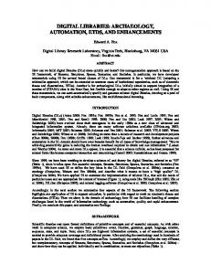
Fig. 2. Immunofluorescence of E1E2-expressing cells stained by human mAbs A8 (Top), 1:7 (Middle), or L1 (Lower). Huh-7 cells expressing E1E2 of gt1– 6 (see Table 1) were incubated with human mAbs. Bound antibody was detected by using an FITC-labeled anti-human antisera.
mAbs 1:7 and A8 Display a Broad Reactivity. The reactivity of mAbs
1:7, A8, and L1 against different genotypes was first determined by immunofluorescence. HCV envelope glycoproteins from different genotypes were expressed in Huh-7 cells, and the permeabilized cells were incubated with these mAbs. As previously observed, HCV-infected cells stained with mAbs 1:7, A8, and L1 displayed a pattern of specific fluorescence in a network of cytoplasmic membranes and the nuclear envelope, which likely correspond to endoplasmic reticulum membranes (Fig. 2) (25). Clone L1 stained cells expressing E1E2 of gt1b, gt2a, gt2b, gt5, and gt6 (Fig. 2) but not gt1a, gt3, and gt4 (isolates gt1a:1, gt3:1, and gt4:1) (Table 1) (data not shown). Interestingly, both mAbs 1:7 and A8 immunostained cells expressing E2 of all of the tested isolates, indicating a broad reactivity for these mAbs (Fig. 2). The broad reactivity of mAbs 1:7 and A8 was confirmed by immunoprecipitation. Indeed, mAbs 1:7 and A8 immunoprecipitated E2 of all isolates tested (Fig. 3). However, gt3 and gt4 gave significantly weaker signals in the final Western blot than the other genotypes. This is probably because of differences in affinity of the antibodies used to develop the blotted
Fig. 1. Binding curves for the three antibody clones. The respective antibodies were incubated in microtiter wells where E1E2 of isolate H77 (gt1a) had been captured on GNA. Detection of bound antibody was performed with enzyme-conjugated anti-human antiserum. 16270 兩 www.pnas.org兾cgi兾doi兾10.1073兾pnas.0705522104
Johansson et al.
Fig. 3. Immunoprecipitations of E1E2 of all genotypes using antibodies 1:7 and A8. Immunoprecipitations of cell lysates from Huh-7 cell expressing E1E2 of gt1– 6 (Table 1) were analyzed by Western blot and detected with a mixture of anti-HCV-E2 antibodies. Still, this antibody mix did not equally well detect all isolates; especially gt4 detection was weaker, as shown by a direct Western blot on lysates from E1E2-expressing cells (far right panel). ‘‘cl3’’ is a human anti-HIV-1 gp120 antibody used as negative control. HC indicates the heavy chain of the mAb used for immunoprecipitation.
mAb Binding to GNA Captured HCV Envelope Glycoprotein from All Genotypes. To further characterize the capacity of our mAbs to
recognize different isolates, the relative binding of mAbs 1:7, A8, and L1 to different genotypes was tested in an ELISA with E1E2 representing different genotypes (Fig. 4) (18, 26). mAbs 1:7 and A8 showed binding to all genotypes whereas mAb L1 had a more limited binding range. Interestingly, mAb L1 did not bind gt1a:1 (H77c) but did bind gt1a:3 (UKN1A20.8), emphasizing the diversity within genotypes. In a similar fashion, mAb L1 bound to gt2a:1 (UKN2A1.2) but did not bind gt2a:2 (JHF-1). mAbs 1:7 and A8 Are Broadly Neutralizing Antibodies. To establish additional characteristics about the human mAbs, we analyzed their potential neutralizing activity. We first tested the ability of our antibodies to neutralize HCVpp bearing the HCV E1 and E2 envelope glycoproteins of HCV gt1a, gt1b, gt2a, gt2b, gt3a, gt4, gt5, and gt6 (13, 18). Interestingly, both 1:7 and A8 neutralized HCVpp bearing E1 and E2 from all genotypes tested (Fig. 5). However, some variations were observed, and a lower neutralizing activity was observed for mAb A8 against the 1b isolates tested (gt1b:1 and gt1b:2). In contrast to mAbs 1:7 and A8, mAb L1 had a more varied behavior (Fig. 5). Indeed, this antibody showed a neutralizing activity against gt1b:1, gt1b:2, gt2a:2 (JFH-1), gt3:1, gt4:2, gt5:1, and gt6:2 isolates, but no neutralizing activity was detected against gt1a:4, gt2b:1, and gt4:1 isolates. It is worth noting that differences in the neutralizing activity of mAb L1 could be observed between gt4:1 and gt4:2, which belong to the same genotype, again indicating different reactivities within the same genotype. We also assessed the neutralizing capability of our mAbs using the recently described JFH-1 HCVcc system (gt2a) (15). Both mAbs 1:7 and A8 neutralized HCVcc JFH-1 in a dose-dependent manner (Fig. 6). This was shown by analyzing the level of E2 in the lysate of infected cells 2 days after infection. The approximate IC50 values were estimated by performing density measurements of E2 after Western blotting. This indicated approximate IC50 values of 560 ng/ml and 60 ng/ml for clones A8 and 1:7, respectively (Fig. 6). Surprisingly, L1 had no neutralizing activity in this HCVcc system, whereas it neutralized HCVpp containing the envelope glycoproteins of the JFH-1 isolate. Because these particles differ in their
assembly process, it is possible that the epitope recognized by L1 is accessible at the surface of HCVpp but not on HCVcc. Differences between HCVpp and HCVcc have indeed already been reported for HCV entry (27). Altogether, our results indicate that mAbs 1:7 and A8 are broadly potent neutralizing antibodies. Epitope Mapping of mAbs 1:7 and A8. We have earlier observed that
mAbs 1:7 and A8 bind to conformational epitopes on E2 and block the interaction with CD81 (22). Therefore, to map their epitope(s) we used a panel of full-length E1E2 mutants of the gt1a H77c isolate, containing single-point alanine substitutions of conserved residues within regions of E2 shown to be involved in CD81 binding (28). Reactivity of the antibodies to the E2 mutants was measured to GNA-captured E1E2 in an ELISA. Both mAbs showed a similar pattern with residues critical for binding being located within region 523–535 (annotated according to the amino acid positions occurring in the H77 sequence). Substitutions G523A, W529A, G530A, and D535A all distinctly abolished binding of these two mAbs to E2 (Fig. 7). Interestingly, three of these residues are also critical for CD81 binding and are conserved over a wide range of isolates (Fig. 8) (28). Discussion In the present article we have assessed the breadth of the antiviral response in vitro of three human mAbs derived from an infected individual (22). Importantly, two of these antibodies bound to E1E2 proteins and neutralized pseudoparticles bearing the HCV envelope glycoproteins of all of the six major genotypes. The third clone, representing a dominant population in the initial screen for these antibodies, reacted to some but not all isolates/proteins of gt1a, gt1b, gt2b, gt4, and gt5. The two broadly neutralizing antibodies were titered for neutralization of the cloned JFH isolate (gt2a), and their epitopes were at least partially mapped by using a panel of single-point mutated E2 proteins based on the H77 isolate (gt1a). In the binding studies we observed a slightly higher affinity for clones 1:7 and A8 for binding to E1E2 of the H77 isolate (gt1a) than the previous assessment using E2 of the HCV-1 strain (also gt1a) (22). Although we cannot exclude that this is related to isolatespecific differences, our antibodies likely recognize E2 with a higher affinity in our GNA-captured E1E2 complexes, which are likely to have a more native conformation than the E2 alone. Indeed, it has been shown that the binding of CD81 is better to E1E2 than to E2
Fig. 4. Binding of human mAbs to E1E2 of different genotypes. Antibodies 1:7, A8, and L1 were incubated with saturating amounts of GNA-captured E1E2 proteins representative of all genotypes of HCV (see Table 1). Bound antibody was detected with alkaline phosphatase-conjugated anti-human IgG antiserum.
Johansson et al.
PNAS 兩 October 9, 2007 兩 vol. 104 兩 no. 41 兩 16271
MEDICAL SCIENCES
membrane for the different E2 genotypes, as shown by a direct Western blot on lysates from E1E2-expressing cells (Fig. 3 Far Right).
Fig. 5. Neutralization of HCVpp bearing E1E2 from different genotypes by human mAbs A8, 1:7, and L1. HCVpp bearing E1E2 of gt1– 6 (depicted as in Table 1) were incubated with 15 g/ml human mAbs A8, 1:7, and L1 for 1 h at 37°C before 2 h of contact with target cells. The amount of infected particles was measured after 2 days as luciferase activity. Results are given as percentages of neutralization relative to infection in the absence of antibody for each genotype (mean ⫾ SD of three independent experiments).
alone (29), and there are reports of anti-E2 antibodies (e.g., clone CBH2) that recognize their epitope only when E2 is coexpressed with E1 (30, 31). Accordingly, we regard the present data as more accurate. A panel of pseudoparticles bearing the HCV envelope glycoproteins of different genotypes allowed us to determine the neutralization profile in vitro for the three mAbs. The results agreed well with the biochemical binding data as well as the cell-staining patterns for cells transiently expressing E1E2 constructs. The pseudoparticle neutralization data provided the antiviral genotype specificity, and the sensitivity was determined by using the HCVcc
Fig. 6. Neutralization of HCVcc by human mAbs A8, 1:7, and L1. Neutralization assays were performed by preincubating HCVcc (clone JFH-1) with increasing amounts of human mAbs A8, 1:7, and L1 for 1 h at 37°C before contact with target cells for 2 h. At 2 days after infection, the level of E2 in the supernatant was assessed by Western blotting with mAb 3/11. The results presented are representative of three independent experiments. 16272 兩 www.pnas.org兾cgi兾doi兾10.1073兾pnas.0705522104
Fig. 7. Mapping of mAbs 1:7 and A8 epitopes by E2 alanine-substitution scanning. Conserved amino acids suggested to be part of the CD81 binding domain on E2 were assessed for their role in interaction with the two mAbs. Binding intensity to a panel of GNA-bound E1E2 proteins containing single alanine substitutions was measured. Binding is expressed as a percentage relative to H77c wild type. Residues G523, W529, G530, and D535 are critical for recognition by both mAbs.
system. Here the titers observed in the neutralization of the cloned JFH-1 virus showed that clone 1:7 was ⬇10 times more active than clone A8 despite the fact that they had similar binding affinities to H77 proteins. In a final set of experiments, we wanted to map the binding site for the three antibodies. From earlier experiments, we knew that they bound to conformational epitopes (22). Therefore, epitope mapping using overlapping peptides was unlikely to provide conclusive results. Instead, we assessed antibody binding to a panel of full-length E1E2 molecules derived from the H77 isolate carrying single-point alanine mutations in regions of E2-implicated CD81 binding (28). Given that one antibody (L1) was not broadly reactive and did not react well with the H77c wild-type sequence, we did not attempt to map the epitope of this antibody. However, the broadly neutralizing clones 1:7 and A8 were mapped. Within the constraints of the amino acids investigated, they appeared to share the same epitope, with E2 residues 523, 526, 527, 529, 530, and 535 critical for the antibody–E1E2 interaction. However, because the two clones react somewhat differently with different patient isolates (Figs. 2 and 5), it is likely that other residues outside the conserved amino acids play a role in recognition by these antibodies. It is possible that these two antibodies have slightly different epitopes and/or different structural features affecting binding. The region critical for binding of clones 1:7 and A8 to H77 has been shown to be equally critical for the E1E2–CD81 interaction, supporting our earlier results (22, 28). The residues now found to be involved are conserved in all genotypes (Fig. 8). However, antibodies binding to this region have been shown to be affected by the presence of high-density lipoprotein (32–34). In preliminary tests, the neutralization of pseudoparticles by clones 1:7 and A8 was ⬇10–20% reduced in the presence of serum (data not shown). Additional work has to be performed to elucidate the extent of this reduction, if any. Still, we note that mAb 1:7 demonstrated a high titer compared with other neutralizing antibodies found in the literature. Johansson et al.
In summary, the broad reactivity of human antibody clones 1:7 and A8 was observed in biochemical binding assays, cell staining, and neutralization of pseudoparticles and HCVcc. Their binding depended on residues in the amino acids 523–535 region of E2, but we note that they had slightly different profiles of reactivity to patient isolates, indicating that they have overlapping but distinct epitopes. In this region of E2 there are several residues conserved across all genotypes. Taken together, our combined data support the existence of a conserved, neutralizing epitope in the mentioned region of E2 and the idea that antibodies to this region can be raised in the course of a natural infection. Broadly neutralizing antibodies have been suggested as important goals for prophylactic immunizations against other viruses, and anti-E2 antibodies correlated with protection in HCV vaccine experiments in chimpanzees (12, 35). Accordingly, this region of E2 may be of interest to explore for vaccine design. In addition, the human antibodies characterized in this article are potential candidates, single or in combination with other human anti-HCV antibodies, for passive immunization to complement other pharmaceutical measures against HCV infection. Materials and Methods Cell Culture. Human Huh-7 hepatoma cells (36) were grown in DMEM supplemented with 10% FCS and 1% PEST (100 units/ml penicillin, 100 g/ml streptomycin) (Gibco BRL, Paisley, U.K.). Transfections and Plasmids for Expression of HCV E1E2 Glycoproteins.
Huh-7 cells were transfected by using FuGENE 6 (Roche, Mannheim, Germany) according to the manufacturer’s recommendations. Plasmids used for expression of HCV E1E2 protein in the immunofluorescence and immunoprecipitation assays were H77c (gt1a; GenBank accession no. AF011751), UKN1B5.23 (gt1b; GenBank accession no. AY734976), UKN2A1.2 (gt2a; GenBank accession no. AY734977), UKN2B1.1 (gt2b; GenBank accession no. AY734982), UKN3A1.28 (gt3; GenBank accession no. AY734984), UKN4.11.1 (gt4; GenBank accession no. AY734986), UKN5.14.4 (gt5; GenBank accession no. AY785283), UKN5.15.7 (gt5; GenBank accession no. EF427672), and UKN6.5.8 (gt6; GenBank accession no. EF427671) (18). To facilitate reading, the E1E2 clones used have been renamed in our figures. A list of the full names of the isolates, accession numbers (if any), and temporary names used in our figures can be found in Table 1. Production of Antibodies. cDNA encoding the human mAbs 1:7, A8, and L1 were subcloned from the original Fab-expressing vector (22) into the vector pMThIgG1 allowing expression of full-length human IgG1 in Drosophila S2 cells (37) as previously described (24). In brief, stable cell lines were established by using the pCoBlast vector (Invitrogen, Paisley, U.K.) conferring blasticidin resistance to stably Johansson et al.
transfected cells. Stable cell lines were grown in Drosophila serumfree media supplemented with 16.5 mM L-glutamine and 25 g/ml blasticidin (all from Invitrogen). mAb expression was induced by the addition of 500 M CuSO4. Ten days after induction, medium was harvested and Igs were purified by using HiTrap Protein A column (Amersham Pharmacia, Uppsala, Sweden). Immunofluorescence. Huh-7 cells expressing E1E2 proteins repre-
senting a range of genotypes were immunostained by using mAb 1:7, A8, or L1 as described previously (24). All antibodies used were diluted to 1 g/ml in 1% BSA in PBS. Immunoprecipitation. Forty-eight hours after transfection with
E1E2 cDNA, 2 ⫻ 106 Huh-7 cells were washed in PBS and lysed in 1.5 ml of RIPA buffer [1% Nonidet P-40, 0.5% sodium deoxycholate, 0.1% SDS (all from Sigma, St. Louis, MO) in PBS with protease inhibitors added (30 l/ml aprotinin, 1 mM sodium orthovanadate (both from Sigma)] for 10 min at 4°C. The chromosomal DNA was aggregated by agitation of the culture dish, and the lysate was centrifuged for 10 min at 10,000 ⫻ g at 4°C to remove cell debris. To reduce background, the supernatant was precleared by the addition of 15 l of Protein G Dynabeads 100.4 (Invitrogen). After 1 h of rotation at 4°C, the beads were removed by using a magnetic rack. A total of 1 g of mAb 1:7, A8, or Clone3 [anti-HIV negative control (38)] was added to the precleared supernatant and incubated with rotation at 4°C overnight. A total of 15 l of protein G Dynabeads 100.4 (Invitrogen) was added and incubated for 4 h at 4°C. The beads were washed three times in RIPA buffer and once in PBS. The beads were prepared as a normal reduced protein sample and run on a 4–12% NuPAGE gel in Mes buffer (Invitrogen) followed by electroblotting onto an Immobilon-P Transfer Membrane (Millipore, Bedford, MA) according to the manufacturer’s recommendations. The membrane was incubated with mAbs H52 (39), ALP98, and AP33 (40) for 1 h at room temperature, washed in PBS twice for 5 min, and incubated with alkaline phosphatase-conjugated goat anti-mouse IgG F(ab⬘)2 antibody (catalog no. 31324; Pierce, Rockford, IL) for 1 h at room temperature. All antibodies were diluted 1:500–1:2,000 in 1% BSA (Sigma) in PBS with 0.05% Tween 20. After washing the membrane as described above, enzyme activity was detected by using 5-bromo4-chloro-3-indolyl phosphate and nitroblue tetrazolium (Sigma) as described in ref. 24. HCV Retroviral Pseudoparticle Neutralization Assay. HCVpp were
produced as described in ref. 41 with plasmids kindly provided by B. Bartosch and F. L. Cosset (Institut National de la Sante´ et de la Recherche Me´dicale, Unite´ 758, Lyon, France). Plasmids encoding HCV envelope glycoproteins of gt1a plasmid (strain H), gt1b (UKN1B5.23), gt2b (UKN2B1.1), gt3a (UKN3A1.28), gt4 PNAS 兩 October 9, 2007 兩 vol. 104 兩 no. 41 兩 16273
MEDICAL SCIENCES
Fig. 8. Conserved nature of contact residues for 1:7 and A8 epitopes across functional E2 genes. Residues G523, W529, G530, and D535 were observed to be critical for mAb binding in the alanine-substitution epitope mapping. In agreement with the broad reactivity of these mAbs, all identified contact residues are completely conserved across functional clones representative of the six HCV genotypes (highlighted with boxes).
(UKN4.11.1), gt5 (UKN5.14.4), and gt6 (UKN6.5.340) were used to create HCVpp displaying E1E2 of the major genotypes (Table 1). These HCV envelope glycoprotein-encoding plasmids were described in refs. 13 and 18. The gt2a plasmid (strain JFH-1) was kindly provided by T. Pietschamnn and R. Bartenschlager (University of Heidelberg, Heidelberg, Germany). Supernatants containing the pseudotyped particles were harvested 48 h after transfection and filtered through 0.45-m-pore membranes. HCVpp were incubated with 15 g of mAb per milliliter for 1 h at 37°C and added to Huh-7 cells seeded the day before in 24-well plates and incubated for 2 h at 37°C. The supernatants were then removed, and the cells were incubated at 37°C. At 48 h after infection, luciferase assays were performed as indicated by the manufacturer (Promega, Madison, WI). Infectious HCV Neutralization Assay. The plasmid pJFH-1, containing the full-length cDNA of JFH-1 isolate and kindly provided by T. Wakita (National Institute of Infectious Diseases, Tokyo, Japan), was used to generate HCVcc as previously described (15, 25). Briefly, the pJFH1 plasmid was linearized and used as a template for in vitro transcription with the MEGAscript kit from Ambion. In vitro transcribed RNA was delivered to Huh-7 cells by electroporation, and viral stocks were obtained by harvesting cell culture supernatants at 3–4 days after transfection. HCVcc were incubated with antibodies for 1 h at 37°C and then added to Huh-7 cells seeded the day before in 24-well plates and incubated for 2 h at 37°C. The supernatants were then removed, and the cells were incubated at 37°C. At 48 h after infection, HCVcc infection level was analyzed by Western blot with anti-E2 mAb 3/11 (39).
assay plates (Nunc Roskilde, Denamrk). Antibody titrations were performed on the H77c glycoproteins at concentrations of 50– 0.016 g/ml mAb 1:7, A8, or L1. For cross-genotype detection, the 50% binding concentration to H77c was used. Bound antibody was detected with anti-human IgG antibody conjugated to alkaline phosphatase (Sigma) and p-nitrophenyl phosphate substrate (Sigma). Absorbance values were determined at 405 nm. Kd values were inferred from antibody binding curves by nonlinear regression using Prism 4 software (GraphPad). Antibody Epitope Mapping by Alanine Scanning. Alanine substitution mutants of gt1a E1E2 glycoprotein (isolate H77c) were captured by using GNA-coated plates and detected with mAbs, as described above. Protein amounts were normalized by using Western blot analysis of monomeric E2 proteins as described previously (28). Captured proteins were detected by using human mAbs at concentrations that gave 50% binding of the wild-type H77c. Mutant E2 glycoproteins are annotated according to the amino acid position in the H77c sequence.
GNA Capture ELISA. An ELISA to detect mAb binding to E2 was performed as previously described (26). In brief, HEK293FT cells were transfected with plasmids encoding the glycoproteins E1 and E2 from all genotypes. Clarified lysates from transfected cells were captured on to GNA (Sigma)-coated Maxisorp enzyme immuno-
The generous gifts of reagents by Arvind Patel (MRC Virdogy unit, Glasgow, U.K.), Jane McKeating (University of Birmingham, Birmingham, U.K.), Birke Bartosch, Franc¸ois-Loı¨c Cosset, Thomas Pietschmann (University of Heidelberg, Heidelberg, Germany), Ralf Bartenschlager, and Takaji Wakita are gratefully acknowledged, as are valuable discussions with all members of the ENHCV Research consortium, Tobias Allander, and Anders Widell. We thank Yves Rouille for immunofluorescence experiments and Ange´line Bilheu, Pauline Horellou, Sabah El barkani, and Sophana Ung for their technical assistance. This work was supported by the European Union Commission (Grants QLK2-CT-2001-01120 and MRTNCT-2006-035599), the Swedish Research Council, the Swedish Foundation for Strategic Research (Cell Factory Program), the Department of Medicine of Karolinska Institutet, the Centre National de la Recherche Scientifique, and the Agence Nationale de Recherche sur le Sida et les He´patites Virales. C.V. was supported by a fellowship from the Agence Nationale de Recherche sur le Sida et les He´patites Virales. J.D. is an International Scholar of the Howard Hughes Medical Institute.
1. Choo QL, Kuo G, Weiner AJ, Overby LR, Bradley DW, Houghton M (1989) Science 244:359–362. 2. Major ME, Rehermann B, Feinstone SM (2001) in Fields Virology, eds Knipe DM, Howley PM (Lippincott Williams & Wilkins, Philadelphia), 4th Ed, pp 1127–1162. 3. Weiner AJ, Brauer MJ, Rosenblatt J, Richman KH, Tung J, Crawford K, Bonino F, Saracco G, Choo QL, Houghton M, et al. (1991) Virology 180:842–848. 4. Bukh J, Purcell RH, Miller RH (1993) Proc Natl Acad Sci USA 90:8234–8238. 5. Simmonds P, Holmes EC, Cha TA, Chan SW, McOmish F, Irvine B, Beall E, Yap PL, Kolberg J, Urdea MS (1993) J Gen Virol 74:2391–2399. 6. Simmonds P, Bukh J, Combet C, Deleage G, Enomoto N, Feinstone S, Halfon P, Inchauspe G, Kuiken C, Maertens G, et al. (2005) Hepatology 42:962–973. 7. Forns X, Bukh J (1999) Clin Liver Dis 3:693–716. 8. Ahn J, Flamm S (2004) Expert Rev Anti Infect Ther 2:17–25. 9. Houghton M, Abrignani S (2005) Nature 436:961–966. 10. Krawczynski K, Alter MJ, Tankersley DL, Beach M, Robertson BH, Lambert S, Kuo G, Spelbring JE, Meeks E, Sinha S, et al. (1996) J Infect Dis 173:822–828. 11. Feray C, Gigou M, Samuel D, Ducot B, Maisonneuve P, Reynes M, Bismuth A, Bismuth H (1998) Ann Int Med 128:810–816. 12. Youn JW, Park SH, Lavillette D, Cosset FL, Yang SH, Lee CG, Jin HT, Kim CM, Shata MT, Lee DH, et al. (2005) Hepatology 42:1429–1436. 13. Bartosch B, Dubuisson J, Cosset FL (2003) J Exp Med 197:633–642. 14. Hsu M, Zhang J, Flint M, Logvinoff C, Cheng-Mayer C, Rice CM, McKeating JA (2003) Proc Natl Acad Sci USA 100:7271–7276. 15. Wakita T, Pietschmann T, Kato T, Date T, Miyamoto M, Zhao Z, Murthy K, Habermann A, Krausslich HG, Mizokami M, et al. (2005) Nat Med 11:791–796. 16. Lindenbach BD, Evans MJ, Syder AJ, Wolk B, Tellinghuisen TL, Liu CC, Maruyama T, Hynes RO, Burton DR, McKeating JA, et al. (2005) Science 309:623–626. 17. Zhong J, Gastaminza P, Cheng G, Kapadia S, Kato T, Burton DR, Wieland SF, Uprichard SL, Wakita T, Chisari FV (2005) Proc Natl Acad Sci USA 102:9294–9299. 18. Lavillette D, Tarr AW, Voisset C, Donot P, Bartosch B, Bain C, Patel AH, Dubuisson J, Ball JK, Cosset FL (2005) Hepatology 41:265–274. 19. Owsianka A, Tarr AW, Juttla VS, Lavillette D, Bartosch B, Cosset FL, Ball JK, Patel AH (2005) J Virol 79:11095–11104. 20. Yu MY, Bartosch B, Zhang P, Guo ZP, Renzi PM, Shen LM, Granier C, Feinstone SM, Cosset FL, Purcell RH (2004) Proc Natl Acad Sci USA 101:7705–7710. 21. Logvinoff C, Major ME, Oldach D, Heyward S, Talal A, Balfe P, Feinstone SM, Alter H, Rice CM, McKeating JA (2004) Proc Natl Acad Sci USA 101:10149–10154.
22. Allander T, Drakenberg K, Beyene A, Rosa D, Abrignani S, Houghton M, Widell A, Grillner L, Persson MAA (2000) J Gen Virol 81:2451–2459. 23. Pileri P, Uematsu Y, Campagnoli S, Galli G, Falugi F, Petracca R, Weiner AJ, Houghton M, Rosa D, Grandi G, et al. (1998) Science 282:938–941. 24. Johansson DX, Drakenberg K, Hopmann KH, Schmidt A, Yari F, Hinkula J, Persson MAA (2007) J Immunol Methods 318:37–46. 25. Rouille Y, Helle F, Delgrange D, Roingeard P, Voisset C, Blanchard E, Belouzard S, McKeating J, Patel AH, Maertens G, et al. (2006) J Virol 80:2832–2841. 26. Patel AH, Wood J, Penin F, Dubuisson J, McKeating JA (2000) J Gen Virol 81:2873– 2883. 27. Kapadia SB, Barth H, Baumert T, McKeating JA, Chisari FV (2007) J Virol 81:374–383. 28. Owsianka AM, Timms JM, Tarr AW, Brown RJ, Hickling TP, Szwejk A, BienkowskaSzewczyk K, Thomson BJ, Patel AH, Ball JK (2006) J Virol 80:8695–8704. 29. Cocquerel L, Kuo CC, Dubuisson J, Levy S (2003) J Virol 77:10677–10683. 30. Cocquerel L, Quinn ER, Flint M, Hadlock KG, Foung SK, Levy S (2003) J Virol 77:1604–1609. 31. Keck ZY, Li TK, Xia J, Bartosch B, Cosset FL, Dubuisson J, Foung SK (2005) J Virol 79:13199–13208. 32. Bartosch B, Verney G, Dreux M, Donot P, Morice Y, Penin F, Pawlotsky JM, Lavillette D, Cosset FL (2005) J Virol 79:8217–8229. 33. Voisset C, Op de Beeck A, Horellou P, Dreux M, Gustot T, Duverlie G, Cosset FL, Vu-Dac N, Dubuisson J (2006) J Gen Virol 87:2577–2581. 34. Dreux M, Pietschmann T, Granier C, Voisset C, Ricard-Blum S, Mangeot PE, Keck Z, Foung S, Vu-Dac N, Dubuisson J, et al. (2006) J Biol Chem 281:18285–18295. 35. Burton DR (2002) Nat Rev Immunol 2:706–713. 36. Nakabayashi H, Taketa K, Miyano K, Yamane T, Sato J (1982) Cancer Res 42:3858–3863. 37. Schneider I (1972) J Embryol Exp Morph 27:363–365. 38. Samuelsson A, Yari F, Hinkula J, Ersoy O, Norrby E, Persson MAA (1996) Eur J Immunol 26:3029–3034. 39. Flint M, Maidens C, Loomis-Price LD, Shotton C, Dubuisson J, Monk P, Higginbottom A, Levy S, McKeating JA (1999) J Virol 73:6235–6244. 40. Owsianka A, Clayton RF, Loomis-Price LD, McKeating JA, Patel AH (2001) J Gen Virol 82:1877–1883. 41. Op De Beeck A, Voisset C, Bartosch B, Ciczora Y, Cocquerel L, Keck Z, Foung S, Cosset FL, Dubuisson J (2004) J Virol 78:2994–3002.
16274 兩 www.pnas.org兾cgi兾doi兾10.1073兾pnas.0705522104
Johansson et al.