94121. Received for publication 19 October 1989 and in revisedform 2. October 1990. .... dehydrated in 2'2-dimethoxypropane and embedded in polybred 8/2.
Human Immunodeficiency Virus-infected Macrophages Produce Soluble Factors that Cause Histological and Neurochemical Alterations in Cultured Human Brains Hemdier,f Norina M. Tang,* and Michael S. McGrath *Department of Laboratory Medicine, San Francisco Veterans Administration Medical Center and University of California, San Francisco, California 94121; tDepartment ofPathology, San Francisco General Hospital and University of California, San Francisco, California 94110; and the AIDS Program, San Francisco General Hospital and University ofCalifornia, San Francisco, California 94110 Lynn Pulliam,* Brian G.
Abstract We wanted to establish an in vitro human model for AIDS-associated dementia and pursue the hypothesis that this disease process may be a result of soluble factors produced by HIV-infected macrophages. Human brain aggregates were prepared from nine different brain specimens, and were treated with supernatants from in vitro HIV-infected macrophages (SI), uninfected macrophages (SU), infected T cells, or macrophage-conditioned media from four AIDS patients. Seven of nine treated brains exposed to SI showed peripheral rarefaction after 1 wk of incubation that by ultrastructural analysis showed cytoplasmic vacuolation. Aggregates from two of three brain cultures treated with SI for 3 wk became smaller, an - 50% decrease in size. The degree of apparent toxicity in brains exposed to patient-derived macrophage supernatants paralleled the proportion of macrophages found to be expressing HIV p24. Ultrastructural abnormalities were not observed in brains treated with supernatants from HIV-infected T cells, uninfected macrophages, or LPS-activated macrophages. Levels of five neurotransmitter amino acids were decreased in comparison to the structural amino acid leucine. These findings suggest that HIV-infected macrophages, infected both in vitro as well as derived from AIDS patients' peripheral blood, produce factors that cause reproducible histochemical, ultrastructural, and functional abnormalities in human brain aggregates. (J. Clin. Invest. 1991.87:503-512.) Key words: AIDS - dementia * cyclic nucleotide phosphohydrolase aggregate * neuropathology -
Introduction Neurological involvement, unrelated to tumors or opportunistic infections, frequently accompanies AIDS and has been referred to as the AIDS dementia complex. Clinically, AIDS dementia presents with cognitive impairment usually followed by motor abnormalities (1). Progression of this dementia can be mild or fulminant and has been shown to be variable among AIDS patients. In most cases of AIDS dementia, computed tomography (CT)' scanning and magnetic resonance imaging Address correspondence to Dr. Lynn Pulliam, Veterans Administration Medical Center, 11 3A, 4150 Clement St., San Francisco, CA 94121. Received for publication 19 October 1989 and in revised form 2 October 1990. 1. Abbreviations used in this paper: CNP, cyclic neocleotide phosphohydrolase; CT, computed tomography; EAA, excitatory amino acids; LDH, lactate dehydrogenase; MOI, multiplicity of infection; MRI, magnetic resonance imaging; NAA, neurotransmitter amino acids; SI, infected supernatant; SU, uninfected supernatant; TNF, tumor necrosis factor. The Journal of Clinical Investigation, Inc. Volume 87, February 1991, 503-512
(MRI) analysis reveal cerebral atrophy (1). Neuropathologic findings in brains of patients with AIDS dementia complex include focal rarefaction or spongiosis, vacuolation, and microglial nodule and multinucleated giant cell formation (2). Evidence to suggest direct HIV infection of neural cells has been reported. HIV DNA and RNA have been detected in brains of AIDS patients and infectious HIV has been isolated from brain tissue and cerebrospinal fluid (3-6). These data coupled with the neurologic symptoms experienced by 60% of patients with AIDS (7) strongly suggest that HIV is neurotropic. Productive HIV infection of primary cultured neural cells (8, 9) has been difficult, and in the absence of cytotoxic infection of neurons, astrocytes, and oligodendrocytes, the pathophysiology of central nervous system (CNS) dysfunction in AIDS may not be related to direct cellular infection. This evidence supports the notion that an indirect mechanism may be responsible for the neurological damage seen in AIDS dementia. Three possible explanations have been suggested. First, products of virus replication may cause damage to surrounding cells. The envelope glycoprotein (gp 120) of HIV has been shown to cause neuronal cell death in hippocampal cultures of fetal mice (10) and stimulate monocyte function resulting in damaging physiological metabolites (1 1). Secondly, HIV gene products may interfere with factors necessary for neural cell function. The HIV gp l 20 has been shown to inhibit the function of neuroleukin, a neurotropic factor that enhances neuronal survival in culture (12). Finally, HIV-infected cells may produce factors that alter neural cell function. In AIDS patients, the cell type that appears to be an in vivo reservoir of HIV infection is the monocyte/macrophage (13, 14). In the brain, the CNS macrophage or microglial cell may also serve as a reservoir for HIV. Unlike the CD4' lymphocyte, the macrophage appears to survive HIV infection. Recently, HIV-infected macrophages were found to produce a potent cytokine termed contra IL- 1 that blocked IL- 1-mediated T cell activation ( 15). The current studies were conducted to test the hypothesis that HIV-infected macrophages might produce soluble factors capable of altering normal brain structure and function in vitro.
Methods Brain aggregate cultures. These experiments used a normal human brain aggregate culture system that contained all the cells of the central nervous system with accompanying myelin (16). This system allows for easy treatment and sampling of brain over time in culture. Human fetal brain tissue between 15 and 18 wk gestation was obtained in accordance with the University of California, San Francisco Committee on Human Research and was gently dissociated through nylon screens to obtain single cells. Suspensions with > 50% viability as determined by trypan blue exclusion were counted on a hemocytometer, and 4 x 10⁷ cells within 4 ml DME supplemented with 0.6% dextrose, 50 ,g/ml gentamicin, and 10% FCS were distributed into 25-ml DeLong flasks
Human Immunodeficiency Macrophages Produce Factors that Alter Brain Cultures
503
(Beilco Biotechnology, Vineland, NJ). Aggregate cultures were constantly rotated throughout the experiments and incubated at 370C in an atmosphere of 10% CO2. After 2-3 d, the aggregate cultures were transferred to 50-ml DeLong flasks and 5 ml of DME supplemented with 15% FCS was added to each flask. 5-ml of medium was exchanged every 2-3 d and replaced with fresh medium alone or with added treatments. After 10 d in culture and before each experiment, aggregates were sampled for histology. Brain aggregates contain all the cells ofthe central nervous system including neurons, astrocytes, oligodendrocytes, microglial cells, and rare ependymal cells and macrophages. Neurons were recognized immunocytochemically by antibodies to a-tubulin or neuron-specific enolase, astrocytes by antibodies to glial fibrillary acidic protein, oligodendrocytes by antibodies to j galactocerebroside, and antibodies to myelin basic protein (16). Electron microscopic ultrastructure analysis was used to confirm findings at the light microscopy level and to identify other cells present. Astrocytes and neurons each make up > 40% of the cells present in aggregates. Neurons are irregular in size; their nuclei are often bilobed and usually contain a prominent nucleolus. The cytoplasm contains endoplasmic reticulum, ribosomes, Golgi apparatus, mitochondria, and small liposomes. Astrocytes contain cytoplasmic fibrils with glycogen granules, mitochondria, and a large irregularly shaped nucleus. Oligodendrocytes represent 10% of cells present in aggregates and are identified by spherical or oval nuclei with dark irregular nucleoplasm. The surrounding perikaryon contain rough endoplasmic reticulum, mitochondria, and often electron dense bodies. Microglia are identified as cells smaller than astrocytes and oligodendrocytes with irregular nuclei containing dense nucleoplasm and surrounded by scant cytoplasm. Cells the size of astrocytes that have an oval nucleus with abundant dense chromatin and a cytoplasm containing phagocytized cell debris, myelin figures, and lipid droplets are considered macrophages. Rarely is a cell observed that is characterized by a large nucleus, scant cytoplasm, and cilia, which are consistent with an ependymal cell. Cyclic nucleotide phosphohydrolase (CNP) levels were determined to quantify possible demyelination in the brain aggregate system. The enzyme occurs in high levels in myelin and oligodendrocytes; thus, it has been used as a biochemical marker for myelination/demyelination processes (17). Brain aggregates can remain in culture and maintain their cell surface markers without added growth factors for up to 2 mo (16). Supernatant preparation and treatment. Supernatants were pools prepared from seven different normal macrophage donors. Macrophages were isolated from peripheral blood of normal blood donors as previously described ( 1 3) and one-half of the macrophages in suspension culture were infected at a multiplicity of infection (MOI) of 1 with HIVDv strain. Both infected (SI) and uninfected (SU) macrophage culture supernatants were changed weekly with cells seeded at a concentration of 2 X IO' cells/ml in RPMI-1640 supplemented with 10% human serum. Infected and uninfected macrophages were maintained in Teflon culture vessels (Savillex Corp., Minnetonka, MN) in suspension culture. 2 wk after infection, up to 30% of the cells from each donor expressed HIV p24 as analyzed by flow cytometry using a p24-specific murine MAb (13). All supernatants were ultracentrifuged in an SW 28 rotor at 25,000 rpm for 24 h (4°C) through a 25% sucrose cushion to insure complete removal of whole virus particles. Supernatants from HIV-infected macrophages were tested in a standard HIV titration system and found to be free of all infectious HIV. Pooled SI were found to contain 30,000 pg HIV p24/ml, as measured by the Abbott HIV antigen capture assay. Tumor necrosis factor a (TNF-a) and y interferon were not detected above background levels (assays performed courtesy of Dr. Arthur Ammann, Genentech Corp., So. San Francisco, CA). For T cell infections, a T lymphoblastoid cell line (VB, also known as SUPT- I) was used. This is a CD4+ T lymphoma cell line (human T lymphotropic virus I-negative) that replicates HIV- I to very high levels within 3-4 d of infection (14). Cells were inoculated with the isolate HIVDv at an MOI of 0.005. Cells were incubated for 60 min at 37°C and then washed to remove unbound virus. Cells were resuspended at -
-
504
L. Pulliam, B. G. Herndier, N. M. Tang, and M. S. McGrath
1.0 X I0 cells/ml in RPMI-1640 supplemented with 10% FCS. Virusmediated cytopathic effects as well as HIV p24 levels peaked at 4 d after infection, at which time the supernatants were harvested. Pooled supernatants from HIV-infected VB cells were found to contain 350,000 pg/ml of HIV p24/ml as measured by the Abbott HIV antigen capture assay after removal of infectious particles by ultracentrifugation. Brain aggregates were incubated in media alone (described above), 20% uninfected macrophage supernatant (SU), 20% HIV-infected macrophage supernatant (SI), or 20% HIV-infected T cell supernatant. Treatments were begun 12-13 d after initiation of cultures and aggregates were sampled weekly; media with or without 20% supernatant were exchanged three times weekly. Seven human brains (HB) were studied. Patient HIV-infected macrophage supernatant preparation and treatment. 30 ml of blood were obtained (unlinked) from four patients with AIDS in accordance with the University of California, San Francisco Committee on Human Research. The PBMC were separated through a Ficoll-Hypaque gradient, washed, and then were allowed to adhere to glass overnight as described for the uninfected macrophages above. After washing free any nonadherent cells, the adherent macrophages were scraped free, counted, and placed into teflon suspension culture at 5 X I0O cells/ml in the above described macrophage medium. After 5 d of cultivation, the cells were spun out oftheir culture supernatant, which was frozen at -70'C until removal of virus particles by ultracentrifugation and subsequent use in brain aggregate culture systems. The harvested patient macrophages were then split into two aliquots after fixation, permeablization, and were stained with anti-HIV p24 MAb or irrelevant control antibodies as described above (13). Immunocytofluorometric analysis was then performed and the percent of cells that stained above background was enumerated for each specimen. A background fluorescence gate was arbitrarily set at 5%; 5% of the isotype-matched control-stained macrophages fell within this gate. The percent p24' cells was that percent above the 5% negative control with values of 1.5% and less considered no different than background. Supernatants from each of the patient specimens were used as described above; treatment began 12-13 d after initiation of brain aggregate culture and the specimens were harvested after I wk. Brain aggregates were incubated in media alone, 20% macrophage supernatant (SU), or 20% supernatant from patients A, B, C, and D. Supernatants from normal macrophages treated with LPS (2 ug/ml, 48 h, 106 cells/ ml) were tested in parallel. Two human brains were studied.
Trypan blue and lactate dehydrogenase (LDH) determinations. Each day after treatment, 10-20 brain aggregates were put in trypan blue for 1 min and viewed as a wet mount using light microscopy. Exclusion of trypan blue indicated viable cells. Parallel with this examination, 200 1AI of supernatant fluid was removed, centrifuged at 2,000 rpm for 10 min at 40C, and 100 ,d of supernatant was removed for LDH assay using spectrophotometric methodology (18). LDH was used as a biochemical index of neural cell injury. Electron microscopy. Approximately 100 treated and untreated aggregates were sampled after 1 wk and fixed in Karnovsky solution overnight. After fixation, aggregates were placed in cacodylate buffer until further processing. Aggregates were postfixed in 1% osmium tetroxide dehydrated in 2'2-dimethoxypropane and embedded in polybred 8/2. Thin sections were stained with uranyl acetate and lead citrate and cells within 10 aggregates per experimental condition were examined in a JEOL 100 SX electron microscope. Histology and immunocytochemistry. Approximately 100 brain aggregates were removed weekly and fixed for 18-24 h in 2% paraformaldehyde, washed with PBS, stained with eosin, and embedded in 1% low temperature melting agarose followed by paraffin. Serial sections (6 um thick) were stained with hematoxylin and eosin (H & E), or were used in an indirect avidin biotin immunoperoxidase technique from Vector Laboratories (Burlingame, CA). Astrocytes were identified by antibodies to glial fibrillary acidic protein (GFAP) purchased from Dako Corp. (Santa Barbara, CA) diluted 1:80 and neurons by antibodies to a-tubulin diluted 1:80 and purchased from Immunobiologicals (Lisle, IL). Diaminobenzidine from Sigma Chemical Co. (St. Louis, MO) was used
as the chromogen substrate and sections were counterstained with hematoxylin. Brain aggregate diameters were determined on 20 aggregates from three different brain specimens held for 3 wk in SU or SI. Brain aggregate diameters in microns were determined on H & E stained slides using a Leitz TAS Image Analyzer at the Laboratory for Cell Analysis (University of California, San Francisco, CA). Statistics were done using the standard Student t-test. Protein, CNP, and neurotransmitter amino acid (NAA) Assays. Flasks containing several thousand aggregates were harvested after 1 wk of culture. Aggregates were solubilized in water, sonicated, and the lysate was aliquoted for protein, CNP, and NAA analyses. Protein was assayed spectrophotometrically in duplicate according to a Micro BCA procedure (Pierce Chemical Co., Rockford, IL). CNP (19) activity was assayed in duplicate on whole cell lysates after protein determination. Approximately 300 .g protein was added to the substrate 2',3'-cAMP (Sigma Chemical Co.) and allowed to react for 30 min. Alkaline phosphatase was added to hydrolyze the reaction product 2'-AMP. The liberated inorganic phosphate was combined with ammonium molybdate to generate a molybdic acid complex which was measured spectrophotometrically. I U of enzyme activity was expressed as nanomoles substrate converted to product per minute of incubation at 30'C per milligram protein. Statistics were done using analysis of variance. NAA levels were analyzed in duplicate at the Protein Structure Core Facility (University of California, Davis, CA) and determined on whole cell lysates by using HPLC. A ninhydrin-based analyzer (model 6300; Beckman Instruments, Fullerton, CA) was used and concentrations were expressed as nanomole/milligram protein. Data for CNP and NAA were initially analyzed by a standard analysis of variance. Paired comparisons between means utilized Newman-Keuls methodology. Western blot analysis. Western blot analysis using sheep anti-HIV gp 120 polyclonal antiserum (courtesy of Dr. John Mills, San Francisco General Hospital, San Francisco, CA) and control sheep serum was performed on equal volumes of supernatant from both HIV-infected T cells as well as HIV-infected macrophages after SDS-PAGE and electroblot transfer to nitrocellulose. An Immunetics (Cambridge, MA) Microblotter 28 was used in a standard Western blot protocol using immune and control sheep serum at a concentration of 1:100 to detect HIV gp l 20. Biotinylated rabbit anti-sheep IgG (Zymed Laboratories, South San Francisco, CA) was used as a second stage antibody and avidin conjugated to horseradish peroxidase with 4-chloronapathol used as a chromogen for the final step to visualize the Western blot bands.
Results In vitro infected macrophages produce factors toxic to human brain cells. All brains exposed to supernatants from HIV-infected macrophages showed similar morphologic changes. No changes from control aggregates were noted in any cultures containing uninfected macrophage supernatant (SU) or supernatant from HIV-infected T cells (Fig. 1 a). Five of seven treated (SI) brain cultures contained aggregates with profound disruption of cellular morphology manifested as peripheral rarefaction or spongiosis after I wk in SI (Fig. I b). From 20 to 90% of the aggregates from these brains were affected. In two of three brain aggregate preparations that were cultivated for 3 wk, the spherical diameters in microns ofaggregates cultured in SI were significantly smaller than those cultured in SU (P < 0.001). (SU- I mean 458±135, SI- 1 194±52; SU-2 mean 411±132, SI-2 204±66, mean±SD with 20 aggregates measured/culture.) To test whether the toxic effects observed in Fig. I would be specific for a subtype of cell, all brain aggregates were analyzed
irkX, Figure 1. (a) Brain aggregates treated with uninfected macrophage supernatant. H & E. X 100. (b) Brain aggregates treated with HIVinfected macrophage supernatant for I wk. Note peripheral rarefaction (arrow). H & E. x 100. The percentage of aggregates affected from each brain sample treated with in vitro infected macrophage supernatants: brain 1, 90%; brain 2, 20%; brain 3, 50%; brain 4, 55%; brain 5, 40%; brain 6, 20%; brain 7, 70%.
for neuronal and astrocytic markers as compared with controls. Fig. 2 shows side-by-side comparison ofSI exposed aggregates (Fig. 2 c, d) as compared with control aggregates (Fig. 2 a,
b). Using this method of analysis, no specific cellular subtype appeared most sensitive to SI-mediated toxicity although two of seven brain aggregates showed a decrease in neurons (Fig. 2 b, d). By electron microscopic ultrastructural examination, the peripheral cells within aggregates were unrecognizable as to cellular type and contained multiple cytoplasmic vacuoles. Cells recognizable as neurons, astrocytes, or oligodendrocytes also contained cytoplasmic vacuoles. Fig. 3 demonstrates the ultrastructural differences between normal (Fig. 3 a) as compared with SI exposed brain aggregates (Fig. 3 b).
Human Immunodeficiency Macrophages Produce Factors that Alter Brain Cultures
505
d
c
*~
_
0^,, .................*
A.'~ ~ ~ ~ ~ ~4
Figure 2. Brain aggregates treated with uninfected macrophage supernatant (a and b) or HIV-infected macrophage supernatant (c and d) and stained immunocytochemically with antibody to glial fibrillary acidic protein (GFAP) (a and c) or a-tubulin (b and d). Arrows indicate positively staining cells that were seen throughout the aggregates treated with uninfected macrophage supernatant. Brain aggregates treated with HIV-infected macrophage supernatant and stained with anti-a-tubulin showed a diffuse peripheral staining pattern with no cells staining positive (d) and the central cells staining with GFAP (c). Brain aggregate controls included staining immunocytochemically with normal rabbit serum (e). A diaminobenzidine substrate was used and the aggregates were counterstained with hematoxylin. x 100. 506
L. Pulliam, B. G.
Herndier, N. M. Tang, and M. S. McGrath
*.'
'r,
.a.:
.
Figure 3.ris (a) Electron fad4. :;-,j* A -amicrograph of a normal neuron 6.. .*~~~~~~~ . ~ L
~
~
~
~
~
~ ~ ~
with a prominent nucleolus from
~an untreated brain aggregate. cytoplasm contains ~~~~~~~~~~~~~~~~~~~The
~'~~ numerous organelles including 0 t ~a;*21 myelin. x9,765. (b) Electron X .A~~~~~~~
~micrograph of an astrocyte with
a process containing filaments from treated with , aggregates 4iJs~y~e b22d^@s.:@ ',~et_.{' -_ 2_ ;.'r.... ;:
tii
kad
*
510
L. Pulliam, B. G.
Herndier, N. M. Tang, and M. S. McGrath
Figure 5.
.
NC A:
"
will be required to identify factors that resulted in the CNP
activity increases. The increase in CNP activity from in vivo infected macrophages may have been secondary to a stress response, and may suggest active attempts at repair of damage; increased mito-
i
*'.,
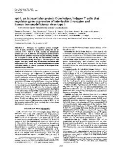
cells exposed to patient macrophage supernatants. Electron micrographs a-d are representative of the morphology of the major cellular phenotype present in patient supernatant exposed aggregates after 100 cells/specimen were examined. (a) Electron micrograph of a cell from a brain aggregate treated with supernatant from patient B et(8.8% p24k macrophages). All cells showed final stages of cell ' death. Cytoplasms were lacking organelles and nuclei were devoid of chromatin. x9,675. (b) Electron micrograph of an unidentified cell from a brain aggregate treated with tof 4 . i A from patient A supernatant (7.1% p24' macrophages). All .,e, cells contained vacuoles within the cytoplasm and clumped chromatin within the nucleus. X14,175. (c) Electron micrograph of two neurons from aggregates treated with supernatant from patient D (5.7% p24' macrophages). Numerous vacuoles appeared in the cytoplasm with an increase in the number of mitochondria (*), as compared with neurons observed in control aggregates. x4,805. (d) Electron micrograph of an astrocyte with a process _In *ifA, *e>;,* fcontaining filaments from a brain aggregate treated with > Aid 54; supernatant from patient C 9E ;^H,, (< 1.5% p24k macrophages). The U'Adcytoplasm contained more vacuoles than cells in control aggregates although the nucleus appeared normal. x7,955.
-
.1
Figure 5. Transmission electron micrographs of brain aggregate
~..
t
chondrial activity was seen in neurons containing a moderate degree of vacuolation (Fig. 5 c). In other systems, CNP levels are known to increase in response to cyclic AMP and its inducers such as norepinephrine and prostaglandin El (20, 21) and growth factors such as somatomedin C (22). It is possible
Human Immunodeficiency Macrophages Produce Factors that Alter Brain Cultures
511
that the increased CNP activity occurred secondary to growth factor elaboration in response to a toxic insult. Virus gene products such as gp 120 have been implicated in some in vitro experiments to account for HIV-related neurotoxicity (10). However, our T cell supernatant shown by Western blot analysis to be secreting gp 120 at a higher level than the infected macrophage cultures did not cause significant structural and functional changes in the brain aggregates. Similarly, the toxic effects are likely not elaborated merely by macrophage activation, as LPS-activated macrophage supernatants caused no ultrastructural changes in exposed brain aggregates. There were only slight increases in CNP activity and LDH levels in the LPS-activated macrophage supernatant exposed brain aggregates. Therefore, it is unlikely that a factor such as TNF-a was responsible for mediating the toxic effects observed in this study, and in a parallel analysis, no TNF-a was found in supernatants from in vitro HIV-infected macrophages. Fluctuations in inhibitory and excitatory neurotransmitter amino acids were assessed to determine their potential tole in soluble factor induced brain dysfunction. Recent reports have shown that excitatory amino acids (EAA) can cause secondary brain tissue injury (23). All five neurotransmitter amino acid levels showed a slight, not statistically significant decrease relative to the structural amino acid leucine when exposed to HIVinfected macrophage supernatants. It was believed, based on some preliminary data (not shown), that measurements of subtle changes in neurotransmitter amino acids would not be interpretable in the setting of widespread cellular damage seen in brain aggregates exposed to patient derived infected macrophages. All of the brain aggregates exposed to supernatant from AIDS patient macrophages manifested significant changes both morphologically and enzymatically. It is unlikely that all four patients had AIDS-associated dementia. Therefore, further analysis in a controlled fashion of monocytes from patients with AIDS dementia as well as those without dementia will be required in order to test whether levels of toxicity observed in aggregates are related to both proportion of monocytes expressing HIV as well as the patient's neurological status. The in vitro model described in this paper reproduces many of the abnormalities found in brains of patients with AIDS dementia including atrophic changes (a decrease in brain aggregate size), rarefaction, and vacuolation changes within a wide spectrum ofcell types. Furthermore, the lack of a decrease in CNP activity in the model is consistent with the inability to correlate demyelination with AIDS dementia. We believe that the culture system described in this paper approximates a valid model for in vitro study of AIDS-associated dementia.
Acknowledgments We thank Ray Swanson for LDH determinations; John Ziegler, Scott Panther, and Linda Noble for helpful discussions; Karen Chew, Diane George, Isabelle Gaston, and Thomas Kuwahara for excellent technical assistance; and John Flickinger for preparation of this manuscript. This work was supported in part by the California Universitywide AIDS Taskforce and the Veterans Administration Center for AIDS Research and Education.
References 1. Price, R. W., and B. J. Brew. 1988. The AIDS dementia complex. J. Infect. Dis. 158:1079-1083.
512
L. Pulliam, B. G. Herndier, N. M. Tang, and M. S. McGrath
2. Navia, B. A., E. S. Cho, C. K. Petito, and R. W. Price. 1986. The AIDS dementia complex: II Neuropathology. Ann. Neurol. 19:525-535. 3. Vazeux, R., N. Brousse, A. Jarry, D. Henin, C. Marche, C. Vendrenne, J. Mikol, M. Wolff, C. Michon, W. Rozenbaum, J.-F. Bureau, L. Montagnier, and M. Brahic. 1987. AIDS subacute encephalitis. Identification of HIV-infected cells. Am. J. Pathol. 126:403-410. 4. Shaw, G. M., M. E. Harper, B. H. Hahn, L. G. Epstein, D. C. Gaidusek, R. W. Price, B. A. Navia, CK. Petito, C. J. O'Hara, J. E. Groopman, E. S. Cho, J. M. Leske, F. Wong-Staal, and R. C. Gallo. 1985. HTLV-III infection in brains of children and adults with AIDS encephalopathy. Science (Wash. DC). 227:177180. 5. Levy, J. A., J. Shimabukuro, H. Hollander, J. Mills, and L. Kaminsky. 1985. Isolation of AIDS-associated retroviruses from cerebrospinal fluid and brain of patients with neurological symptoms. Lancet. ii:586-588. 6. Ho, D. D., T. R. Rota, R. T. Schooley, J. C. Kaplan, J. D. Allan, J. E. Groopman, L. Resnick, and D. Felsenstein. 1985. Isolation of HTLV-III from cerebrospinal fluid and neural tissues of patients with neurologic syndromes related to the acquired immunodeficiency syndrome. N. Engi. J. Med. 313:14931497. 7. Hollander, H., and J. A. Levy. 1987. Neurologic abnormalities and recovery of human immunodeficiency virus from cerebrospinal fluid. Ann. Intern. Med. 106:692-695. 8. Cheng-Mayer, C., J. Rutka, M. Rosenblum, C. McHugh, D. Stites, and J. Levy. 1987. Human immunodeficiency virus can productively infect cultured human glial cells. Proc. NatL. Acad. Sci. USA. 84:3526-3530. 9. Chiodi, F., S. Fuerstenberg, M. Gidlund, B. Asjo, and E. M. Fenyo. 1986. Infection of brain-derived cells with the human immunodeficiency virus. J. Virol.
61:1244-1247. 10. Brenneman, D. E., G. L. Westbrook, S. F. Fitzgerald, D. L. Ennist, K. L. Elkins, M. R. Ruff, and C. B. Pert. 1988. Neuronal cell killing by the envelope protein of HIV and its prevention by vasoactive intestinal peptide. Nature
(Lond.). 335:639-642. 11. Wahl, L. M., M. L. Corcoran, S. W. Pyle, L. 0. Arthur, A. Harel-Bellan, and W. L. Farrar. 1989. Human immunodeficiency virus glycoprotein (gpl2O) induction of monocyte arachidonic acid metabolite and interleukin. Proc. Natl. Acad. Sci. USA. 86:621-625. 12. Gurney, M. E., S. P. Heinrich, M. R. Lee, and H.-S. Yin. 1986. Molecular cloning and expression of neuroleukin, a neurotropic factor for spinal and sensory neurons. Science (Wash. DC). 23:566-574. 13. Crowe, S., J. Mills, and M. S. McGrath. 1987. Quantitative immunocytofluorographic analysis of CD4 surface antigen expression and HIV infection of human peripheral blood monocyte/macrophages. AIDSRes. Hum. Retroviruses. 3:135-145. 14. McGrath, M. S., K. M. Hwang, S. E. Caldwell, I. Gaston, K.-C. Luk, P. Wu, V. L. Ng, S. Crowe, J. Marsh, T. Deinhart, P. V. Lekas, J. C. Vennari, H.-W. Yeung, and J. D. Lifson. 1989. GLQ223: an inhibitor of human immunodeficiency virus replication in acutely and chronically infected cells of lymphocyte and mononuclear phagocyte lineage. Proc. Natl. Acad. Sci. USA. 86:2844-3848. 15. Locksley, R. M., S. Crowe, M. D. Sadick, F. P. Heinzel, K. D. Gardner, Jr., M. S. McGrath, and 3. Mills. 1988. Release of interleukin I inhibitory activity (contra IL-I) by human monocyte-derived macrophages infected with human immunodeficiency virus in vitro and in vivo. J. Clin. Invest. 82:2097-2105. 16. Pulliam, L., M. E. Berens, and M. L. Rosenblum. 1988. A normal human brain cell aggregate model for neurobiological studies. J. Neurosci. Res. 21:521530. 17. Kurihara, T., and Tsukada, Y. 1967. The regional and subcellular districution of 2',3'-cyclic nucleotide 3'-phosphohydrolase in the central nervous system. J. Neurochem. 14:1167-1174. 18. Koh, J. Y., and D. W. Choi. 1987. Quantitative determination of glutamate mediated cortical neuronal injury in cell culture by lactate dehydrogenase efflux assay. J. Neurosci. Res. 20:83-90. 19. Prohaska, J. R., D. A. Clark, and W. W. Wells. 1973. Improved rapidity and precision in the determination of brain 2'3'-cyclic nucleotide 3'-phosphohydrolase. Anal. Biochem. 56:275-282. 20. McMorris, F. A., T. M. Smith, T. J. Sprinkle, and J. S. Auszmann. 1985. Induction of myelin components: cyclic AMP increases the synthesis rate of 2'3'-cyclic nucleotide 3'-phosphohydrolase in C6 glioma cells. J. Neurochem. 44:1242-1251. 21. McMorris, F. A. 1983. Cyclic AMP induction of the myelin enzyme 2',3'cyclic nucleotide 3'-phosphohydrolase in rat oligodendrocytes. J. Neurochem. 41:506-515. 22. McMorris, F. A., T. M. Smith, S. Desalvo, and R. W. Furlanetto. 1986. Insulin-like growth factor l/somatomedin C: A potent inducer of oligodendrocyte development. Proc. Nat!. Acad. Sci. USA. 83:822-826. 23. Faden, A. I., P. Demediuk, S. S. Panter, and R. Vink. 1989. The role of excitatory amino acids and NMDA receptors in traumatic brain injury. Science (Wash. DC). 244:798-800.