Cathepsin L was purified to apparent homogeneity from human liver obtained post ..... (c) and (d) cathepsin L, reduced; (b) and (e) reference proteins. Vol. 226.
233
Biochem. J. (1985) 226, 233-241 Printed in Great Britain
Human liver cathepsin L Robert W. MASON, George D. J. GREEN* and Alan J. BARRETT Department of Biochemistry, Strangeways Laboratory, Worts Causeway, Cambridge CBR 4RN, U.K.
(Received 28 August 1984/Accepted 19 October 1984) Cathepsin L was purified to apparent homogeneity from human liver obtained post mortem. It was necessary to treat the homogenate at pH4.2 and 37°C to release active enzyme. The purification procedure involved ion-exchange chromatography on carboxymethyl-Sephadex and the Mono S column of a Pharmacia fast-protein-liquidchromatography system. The enzyme was found to consist of two polypeptide chains of M, 25 000 and 5000. The larger chain was shown to contain the active-site cysteine residue. Human cathepsin L proved to be similar to the rat and rabbit enzymes in regard to kinetic constants for the substrate benzyloxycarbonylphenylalanylarginine 7-(4-methyl)coumarylamide and rates of inactivation by the active-site-directed reagents benzyloxycarbonylphenylalanylphenylalanyldiazomethane and benzyloxycarbonylphenylalanylalanyldiazomethane. Thus clear characteristics of cathepsin L are now emerging, and these should simplify the identification of the enzyme in other tissues and species. Cathepsin L is of particular interest because it has a much higher specific activity in the degradation of a variety of physiological protein substrates, Abbreviations used: names of amino acids, peptides and their derivatives are abbreviated in accordance with IUPAC-IUB Recommendations [Biochem. J. (1972) 126, 773-780]. Additional abbreviations are: Cit, L-citrulline; Dns, 5-dimethylaminonaphthalene-lsulphonyl; f.p.l.c., fast protein liquid chromatography (Pharmacia system); N HMec, 4-methyl-7-coumarylamide; SDS, sodium dodecyl sulphate. The inhibitors are: E-64, L-3-carboxy-trans-2,3-epoxypropyl-leucylamido-(4guanidino)butane (also D-isomer where indicated); Ep420. DL-3-benzoxy-trans-2,3-epoxypropyl-isoleucyltyrosine methyl Ep-459, L-3-carboxy-transester; 2,3-epoxypropyl-leucylamido-(4-amino)butane; Ep-460, L 3 carboxy trans - 2,3 epoxypropyl - leucylamido (4 benzyl-oxycarbonylamino)butane; Ep-475, L-3-carboxy-
-
-
-
-
-
trans-2,3-epoxypropyl-leucylamido-(3-methyl)butane. The abbreviations used for enzyme kinetic parameters are: [I]t, total inhibitor concentration; kca,., catalytic constant; kI2, rate constant for irreversible inhibition of enzyme-inhibitor complex; k'+2, apparent second-order rate constant for inactivation, independent of substrate concentration; k'+2, apparent second-order rate constant for inactivation in the presence of substrate; Ki(app.), apparent inhibition constant in the presence of substrate; vo, rate of reaction in absence of inhibitor; vi, rate of reaction in presence of inhibitor. * Present address: Friedrich Miescher Institut, P. 0. Box 273, CH-4002 Basel, Switzerland.
Vol. 226
including collagen, than the other lysosomal proteinases (Barrett & Kirschke, 1981 ; Kirschke et al., 1982). There are difficulties in the isolation of human cathepsin L, however, and there has been no full report of this. Thomas et al. (1980) briefly reported the detection of a cathepsin L-like enzyme in human kidney. The enzyme was separated from cathepsin B and shown to degrade azocasein and collagen, but no further characterization was reported. An enzyme called 'collagenolytic cathepsin' (later named 'cathepsin N'), has been found in human placenta (Evans & Etherington, 1978) and in human liver biopsies (Murawaki & Hirayama, 1980); it differed from cathepsin B and resembled cathepsin L in its low activity towards a range of synthetic substrates. Cathepsin N was shown to be a cysteine proteinase by its sensitivity to inhibition by iodoacetic acid, HgCl2 and leupeptin, but, since collagen was the only substrate used, a full comparison of properties with cathepsin L is not possible. Some characteristics of an enzyme described as human liver cathepsin L have been reported (Pagano & Engler, 1982). The purification procedure, not described fully, seems to have been similar to that used for the purification of cathepsin N (Evans & Etherington, 1978). Like human kidney cathepsin L, the enzyme was shown to hydrolyse azocasein, but had little activity against low-Mr synthetic substrates.
234
R. W. Mason, G. D. J. Green and A. J. Barrett
The purpose of the present investigation was to purify cathepsin L from human liver and to compare its properties with those of the rabbit and rat liver enzymes. Preliminary reports of the results have been presented (Mason & Barrett, 1984a,b).
Enzyme assays Enzyme assays and active-site titrations were performed essentially as described by Barrett & Kirschke (1981). Thus, cathepsin L was routinely assayed with Z-Phe-Arg-NHMec (5yM) at 30°C and pH5.5, and cathepsins B and H with Z-ArgArg-NHMec (5M) and Arg-NHMec (5M) respectively. The molar concentration of active cysteine proteinase was measured by titration with E-64 or Ep-475. Km values were determined by the method of Wilkinson (1961). Activity against azocasein was assayed in reaction mixtures (0.2 ml) containing 3 M-urea, stopped with 1 ml of 3% (w/v) trichloroacetic acid. All other conditions were exactly as described previously.
Experimental Materials Aminomethylcoumarin substrates were from Cambridge Research Biochemicals, Cambridge, U.K., Z-Phe-Cit-NHMec, Z-Phe-Ala-NHMec and Z-Phe-Met-NHMec being custom-synthesized. Z-Phe-Phe-CHN2 and Z-Phe-Ala-CHN2 were kindly given by Dr. E. N. Shaw, Friedrich Miescher Institute, Basel, Switzerland. Iodoacetic acid (specially purified for biochemical work) and Brij 35 (polyoxyethylene dodecyl ether) were supplied by BDH Chemicals, Poole, Dorset, U.K. 2-Hydroxyethyl disulphide was from Aldrich Chemical Co., Gillingham, Dorset, U.K. a-Methyl D-mannoside (grade III), cytochrome c (ox heart, type Ha), carbonic anhydrase (bovine erythrocyte), transferrin (human), albumin (bovine serum, crystallized), phosphorylase a (rabbit muscle, twice crystallized), insulin (bovine pancreas), soya-bean trypsin inhibitor (type II-S), E64, mersalyl acid and Brilliant Blue G were purchased from Sigma Chemical Co., Poole, Dorset, U.K. Aprotinin was given by Dr. E. Philip, Bayer A.G., Wuppertal, Federal Republic of Germany, and sheep IgG by Dr. M. E. Davies of this Department. Ep-475 and other epoxide analogues were given by Dr. K. Hanada, Taisho Pharmaceutical Co., Saitama, Japan. 6N-[3H]Acetyl-Ep-459 was prepared in this laboratory by Dr. C. Parkes and Dr. D. H. Rich. Leupeptin was supplied by the Peptide Research Foundation, 476 Ina, Minoh-shi, Osaka 562, Japan. Ampholines were purchased from LKB Instruments Ltd., South Croydon, Surrey CR2 9PX, U.K. Sephadex G-25, CM-Sephadex C-50, phenyl-Sepharose and concanavalin A-Sepharose were from Pharmacia (G.B.) Ltd., Milton Keynes, Bucks. MK9 3HP, U.K., who also supplied the f.p.l.c. system. Rat liver cathepsin L was a gift from Dr. H. Kirschke, Physiological Chemistry Institute, Halle (Saale), German Democratic Republic. Rabbit liver cathepsin L was prepared as described previously (Mason et al., 1984). Azo-casein was prepared as described by Barrett & Kirschke (1981). Samples of human liver removed at autopsy, free from disease, were kindly contributed by Dr. P. Stovin, Papworth Hospital, Cambridge, U.K.; they were stored at - 20°C until required for use.
Measurement of rate constants for irreversible inhibition Inhibition rate constants were determined by use of continuous assays in the presence of inhibitor and substrate as described by Tian & Tsou (1982). The reaction scheme is assumed to be as in eqn. (1): +S
K,
-+ ES EE+P
(1)
E K,I
-EI +2 ) El
Enzymes were preincubated with 1 mM-dithiothreitol for 2 h at 4°C. Reaction mixtures, containing substrate (1 M-Z-Phe-Arg-NHMec), inhibitor, 100mM-sodium acetate buffer, pH5.5, 0.01% Brij 35, 1 mM-EDTA and 1 mM-dithiothreitol, were preincubated for 5 min in a cuvette placed in a holder thermostatically controlled at 30°C. Reactions were started by the addition of activated enzyme (1.OnM final concn.). Appearance of product was recorded continuously in a Perkin-Elmer LS-3 spectrofluorimeter standardized with 0.2 JUMaminomethylcoumarin, with excitation at 360nm and emission at 460nm, and the curves were analysed by the Guggenheim method as described by Jencks (1969) to determine the half-life of the enzyme. Linear-regression coefficients were usually greater than 0.998 and total times greater than four half-lives. Fast-reacting inhibitors were used at 100-1000nM final concentration, and the total amount of substrate consumed during assays was below 15%. The observed (pseudo-first-order) rate of inactivation, k0b., was calculated as 0.695/to.5. At a fixed substrate concentration, the apparent second-order rate constant for inactivation, k'+ 2, (eqn. 2) was calculated as kobS/[I]t (Thompson & Blout, 1973). Provided that the substrate concentration did not vary greatly during the course of reaction, the effect of substrate on k+2 would be 1985
Human liver cathepsin L constant (eqn. 3 below; Tian & Tsou, 1982]: kS
kL
> EI E+I (2) (3) k+2 = k+2/(1 +[S]IKm) Measurement of Ki for leupeptin Ki values for the inhibition of cathepsin L by leupeptin were determined as described by Green et al. (1984). Enzymes were used at a concentration of O.Olnm and leupeptin in the range 1-15nM. Apparent K, values were determined from replots of the form [I],/[I - (v I/vo)] versus vo/v1 (Henderson, 1972). The substrate concentration used was 10 /M, therefore corrections we made by use of the relationship:
Ki = Ki(app)I(1 + [SI/Km) for simple competition. All measurements were made with less than 2% hydrolysis. The incubation temperature was lowered to 20°C because cathepsin L was too unstable at higher temperatures in the low concentrations needed for these measurements. At 20°C, 95% of enzymic activity remained after 40min incubation of enzyme with activator and substrate. Determination of protein Protein concentrations were determined by the method of Bradford (1976), with bovine serum albumin as standard, and during the course of enzyme purification they were determined by measurement of A280Isoelectric focusing Samples of human liver cathepsin L were subjected to isoelectric focusing in agarose gel and subsequently stained for Z-Phe-Arg-NHMechydrolysing activity as described previously (Mason et al., 1984). Immediately after focusing, a strip of gel was cut into 5 mm segments, each of which was immersed overnight at 4°C in 1 ml of water, and the pH then determined by use of a glass micro-electrode. The activity of the eluates towards Z-Phe-Arg-NHMec was measured to confirm the results obtained by staining the gels directly.
SDS/polyacrylamide-gel electrophoresis SDS/polyacrylamide-gel electrophoresis was performed as described by Bury (1981), with the same reference proteins. Separating gels of 12.5% total acrylamide concentration (2.6% of this as methylenebisacrylamide) were used. Gels were stained for protein with Brilliant Blue G as described previously (Bury, 1981). For active-site labelling of cathepsin L with 6,N[3H]acetyl-Ep-459, samples were preincubated with an equimolar concentration of inhibitor in the Vol. 226
235 presence of 1 mM-dithiothreitol for 1 h at 37°C. Protein was then precipitated with 10% (w/v) trichloroacetic acid, and run on SDS/polyacrylamide gels as usual. Bands were cut from the stained gels for determination of radioactivity as described by Goodman & Martzura (1971).
Purification of human cathepsin L Frozen liver (1.4kg) was partially thawed, cut into small pieces and homogenized in 2vol. of extraction buffer [1% NaCl/0.1% EDTA/2% (v/v) butan-l-ol]. Insoluble material was removed by centrifugation (1350g for 30min) and the supernatant was adjusted to pH4.2, incubated at 37°C for 4h, and left overnight at 4°C. Precipitated protein was removed by centrifugation as described above, and then a 20-80%-satn.(NH4)2SO4 fraction was taken. All operations (except centrifugation) up to and including this step were performed in a class I microbiological safety cabinet. The pellet was resuspended in 200ml of 20mM-sodium acetate buffer, pH5.5, containing 1 mM-EDTA (buffer A), and insoluble material was removed by centrifugation (2000Og for 30min). The supernatant was transferred into buffer A by gel chromatography through Sephadex G-25. The sample was applied to a column (25 cm x 4.4cm) of CM-Sephadex C-50, which was washed with 2 bed volumes of buffer and eluted with a linear salt gradient to 1.OM-NaCl (1900ml total volume). Typically, cathepsin H was eluted at 80mM-, cathepsin B at 150mM- and cathepsin L at 400mM-NaCl. Fractions containing cathepsin L were combined and adjusted to 25% saturation with (NH4)2S04. The enzyme was then adsorbed to a column (10 cm x 1.6cm) of phenyl-Sepharose, which was washed with 25%-satd. (NH4)2 SO4 in buffer A before cathepsin L was eluted with 25% (v/v) ethylene glycol in buffer A. Active fractions were combined and equilibrated into 20mM-sodium malonate buffer, pH5.5, containing 1 mM-EDTA, and then chromatographed on the Mono S (HR5/5) column of a Pharmacia f.p.l.c. system, with a linear gradient (0-O.5M; 25 ml) of NaCl in the same buffer. Active fractions were diluted 5-fold in 20mM-sodium acetate buffer, pH 5.0, containing 1 mM-EDTA, and reapplied to the Mono S column equilibrated with this buffer. A similar NaCl gradient was applied. Peak activity from the two column runs was eluted at 360 and 380mM-NaCl respectively. Results and discussion
Purification Human cathepsin L was purified 3500-fold, on the basis of total activity against Z-Phe-Arg-
236
R. W. Mason, G. D. J. Green and A. J. Barrett
NHMec after acid extraction (Table 1). Approximately 700pg of purified enzyme was obtained from 1.4 kg of liver; this was found to be 40% active by titration with E-64. The apparent yield of enzyme was 5.1%, but this value is an underestimate, since cathepsin L is responsible for less than 25% of the total Z-Phe-Arg-NHMec-hydrolysing activity in the original extract (see below). Acid treatment. The incubation of a crude homogenate of human liver at low pH before purification of cathepsin L proved to be the most critical step in the procedure. Cathepsin L exists in human liver homogenate in a 'latent' form, and omission of the acid treatment resulted in a negligible yield of the active enzyme. Conditions of acid treatment giving maximal generation of active cathepsin L were determined by two different methods. The first involved measuring the ratio of activities against Z-Phe-Arg-NHMec and Z-Arg-Arg-NHMec. Z-Arg-Arg-NHMec is cleaved by cathepsin B but not by cathepsin L (Barrett & Kirschke, 1981), whereas Z-Phe-ArgNHMec is hydrolysed by both enzymes, so that an increase in the Z-Phe-Arg-NHMec/Z-Arg-ArgNHMec ratio was taken as a measure of increased cathepsin L activity. Later, when it was established that human liver cathepsin L is irreversibly inhibited by Z-Phe-Phe-CHN2 more than two orders of magnitude faster than cathepsin B (Table 3 below), the contribution of cathepsin L to the ZPhe-Arg-NHMec-hydrolysing activity was determined by measuring the decrease in activity caused by this compound. Preincubation of fractions with 0.5 guM-Z-Phe-Phe-CHN2 and 1 mMdithiothreitol for 10min at 30°C before assay inhibited cathepsin L by 95%, whereas cathepsin B was inhibited by less than 10% (determined by inhibition of Z-Arg-Arg-NHMec hydrolysis) by this treatment. Maximal activation of cathepsin L in the homogenate was found to occur at pH4.2, with 90% of this amount at pH 4.0 and 4.5. Below pH 3.9 and above pH 5.0, yields of both cathepsin L and cathepsin B were greatly diminished. The release
of active enzyme was also highly dependent on temperature and incubation time. The conditions of 4h at 37°C followed by 16h at 4°C were chosen for convenience, but a similar amount of activity could be released by incubation for 24h at 25°C. The presence of pepstatin, an inhibitor of aspartic proteinases (1 Mg/ml), or reversible inhibitors of cysteine proteinases (1 mM-mersalyl acid or 1 mM2-hydroxyethyl disulphide) during the acid treatment did not affect the release of active enzyme. Under the chosen conditions of acid treatment, the activity attributable to cathepsin L increased over 10-fold, to account for 25% of the total Z-PheArg-NHMec hydrolysis in the standard assay. Cathepsin B activity also increased, but only by 1.5-2.0-fold. The reason for the inactivity of cathepsin L in the homogenate is not known. Possibly the enzyme is complexed by cystatins A and B, the cytosolic inhibitors of cysteine proteinase that are present in human liver cells (Davies & Barrett, 1984). These inhibitors bind cathepsin L much more tightly than cathepsin B, but would not normally be destroyed by the conditions of acid treatment used here (Green et al., 1984). In view of the results with the inhibitors of proteinases, it also seems unlikely that the liberation of cathepsin L activity is a consequence of the action of cysteine or aspartic proteinases. Working with extracts of rabbit muscle, Matsumoto et al. (1983) have shown a decrease in the activity of inhibitors of cysteine proteinases as a result of incubation at acid pH, but were also unable to demonstrate any enzymic involvement. Further purification. Once the enzyme was extracted, its purification proved relatively simple. Human cathepsin L bound very tightly to cationexchange media, although its pI is in the range 5.76.3 (see below). This property, which has previously been described for cathepsin L of rat and rabbit liver (Kirschke et al., 1977; Mason et al., 1984) led to the use of three cation-exchange columns under different conditions to purify the enzyme. Anionexchange columns could not be used because of the
Table 1. Purification of human liver cathepsin L Activity was measured against Z-Phe-Arg-NHMec as described in the Experimental section. Total protein Total activity Specific activity Purification Step (A280) (Mmol/min) factor (umol/min per A280 unit) Acid extraction (NH4)2SO4 fractionation
CM-Sephadex Phenyl-Sepharose Mono S pH 5.5 pH 5.0
60025 14590 110 60.5
2.73 0.85
254 240 69 50.8
19.1 12.9
0.004 0.016 0.63 0.84
7.01 15.1
I 4 148 210
1656 3565
Yield
(%) 100 94 .27 20 7.5 5.1
1985
Human liver cathepsin L
237
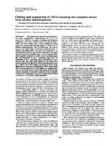
instability of cathepsin L above pH 5.5 (Fig. 2 below). The phenyl-Sepharose column served mainly to concentrate the sample; all protein applied was bound, but some coloured material remained on the column after elution of the enyzme. Physicochemical properties Relative molecular mass. SDS/polyacrylamidegel electrophoresis of the enzyme under nonreducing conditions showed two bands of protein at M, 25000 and 29000 (Fig. 1). After reduction, two bands were seen, at M, 25 000 and 5000. These results suggest that human liver cathepsin L comprises forms of M, 29000 and 25000, of which the larger consists of two chains linked by disulphide bonds. There was no evidence for the existence of a single-chain Mr-29 000 form in any of our preparations. It is not yet clear whether
cathepsin L occurs as a two-chain or single-chain form in vivo, since the isolation procedure requires a step that might permit autolytic cleavage. Having used a different purification procedure for the isolation of an enzyme described as cathepsin L, Pagano & Engler (1982) saw only a single diffuse band of protein on SDS/polyacrylamide-gel electrophoresis with reduction, which corresponded to Mr 30000. We found that only the Mr-25000 and -29000 chains were labelled with 6,N-[3H]acetyl-Ep459 (Fig. 1), indicating that these contain the active site, in agreement with the labelling of the large chain of rat liver cathepsin L by Dns-Ala-Ala-PheCHN2 and Z-Phe-Phe-1 4CHN2 (Kirschke et al., 1984a). Cathepsin H from rat liver has also been shown to occur as a two-chain form, with the active site in the larger chain (Takio et al., 1980). In contrast, cathepsin B of several species has been
iimw
..
AM..
......
L
I
I
..
......
_
600 400 200
c
(a)
(b)
600 400 200
3H radioactivity
3H radioactivity
(d.p.m.)
(d.p.m.)
0
Fig. 1. SDS/polyacrylamide-gel electrophoresis of human cathepsin L SDS/polyacrylamide-gel electrophoresis under reducing and non-reducing conditions was in 12.5% (w/v)acrylamide gels as described in the Experimental section. Active-site labelling with 6,N-[3H]acetyl-Ep-459 was as described in the Experimental section. Incorporation of 3H label is represented graphically alongside each gel. Approx. 0.2 ug of enzyme (determined by active-site titration) was applied to each lane. (a) Cathepsin L, unreduced; (c) and (d) cathepsin L, reduced; (b) and (e) reference proteins.
Vol. 226
238
R. W. Mason, G. D. J. Green and A. J. Barrett
shown to have a light chain containing the catalytic cysteine residue that is formed by cleavage of the single-chain form of the protein near the N-terminus (e.g. Takio et al., 1980). Sequence data have shown that the essential cysteine residues of papain and a number of homologous cysteine proteinases, including cathepsins B, H and S, are near the N-terminus (Barrett et al., 1984). Multiple isoelectric forms. Staining of isoelectricfocusing gels containing human cathepsin L for activity hydrolysing Z-Phe-Arg-NHMec revealed a major band at pH 5.9, with several minor bands in the range pH 5.7-6.3. We conclude that human liver cathepsin L consists of multiple forms with isoelectric points in the range 5.7-6.3. A similar range of isoenzymic forms has been found for both the rat and rabbit liver enzymes (Kirschke et al., 1977; Mason et al., 1984). Behaviour on concanavalin A-Sepharose. When the purified cathepsin L was applied to a column of concanavalin A-Sepharose, 90% passed straight through, and the remaining 10% could be eluted with 200mM-a-methyl D-mannoside. The enzymes from rat liver and rabbit liver have also been shown to bind partially to this gel (Barrett & Kirschke, 1981; Mason et al., 1984). It may be that heterogeneity of glycosylation is a general property of cathepsin L.
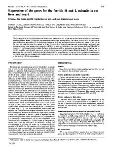
24h in the pH range 4.5-5.5 (Fig. 2a). At pH7.0, however, 85% of activity was lost after only 15 min incubation. Rat and rabbit liver cathepsin L are similarly unstable near to neutral pH (Kirschke et al., 1980; Etherington et al., 1984). This instability was shown to be partially responsible for the decline in activity above pH 6.0 (Fig. 2b). When hydrolysis of this substrate was measured during the first 30s of incubation, a significant amount of activity could be detected at pH 8.0. Similar results have been found for human cathepsin B and rabbit cathepsin L (A. J. Barrett, R. W. Mason & D. J. Etherington, unpublished work). Action on peptidyl-NHMec substrates. The activities of cathepsin L from human, rabbit and rat liver against a range of synthetic substrates were determined (Table 2). The substrate concentrations were low (0.5 /M), so that the activities determined may be expected to be approximately proportional to values for kca./Kmc. Of those tested, Z-Phe-Arg-NHMec was clearly the best substrate, and values of Km and kcat were similar for all three enzymes. The values are in reasonable agreement with those reported previously for the rat and rabbit enzymes (Barrett & Kirschke, 1981; Mason et al., 1984). The specific activity of the enzyme from human liver purified by Pagano & Engler (1982) for the hydrolysis of Z-Phe-Arg-NHMec has not been reported, but it can be determined indirectly. Titration of the preparation with E-64 indicated that it contained 45% of active protein, and yet 1 NM-enzyme was used in assays with 25 /M-Z-Phe-
Catalytic activity Stability. Human liver cathepsin L retained at least 70% of its activity after incubation at 37°C for
100
60
60
.~40
40
20
20
o CZ
0
4
5
6
7
J
8
0
4
5
6
7
8
J
9
pH
Fig. 2. Stability of human liver cathepsin L and its effect on the pH optimum of Z-Phe-Arg-NHMec hydrolysis (a) Human liver cathepsin L was incubated in the absence of thiol activator at 37°C in 100mM-buffer at a range of pH values (see below) for 15min (0), 1h (A) or 24h (EO). Activity was then measured under standard assay conditions. (b) Z-Phe-Arg-NHMec hydrolysis was measured at 30°C over a range of pH values either for a standard 10min assay (A) or by recording continuously over the first 30s of hydrolysis (0). Buffers were sodium formate (pH3.5-4.0), sodium acetate (pH4.0-5.0), sodium malonate (pH5.0-6.0), sodium phosphate (pH6.0-7.5) and glycylglycine hydrochloride (pH7.5-8.5).
1985
Human liver cathepsin L
239
Table 2. Activity of cathepsin L against synthetic substrates All assays were at 30°C, pH 5.5, with 0.5 Mm-substrate. Results are expressed as a percentage of activity for Z-PheArg-NHMec, values in parentheses being Km values (#M) and kcat. values (s-1) respectively. Activity (0/) Substrate
Z-Phe-Arg-NHMec Z-Arg-Arg-NHMec Arg-NHMec Z-Phe-Ala-NHMec Z-Phe-Cit-NHMec Z-Phe-Met-NHMec
Source of enzyme ...
Human liver
Rabbit liver
Rat liver
100 (2.4, 17)
100 (1.8, 20)