Human Monoclonal Antibodies to Pandemic 1957 H2N2 and Pandemic 1968 H3N2 Influenza Viruses Jens C. Krause,a Tshidi Tsibane,c Terrence M. Tumpey,f Chelsey J. Huffman,a Randy Albrecht,c,e David L. Blum,a Irene Ramos,c Ana Fernandez-Sesma,c,d,e Kathryn M. Edwards,a Adolfo García-Sastre,c,d,e Christopher F. Basler,c and James E. Crowe, Jr.a,b Departments of Pediatricsa and of Microbiology and Immunology,b Vanderbilt University Medical Center, Nashville, Tennessee, USA; Department of Microbiology,c Department of Medicine, Division of Infectious Diseases,d and Global Health and Emerging Pathogens Institute,e Mount Sinai School of Medicine, New York, New York, USA; and Influenza Division, Centers for Disease Control and Prevention, Atlanta, Georgia, USAf
Investigation of the human antibody response to the 1957 pandemic H2N2 influenza A virus has been largely limited to serologic studies. We generated five influenza virus hemagglutinin (HA)-reactive human monoclonal antibodies (MAbs) by hybridoma technology from the peripheral blood of healthy donors who were born between 1950 and 1968. Two MAbs reacted with the pandemic H2N2 virus, two recognized the pandemic H3N2 virus, and remarkably, one reacted with both the pandemic H2N2 and H3N2 viruses. Each of these five naturally occurring MAbs displayed hemagglutination inhibition activity, suggesting specificity for the globular head domain of influenza virus HA. When incubated with virus, MAbs 8F8, 8M2, and 2G1 each elicited H2N2 escape mutations immediately adjacent to the receptor-binding domain on the HA globular head in embryonated chicken eggs. All H2N2-specific MAbs were able to inhibit a 2006 swine H2N3 influenza virus. MAbs 8M2 and 2G1 shared the VH1-69 germ line gene, but these antibodies were otherwise not genetically related. Each antibody was able to protect mice in a lethal H2N2 virus challenge. Thus, even 43 years after circulation of H2N2 viruses, these subjects possessed peripheral blood B cells encoding potent inhibiting antibodies specific for a conserved region on the globular head of the pandemic H2 HA.
I
nfluenza pandemics occurred in 1918 (H1N1), 1957 (H2N2), and 1968 (H3N2), and again in 2009 (H1N1) (34). H3N2 viruses have circulated in humans for over 4 decades since the onset of the 1968 pandemic, but H2N2 viruses circulated for only 11 years, from 1957 to 1968. Subjects born after 1968 typically do not possess neutralizing titers against H2N2 viruses because of lack of exposure (20, 27). As herd immunity wanes in the human population, the pandemic potential of this subtype virus increases significantly (12, 40). Virulent pandemic H2N2 virus is still being maintained in countless laboratory freezers across the globe (34). Also, antigenically conserved counterparts of the pandemic strain of 1957 continue to circulate in avian (13, 18, 28) and swine (18) reservoirs. The occurrence of the 2009 H1N1 influenza pandemic showed that an antigenically conserved pandemic influenza virus can reemerge in the human population from an animal reservoir (8), even in the face of widespread immunity to H1 viruses after vaccination or infection with circulating drifted H1N1 viruses. With this in mind, some experts have recommended vaccination against H2N2 viruses, which could cause the next pandemic (20, 32). There is great interest in understanding the humoral response to the hemagglutinin (HA) of influenza viruses, as the presence of HA-neutralizing antibodies has been correlated with protection from infection and/or disease. Many mouse monoclonal antibodies (MAbs) against H3 influenza viruses were derived for the purposes of mapping murine H3 HA B cell epitopes (36, 37, 41, 42). One MAb, designated S139/1, was reported to neutralize not only H3 viruses but also H1, H2, and H13 viruses through binding of antigenic site B (52). Human H3 MAbs have been created using phage display (23, 24, 29), plasmablast single cell cloning (46), or hybridoma technology (50, 53). Murine MAbs against 1957 H2 also have been made (21, 35, 39) and used for antigenic mapping (28, 35, 49), including an antibody directed to an HA stem region epitope that is preserved across influenza virus phylogenetic
6334
jvi.asm.org
Journal of Virology
group 1 HAs (25). A human antibody to H3 subtype virus HA globular head with limited cross-reactivity to other influenza virus subtypes, including H2, was described recently (22). It has been speculated that the H2 HA glycoprotein might be less tolerant of mutations (including the inability to acquire glycosylation sites by genetic point mutations), explaining the short reign of the H2 virus in humans (28, 35). An alternative explanation is the presence of two potentially immunogenic stem epitopes on H2 HA that may have induced immunity on a population level from which the virus could not escape (25, 35, 48). Here, we report on the cloning and characterization of human MAbs, cloned from the peripheral blood of healthy immune donors, against pandemic H2 or H3 HA, including one MAb that exhibits the ability to inhibit both H2 and H3 viruses. MATERIALS AND METHODS Ethics statement. All clinical investigation was conducted according to Declaration of Helsinki principles. Acquisition of human blood samples was approved by the Vanderbilt University Institutional Review Board, and written informed consent was received from participants prior to inclusion in the study. The animal studies were carried out in strict accordance with recommendations in the Guide for the Care and Use of Laboratory Animals of the National Institutes of Health (NIH) (21a). All mouse procedures were approved by Institutional Animal Care and Use Committee (IACUC) of the Centers for Disease Control and Prevention and
Received 19 December 2011 Accepted 19 March 2012 Published ahead of print 28 March 2012 Address correspondence to James E. Crowe, Jr.,
[email protected]. J.C.K. and T.T. contributed equally to this article. Supplemental material for this article may be found at http://jvi.asm.org/. Copyright © 2012, American Society for Microbiology. All Rights Reserved. doi:10.1128/JVI.07158-11
p. 6334 – 6340
June 2012 Volume 86 Number 11
Human Monoclonal Influenza H2N2/H3N2 Antibodies
were conducted in an Association for Assessment and Accreditation of Laboratory Animal Care International-accredited facility. Animal studies were performed in accordance with the IACUC guidelines under protocol 2198TUMMOUC-A7, “Studies on the Pathogenesis of and Immunity to Influenza Viruses in Mice.” Generation and purification of recombinant soluble HA molecules. A cDNA encoding the full-length A/Japan/305⫹/1957 virus HA protein (GenBank accession number AAA64362) was sequence optimized for expression in human cells and synthesized (GenScript). The extracellular domain was amplified using PCR and cloned into a vector containing a thrombin site, a fibritin trimerization domain, and a 6⫻ histidine tag (31). A 1968 H3 HA construct was sequence optimized for expression in human cells and synthesized (GeneArt), based on the extracellular domain of the HA gene from A/Aichi/2/1968, a GCN4 trimerization domain, a tobacco etch virus (TEV) protease recognition site, and a 6⫻ histidine tag. Both constructs were expressed in a pcDNA3.1(⫹) vector (Invitrogen) in 293F cells (Invitrogen), purified over nickel columns using an ÅKTA chromatography instrument (GE), and concentrated with Amicon filters as described above. Enzyme-linked immunosorbent assay (ELISA) for screening hybridoma supernatant. Clear plates (Nunc; 384 wells, catalog no. 242757) were coated with HA at 1 g/ml in Dulbecco’s phosphate-buffered saline (D-PBS) overnight and blocked with 0.5% cow’s milk, 0.2% goat serum, and 0.05% Tween 20 (Sigma; catalog no. P7949) in D-PBS. Five microliters of hybridoma supernatant per well was transferred to 25 l of blocking solution with a multichannel pipettor. Secondary alkaline phosphatase (AP)-conjugated goat anti-human IgG antibodies (Meridian Life Science; catalog no. W99008A) were diluted 1:8,000 in blocking solution and added after four automated washing steps. After another wash, phosphatase substrate (Sigma; catalog no. S0942) was dissolved in substrate buffer per the instructions of the manufacturer and dispensed onto the plates. The optical density of solution in plates was read at 405 nm on a PowerWave HT (BioTek). Hybridoma generation and recombinant antibody expression. Peripheral blood mononuclear cells (PBMCs) were isolated, Epstein Barr virus (EBV) transformed in 384-well plates (Nunc) in the presence of 2.5 g/ml CpG ODN 2006 (InvivoGen), 10 M Chk2 inhibitor II (Sigma; catalog no. C3742), and 1 g/ml cyclosporine (Sigma), essentially as previously described (15, 16, 53, 54). The antibodies described can be obtained for research purposes under a materials transfer agreement. Purification of antibodies from hybridoma cell line supernatants. The hybridoma cell lines were grown in medium E (Stemcell Technologies) until resuspension of the cells in Hybridoma-SFM medium (Invitrogen). Supernatant was harvested after 1 week, fast-performance liquid chromatography (FPLC) purified with protein G (for hybridoma-derived 2G1) or MabSelect SuRe affinity columns (for all other antibodies; both from GE), and concentrated with Amicon Ultra centrifugal filters with a 30-kDa molecular mass cutoff (Millipore). VLP expression and HAI assays. Expression plasmids encoding HA or neuraminidase proteins were coexpressed in 293T cells to produce virus-like particles (VLPs) (4, 54). Two days posttransfection, supernatants were collected. Hemagglutination inhibition (HAI) assays were performed as described previously (44) using VLPs or live virus, as indicated in the tables. We used the numbering scheme from Xu et al. for H2 HA (Protein Data Bank [PDB] identifiers: 3KU3/3KU5/3KU6) (48). Microneutralization assay. Different dilutions of antibody were incubated with 5 log10 50% tissue culture infective doses (TCID50) of each virus for 1 h. The mixture was used to infect Madin-Darby canine kidney (MDCK) cells in triplicate for an hour at 37°C. The plate was harvested 3 days later and read in an HA assay. The endpoint was the lowest concentration that gave no HA activity. Isolation and characterization of antibody escape mutant viruses. We selected new antibody escape mutant viruses by incubating virus with neutralizing antibodies followed by inoculation of the mixture in 10-dayold embryonated chicken eggs, essentially as described previously (3, 51).
June 2012 Volume 86 Number 11
RNA was extracted from virus-infected allantoic fluid, and then cDNA was generated by reverse transcriptase PCR (RT-PCR), cloned molecularly, and sequenced. In vivo antiviral effect of H2N2-specific MAbs. Female 8-week-old BALB/c mice were inoculated intranasally with 5 times the 50% lethal dose (LD50) in a 50-l volume of the virulent A/Albany/6/1958 H2N2 influenza virus (26, 38). At 24 h after inoculation, mice were each administered 200, 20, or 2 g (approximately 10, 1, or 0.1 mg/kg of body weight) of Ab 8F8, 8M2, or 2G1 or an equal volume of 10 mg/kg of polyclonal human IgG (Sigma) by the intraperitoneal (i.p.) route in groups of 10 mice. Mice were observed for weight loss for 14 days. Subsets of four animals treated with Abs were euthanized on day 4 after inoculation, and whole lungs were homogenized in 1 ml of sterile PBS. Virus titers in lung tissue homogenates were determined by plaque titration in Madin-Darby canine kidney cell monolayer cultures and expressed as log10 PFU/ml. Statistics were performed with GNU R 2.13.1 (R Foundation for Statistical Computing). The log rank test was used to compare the survival distributions. The Wilcoxon rank sum test was used to compare the lung virus titers. Review of H2N2 sequences. We queried the Influenza Research Database (IRD; www.fludb.org) (30) on 29 April 2011 for all naturally occurring, nonredundant human H2N2 HA sequences between 1957 and 1968 to identify the variability of key residues. After an alignment with ClustalW (17), complete sequences were pruned to residues 59 to 252 (encoding the globular head) of the HA1 subunit using MacVector 12 software. Redundant sequences were eliminated with a redundancy threshold of 100 in Jalview 2.6.1. A phylogram was generated with MacVector 12 using neighbor joining, best tree, symmetric tie breaking, uncorrected (“p”) distance settings and rooted to A/Japan/305/1957 (CY014976). Nucleotide sequence accession numbers. Antibody nucleotide sequences have been deposited in GenBank under accession numbers JN130388 to JN130397.
RESULTS
Hybridoma generation and molecular cloning. We screened peripheral blood cells from a total of 26 healthy donors born between 1957 and 1968 and from three donors who participated in an NIH-sponsored clinical trial of an experimental monovalent subvirion H5N1 influenza vaccine (1) by testing the supernatants of EBV-transformed B cells for antibodies binding to recombinant A/Japan/305⫹/1957 H2N2 HA or A/Aichi/2/1968 H3 HA by ELISA. Lymphoblastoid cell lines from wells with supernatants containing HA-reactive antibodies were fused with HMMA2.5 myeloma cells to generate hybridomas. Five antibodies were cloned from different donors. MAbs 8F8 and 8M2 reacted with the pandemic H2N2 HA, MAbs 7A13 and 11J19 recognized the pandemic H3N2 HA, and remarkably, MAb 2G1 reacted with both the pandemic H2N2 and H3N2 HA (Table 1). The data generally agreed well with those from the microneutralization assay, except that MAb 2G1 did not neutralize the 1968 virus at the highest concentration tested (Table 2). Molecular cloning of the antibodies and recombinant expression confirmed that the antibody gene sequences coded for HA-reactive antibody proteins. Nucleotide sequence analysis of variable gene sequences (Table 3) using the international ImMunoGeneTics (IMGT) information system (2) revealed that the H2 MAbs 8M2 and 2G1 shared the VH1-69 germ line gene segment with recently published human globular head antibodies derived of one clone (22). Reactivity of H2 antibodies. We tested the three H2 HA-reactive antibodies in hemagglutination inhibition (HAI) assays against a panel of representative influenza virus strains. MAbs 8F8, 8M2, and 2G1 each inhibited several H2 strains, suggesting
jvi.asm.org 6335
6336
jvi.asm.org
TABLE 2 Microneutralization data of H2/H3-specific human MAbs
⬎ ⬎ ⬎ ⬎ ⬎
⬎ ⬎ ⬎ ⬎ ⬎
Neutralization activitya (g/ml) MAb
A/Singapore/1/ 1957 H2
A/Japan/305/ 1957 H2
A/Hong Kong/1/ 1968 H3
8F8 Fab 8M2 Fab 2G1 Fab 7A13 Fab 11J19 Fab
⬍0.2 0.7 1.3 ⬎⬎ ⬎⬎
⬍0.2 ⬎ ⬍0.2 ⬎⬎ ⬎⬎
⬎⬎ ⬎⬎ ⬎⬎ ⬍0.8 6.3
⬎ ⬎ 5 0.7 2.5
⬎ ⬎ ⬎ ⬎ ⬎
⬎ ⬎ ⬎ ⬎ ⬎
⬎ ⬎ ⬎ ⬎ ⬎
a The highest titer that still showed neutralization activity is shown. ⬎, activity was not detected at the highest concentration tested, 20 g/ml. ⬎⬎, activity was not detected at the highest concentration tested, 100 g/ml.
a
The highest titer that still showed HAI activity is shown. NT, not tested. ⬎, activity was not detected at 20 g/ml.
1.3 2.5 1.3 ⬎ ⬎ ⬎ 10 ⬎ ⬎ ⬎ 0.1 0.7 0.05 ⬎ ⬎ 0.32 1.25 0.32 NT NT 0.4 0.2 0.2 ⬎ ⬎ 0.1 0.7 0.025 ⬎ ⬎ 0.4 ⬎ 0.7 ⬎ ⬎ 0.05 0.2 0.1 ⬎ ⬎ 8F8 8M2 2G1 7A13 11J19
H3N2 H2N3A/swine/ A/Singapore/ A/Rockville Missouri/ A/Hong H1N1A/ H5N1A/ 1/1957 A/Japan/ A/Ann Arbor/ Illinois/ A/Albany/ A/Netherlands/ A/Ann Arbor/ 2124514/ Kong/ A/Alabama/ A/Philippines/ A/New York/ California/ Vietnam/ 305/1957 23/1957 5/1957 6/1958 65/1963 7/1967 2006 (VLP) 1/1968 1/1981 2/1982 x PR8 1279/2008 04/2009 1203/2004 MAb (VLP) H2N2
HAI activitya against MAb (g/ml)
TABLE 1 Specific HAI activities of antibodies against influenza viruses
Krause et al.
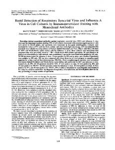
that they targeted the HA globular head. This specificity of 8M2 and 2G1 was surprising given the previously reported strong association of HA stem specificity with the use of the VH1-69 germ line gene segment in antibodies to influenza (7, 9, 33, 45). MAb 8F8 and MAb 2G1 both potently inhibited all H2 strains tested except for a virus circulating in 1967 (Table 1). MAb 8M2 inhibited all strains tested, including the virus from 1967, but did not react with the Japan/305/57 strain (GenBank accession number CY044325). Interestingly, MAbs 8F8, 8M2, and 2G1 each inhibited a swine H2N3 influenza virus strain from 2006 (Table 1). Epitope mapping of H2N2 antibodies. Next, we sought to understand why MAb 8M2 did not neutralize the Japan/305/57 strain. The sequence of the Japan/305⫹/57 virus differs from that of Japan/305/57 mainly in a serine residue at position 228 instead of a glycine (Fig. 1), but four slightly different sequences of Japan/ 305/57 have been published (see Fig. S1 in the supplemental material). This S228G change results in a switch from avian receptor specificity to human receptor specificity (5, 6, 49). To test whether a residue that mediated this receptor specificity also mediated escape from 8M2 inhibition, we made H2N2 virus-like particles (VLPs) using the cDNA from the HA of Japan/305⫹/57 virus. As expected, MAb 8M2 inhibited the HA activity of these VLPs (Table 4). When we reverted this serine back to a glycine in the Japan/ 305⫹/57 HA and used the cDNA to make VLPs, inhibition of those VLPs by MAb 8M2 was reduced markedly (Table 4). To more fully define the antibody epitopes, we generated escape mutant viruses, using the rationale that sequence polymorphisms in escape mutants will reflect the epitopes recognized by the MAb. MAb 8M2 selected for a G135D mutation in the Singapore/57 virus background, a residue located on the edge of the receptor-binding domain (RBD) opposite of residue 228 implicated above (Fig. 1). MAb 8F8 selected for a T193K mutation in the Singapore/57 background and an R137Q mutation in the Japan/305/57 background. Finally, MAb 2G1 elicited a K156E mutation in the Singapore/57 background. These residues at positions 137 and 156/193 also are located at the edge of the RBD on opposite sides of the structure, but at an approximate 90° rotation compared to the axis of the 135/228 residues (Fig. 1). We built those mutations back into VLPs to validate them as escape mutations. Indeed, the G135D mutation in the Japan/305⫹ HA protein context eliminated inhibition by MAb 8M2, and the T193K mutation reduced inhibition (Table 4). The T193K mutation eliminated inhibition by MAb 8F8, and R137Q markedly reduced inhibition for this antibody only. The K156E mutation conferred escape only for 2G1. Sequence analysis of H2N2 HA. We next sought to under-
Journal of Virology
Human Monoclonal Influenza H2N2/H3N2 Antibodies
TABLE 3 Genetic and binding kinetic features of H2/H3 influenza-specific human MAbsa Heavy chain MAb
Birth VH Subject yr
8F8 8M2 2G1
9 16 92
1958 1956 1950
7A13 7 11J19 13
1960 1965
D
Light chain JH
VL/VK
3–33*01 5–12*01 6*02 1–44*01 1–69*01 3–16*02 4*02 3–15*01 1–69*01 1–26*01 5*02 1-33*01 or 1D-33*01 4–4*02 2–21*02 1*01 4–69*01 4–39*01 3–10*01 4*02 1–9*01
Mutation JL/JK
/ VH
N P insertions insertions D JH VL
Kinetics JL (Kd [M])
2*01 or 3*01 1*01 5*01
21 (22) 10 30 (32) 13 23 (24) 8
0 0 0
0 4 0
1 2 1
18 1 12 0 10 (13) 0
2.7 ⫻ 10⫺8 1.3 ⫻ 10⫺7 1.8 ⫻ 10⫺8
28 (29) 19 18 (19) 1
0 0
4 1
2 2
13 0 14 (17) 0
2.5 ⫻ 10⫺8 4.3 ⫻ 10⫺9
3*02 4*01
a 8F8, 8M2, and 2G1 reacted with H2 HA; 2G1, 7A13, and 11J19 reacted with H3 HA. The genetic features were determined using the international ImMunoGeneTics (IMGT) information system (2). Binding kinetics were determined with an Octet Red instrument and anti-PentaHIS tips as described previously (15). Recombinant soluble A/Japan/305⫹/ 1957 H2 HA protein was used as a binding partner for 8F8, 8M2, and 2G1; recombinant soluble A/Hong Kong/1/1968 H3 HA protein was used for 7A13 and 11J19.
stand the influence of naturally occurring H2N2 HA antigenic drift mutations on the activity of our antibodies using previously isolated field strains of H2 viruses. A phylogram of the amino acid sequence of all naturally occurring, nonredundant human H2N2 HAs revealed two distinct populations of early (1957 to 1960) and late (1963 to 1968) H2N2 influenza virus strains (see Fig. S2 in the supplemental material). We performed a multiple sequence alignment of those strains in the order of the phylogram to document the sequence variability, particularly in the key residues of the escape mutations (see Fig. S1 in the supplemental material). Residue G135 was well conserved except for aspartic acid in A/Kumamoto/1/1965 and serine in A/Moscow/1019/1965 (Fig. S1); it is questionable whether the latter is truly a 1965 strain, as it is very similar to the 1957 to 1961 strains. The arginine in position 137 of H2 HA has mutated to a glutamine in two early H2 strains (so the
FIG 1 Space-filling model of 1957 influenza HA (PDB: 3KU3) (48). This is a view onto the RBD of the membrane-distal globular head of a single monomer. Residues that mediate escape from MAbs when mutated are colored: red for 8F8 escape mutations, green for 8M2 escape mutations, and blue for the 2G1 escape mutation. Other residues that are part of the RBD but have not been implicated as escape mutations of 8F8 or 8M2 are colored in dark gray.
June 2012 Volume 86 Number 11
above R137Q escape mutant is present in naturally occurring H2N2 viruses), to a methionine in six late H2 strains, and to a lysine in most other late H2N2 strains (Fig. S1). A T193A mutation is found in almost all late H2N2 strains, although two H2 HAs display a glutamic acid in this position. A K156E mutation is found in occasional early or late H2 strains; glutamine or threonine also was found in this position, but the original lysine predominated overall. Interestingly, the lysine residue at position 156 on HA that is critical for recognition by the heterosubtypic 2G1 antibody is present in the HA of every pandemic virus isolated to date (1918 H1, 1957 H2, 1968 H3, 2009 H1) and even the HA of the H5N1 A/VietNam/1203/2004 strain, which is highly virulent in humans. Residue 228 is split between the serine typical of human receptor specificity and the glycine of avian receptor specificity, although only a serine is found in this position in later strains. It should be noted that human H2N2 viruses have been passaged many times in eggs and that the passage history for some of the strains is unknown; whether viruses with avian receptor specificity truly cocirculated in 1957 or whether those isolates are drift variants from subsequent egg passage is controversial (6, 19, 26). We introduced some of these mutations into the A/Japan/305⫹/1957 background to test the specificity of our H2N2 antibodies in HAI assays against VLPs. MAb 8F8 was very sensitive to changes in position 137, with an R137M mutation leading to loss of inhibition and an R137K mutation to a marked reduction of inhibition (Table 4). This finding might explain why MAb 8F8 does not inhibit late H2N2 strains, although changes at other residues not identified by escape mutations might contribute. In vivo therapeutic efficacy of H2 antibodies. We next tested the H2-reactive MAbs in a therapeutic mouse model of H2N2 influenza virus infection (Fig. 2; Table 5). MAbs 8F8, 8M2, and 2G1 each protected all animals at the highest dose of 200 g; only MAb 2G1 also protected all animals at the intermediate dose and a single animal at the lowest dose (Fig. 2A). This trend was reflected in the animal weight curves, with animals in the high-dose groups gaining weight by day 14 compared to baseline (Fig. 2B). At the highest dose, the antibodies were able to reduce H2N2 lung titers between 2.6 log10 PFU/ml (for 2G1) and 2.2 log10 PFU/ml (for 8M2) compared to the IgG control (Table 5). Reactivity of H3N2 antibodies. Surprisingly, the H2-reactive MAb 2G1 also inhibited the pandemic 1968 H3 virus, but not the later H3 viruses that were tested (Table 1). The H3-specific antibodies 7A13 and 11J19 also inhibited A/Hong Kong/1/1968 H3N2, but they did not inhibit later H3 strains (Table 1), suggest-
jvi.asm.org 6337
Krause et al.
TABLE 4 Specific HAI activities of human H2N2 antibodies against wild-type or mutated VLPs HAI activitya (g/ml) against MAb H2N2 VLPs MAb
WT
G135D
G135S
R137Q
R137 M
R137K
K156E
T193K
T193A
S228G
8F8 8M2 2G1
⬍0.2 0.7 ⬍0.2
0.7 ⬎ 0.7
⬍0.2 1.3 ⬍0.2
10 0.7 ⬍0.2
⬎ 1.3 ⬍0.2
10 0.7 ⬍0.2
0.1 0.4 ⬎
⬎ 10 1.3
⬍0.2 0.7 ⬍0.2
0.7 2.5 0.4
a The highest titer that still showed HAI activity is shown. ⬎, activity was not detected at the highest concentration tested, 20 g/ml. VLPs are based on the HA of A/Japan/305⫹/ 1957 except for the K156 mutant, which is based on A/Singapore/1/1957. WT, wild type.
ing that these antibodies do not display significant cross-reactivity within this influenza virus subtype. DISCUSSION
B cells specifying MAbs to 20th-century pandemic influenza viruses can still be detected in the peripheral blood of humans. The persistence of virus-specific memory B cells in the circulation is remarkable. We previously showed that H1-specific human MAbs to the 1918 H1N1 pandemic virus can be cloned from the peripheral blood of survivors of the pandemic many decades after circulation of that virus (54). Here, we used a similar approach (53) to clone neutralizing human MAbs against 1957 H2N2 or 1968 H3N2 pandemic viruses. Influenza antibodies also can be cloned from plasmablasts of recent vaccinees or those convalescing from disease (46), but this technology is not suitable for a pathogen that is no longer in circulation and for which a routine vaccine is unavailable. In contrast, human hybridoma technology can be used to generate MAbs against an antigen that has not been
in human circulation for at least 43 years, such as H2N2 influenza. In summary, we have shown for all three influenza pandemics of the 20th century (1918, 1957, and 1968) that B cells specific for the pandemic virus can still be found in the peripheral blood of human beings in the 21st century. Antibodies targeting the H2 RBD may have been present on a population level. The first major epitope mapping of H2N2 HA was performed almost 30 years ago using murine MAbs (49). Interestingly, half of these antibodies—like MAb 8M2— did not inhibit virus with avian receptor specificity; the influence of avian receptor specificity was even highlighted by Yamada et al. in the title of their article (49). Receptor specificity can influence antibody inhibition when the epitope includes residues that mediate that receptor specificity (i.e., that are part of the RBD). Unlike H1N1 HA in early epitope mapping studies (3), H2N2 was found not to have discrete murine epitopes but to have overlapping epitopes on its globular head, as the antibodies isolated competed with each other for binding to HA over the RBD (49). These findings are consistent in principle with the epitope mapping of human MAbs in our study that elicited escape mutations immediately adjacent to the RBD. The number of human antibodies in our study is limited but, taken together with the prior work by Yamada et al., suggests that most of the circulating B cells specific for H2N2 influenza are targeted to the RBD. Why both the human and the murine immune responses to H2 are so focused on the relatively conserved RBD is unclear. This may have contributed to the limited circulation of H2 viruses. Understanding this phe-
TABLE 5 Therapeutic efficacies of H2N2-reactive MAbs against virus replication in mice inoculated with A/Albany/6/1958 H2N2 virusa Dose (g/mouse)
Lung virus titer (log10 PFU/ml), mean ⫾ SD
8F8
200 20 2
4.4 ⫾ 0.1 * 5.8 ⫾ 0.1 * 6.5 ⫾ 0.2 *
8M2
200 20 2
4.7 ⫾ 0.3 * 6.3 ⫾ 0.2 * 6.8 ⫾ 0.1
2G1
200 20 2
4.3 ⫾ 0.3 * 5.0 ⫾ 0.5 * 6.3 ⫾ 0.4 *
IgG control
200
6.9 ⫾ 0.1
Antibody
FIG 2 Therapeutic efficacy of MAbs against disease caused by the A/Albany/ 6/1958 H2N2 virus in mice. Mice were inoculated on day 0 and treated on day 1 with the indicated antibody (8F8, 8M2, 2G1, or a human IgG control) and dose. In each group, six mice were monitored for survival (A) and weight (B). At the 8F8 200-g dose (P ⬍ 0.01), the 8F8 20-g dose (P ⬍ 0.05), the 8M2 200-g dose (P ⬍ 0.01), the 2G1 200-g dose (P ⬍ 0.01), and the 2G1 20-g dose (P ⬍ 0.01), treatment conferred a survival advantage by log rank test.
6338 jvi.asm.org
a
Four mice were inoculated intranasally with 5 times the LD50 and administered MAb 8F8, 8M2, 2G1, or human IgG i.p. 24 h later. Mice were euthanized on day 4 after inoculation for the determination of lung titers. *, at the ␣ ⫽ 0.025 level controlling the overall type I error at 7.5%, the lung homogenates differ from the IgG control group by the Wilcoxon rank sum test (P ⬍ 0.05).
Journal of Virology
Human Monoclonal Influenza H2N2/H3N2 Antibodies
nomenon better might help to improve current influenza vaccines. Antibodies that contact amino acid residues that are components of the RBD have been described previously (11, 14, 15, 43). Although our H2N2 antibodies likely make contact within the RBD pocket (the heterosubtypic activity of 2G1 would be difficult to explain otherwise), such contact residues are difficult to identify by the escape mutation method we used since such viruses likely would be reduced in replicative capacity. X-ray crystallography structures of antibody-H2 HA complexes would be helpful to elucidate these interactions on the atomic level. 2G1-like antibodies may have provided relative protection from H3 virus. Neither the H2-specific MAbs 8M2 and 8F8 nor the H3-specific MAbs 7A13 and 11J19 neutralized the other subtype. However, the human H2 antibody 2G1 did inhibit the 1968 H3 influenza virus, which is a very unusual phenotype that has not been described previously, though MAb 2G1 did not neutralize in a microneutralization assay. It is likely that H2 virus was the inciting event for MAb 2G1, since infection with pandemic viruses is substantial and H2 virus circulated before H3, and since this antibody exhibits more potent inhibition of H2 virus. The relative conservation of the critical residue 156 may have been the structural basis for this cross-reactivity, though other residues likely contributed. Heterosubtypic HA globular head domain-specific antibodies are probably rare because of the variability in the dominant antigenic loops, but the presence of 2G1-like antibodies might have contributed to the diminished severity of the 1968 H3N2 pandemic as opposed to the 1957 H2N2 pandemic. This phenomenon has previously been attributed to cross-reactive N2 neuraminidase antibodies (47), but human cross-reactive N2specific MAbs have not been described so far. H2 MAbs might be useful for diagnostic or therapeutic purposes. Experimental H2N2 vaccine candidates exist but may not be protective after a single dose (10). Therefore, passive transfer of antibodies such as MAb 8F8, 8M2, or 2G1 could be used in case of a 1957-like virus pandemic to protect high-risk individuals. Also, these antibodies could be useful as diagnostic reagents or to differentiate H2N2 viruses with human or avian receptor specificity. ACKNOWLEDGMENTS This work was supported by NIH grants P01 AI058113 and R21AI085306, Vanderbilt CTSA grant UL1RR024975, a pilot project from UL1RR029887 to C.F.B., DOD grant HDTRA1-10-1-0067, and CRIP, an NIAID-funded CEIRS Center for Research on Influenza Pathogenesis, contract number HHSN266200700010C. The Genome Sciences Resource was supported by the Cancer Center Support Grant (CA068485), the Vanderbilt Digestive Disease Research Center (DK058404), and the Vanderbilt Vision Research Center (EY008126). The funders had no role in study design, data collection and analysis, decision to publish, or preparation of the manuscript. We thank the anonymous blood donors; M. Posner and L. Cavacini for providing the HMMA2.5 cell line; Jose A. Archuleta, Jr., and Cheryl Kinnard of the Vanderbilt Clinical Trials Center; Belinda G. Johnson of the Vanderbilt Vaccine Trials and Evaluations Unit; and Scott A. Smith, Rui Xu, and Ian A. Wilson for helpful discussions.
REFERENCES 1. Bernstein DI, et al. 2008. Effects of adjuvants on the safety and immunogenicity of an avian influenza H5N1 vaccine in adults. J. Infect. Dis. 197:667– 675. 2. Brochet X, Lefranc MP, Giudicelli V. 2008. IMGT/V-QUEST: the highly customized and integrated system for IG and TR standardized V-J and V-D-J sequence analysis. Nucleic Acids Res. 36:W503–W508.
June 2012 Volume 86 Number 11
3. Caton A, Brownlee G, Yewdell J, Gerhard W. 1982. The antigenic structure of the influenza virus A/PR/8/34 hemagglutinin (H1 subtype). Cell 31:417– 427. 4. Chen BJ, Leser GP, Morita E, Lamb RA. 2007. Influenza virus hemagglutinin and neuraminidase, but not the matrix protein, are required for assembly and budding of plasmid-derived virus-like particles. J. Virol. 81:7111–7123. 5. Choppin PW, Tamm I. 1960. Studies of two kinds of virus particles which comprise influenza A2 virus strains. II. Reactivity with virus inhibitors in normal sera. J. Exp. Med. 112:921–944. 6. Connor RJ, Kawaoka Y, Webster RG, Paulson JC. 1994. Receptor specificity in human, avian, and equine H2 and H3 influenza virus isolates. Virology 205:17–23. 7. Corti D, et al. 2010. Heterosubtypic neutralizing antibodies are produced by individuals immunized with a seasonal influenza vaccine. J. Clin. Invest. 120:1663–1673. 8. Dawood FS, et al. 2009. Emergence of a novel swine-origin influenza A (H1N1) virus in humans. N. Engl. J. Med. 360:2605–2615. 9. Ekiert DC, et al. 2009. Antibody recognition of a highly conserved influenza virus epitope. Science 324:246 –251. 10. Hehme N, Engelmann H, Kunzel W, Neumeier E, Sanger R. 2002. Pandemic preparedness: lessons learnt from H2N2 and H9N2 candidate vaccines. Med. Microbiol. Immunol. 191:203–208. 11. Hensley SE, et al. 2009. Hemagglutinin receptor binding avidity drives influenza A virus antigenic drift. Science 326:734 –736. 12. Hilleman MR. 2002. Realities and enigmas of human viral influenza: pathogenesis, epidemiology and control. Vaccine 20:3068 –3087. 13. Kaverin NV, et al. 2000. Cross-protection and reassortment studies with avian H2 influenza viruses. Arch. Virol. 145:1059 –1066. 14. Knossow M, Skehel JJ. 2006. Variation and infectivity neutralization in influenza. Immunology 119:1–7. 15. Krause JC, et al. 2011. A broadly neutralizing human monoclonal antibody that recognizes a conserved, novel epitope on the globular head of the influenza H1N1 virus hemagglutinin. J. Virol. 85:10905–10908. 16. Krause JC, et al. 2011. Epitope-specific human influenza antibody repertoires diversify by B cell intraclonal sequence divergence and interclonal convergence. J. Immunol. 187:3704 –3711. 17. Larkin MA, et al. 2007. Clustal W and Clustal X version 2.0. Bioinformatics 23:2947–2948. 18. Ma W, et al. 2007. Identification of H2N3 influenza A viruses from swine in the United States. Proc. Natl. Acad. Sci. U. S. A. 104:20949 –20954. 19. Matrosovich M, et al. 2000. Early alterations of the receptor-binding properties of H1, H2, and H3 avian influenza virus hemagglutinins after their introduction into mammals. J. Virol. 74:8502– 8512. 20. Nabel GJ, Wei CJ, Ledgerwood JE. 2011. Vaccinate for the next H2N2 pandemic now. Nature 471:157–158. 21. Nagieva FG, et al. 1989. Preparation and properties of monoclonal antibodies to influenza virus A/Krasnodar/101/59 (H2N2). Vopr. Virusol. 34: 543–547. 21a.National Research Council. 1996. Guide for the care and use of laboratory animals. Office of Laboratory Animal Welfare, National Institutes of Health, Bethesda, MD. 22. Ohshima N, et al. 2011. Naturally occurring antibodies in humans can neutralize a variety of influenza virus strains, including H3, H1, H2, and H5. J. Virol. 85:11048 –11057. 23. Okada J, et al. 2011. Localization of epitopes recognized by monoclonal antibodies that neutralized the H3N2 influenza viruses in man. J. Gen. Virol. 92:326 –335. 24. Okada J, et al. 2010. Monoclonal antibodies in man that neutralized H3N2 influenza viruses were classified into three groups with distinct strain specificity: 1968 –1973, 1977–1993 and 1997–2003. Virology 397: 322–330. 25. Okuno Y, Isegawa Y, Sasao F, Ueda S. 1993. A common neutralizing epitope conserved between the hemagglutinins of influenza A virus H1 and H2 strains. J. Virol. 67:2552–2558. 26. Pappas C, et al. 2010. Receptor specificity and transmission of H2N2 subtype viruses isolated from the pandemic of 1957. PLoS One 5:e11158. 27. Pyhala R. 1985. Antibody status to influenza A/Singapore/1/57(H2N2) in Finland during a period of outbreaks caused by H3N2 and H1N1 subtype viruses. J. Hyg. (Lond.) 95:437– 445. 28. Schäfer JR, et al. 1993. Origin of the pandemic 1957 H2 influenza A virus and the persistence of its possible progenitors in the avian reservoir. Virology 194:781–788.
jvi.asm.org 6339
Krause et al.
29. Shibuya T, et al. 2008. Identification of a human monoclonal Fab with neutralizing activity against H3N2 influenza A strain from a newly constructed human Fab library. Microbiol. Immunol. 52:162–170. 30. Squires B, et al. 2008. BioHealthBase: informatics support in the elucidation of influenza virus host pathogen interactions and virulence. Nucleic Acids Res. 36:D497–D503. 31. Stevens J, et al. 2004. Structure of the uncleaved human H1 hemagglutinin from the extinct 1918 influenza virus. Science 303:1866 –1870. 32. Stöhr K. 2010. Vaccinate before the next pandemic? Nature 465:161. 33. Sui J, et al. 2009. Structural and functional bases for broad-spectrum neutralization of avian and human influenza A viruses. Nat. Struct. Mol. Biol. 16:265–273. 34. Taubenberger JK, Morens DM. 2010. Influenza: the once and future pandemic. Public Health Rep. 125(Suppl 3):16 –26. 35. Tsuchiya E, et al. 2001. Antigenic structure of the haemagglutinin of human influenza A/H2N2 virus. J. Gen. Virol. 82:2475–2484. 36. Underwood PA. 1984. An antigenic map of the haemagglutinin of the influenza Hong Kong subtype (H3N2), constructed using mouse monoclonal antibodies. Mol. Immunol. 21:663– 671. 37. Underwood PA. 1982. Mapping of antigenic changes in the haemagglutinin of Hong Kong influenza (H3N2) strains using a large panel of monoclonal antibodies. J. Gen. Virol. 62(Pt 1):153–169. 38. Viswanathan K, et al. 2010. Determinants of glycan receptor specificity of H2N2 influenza A virus hemagglutinin. PLoS One 5:e13768. 39. Walls HH, Harmon MW, Slagle JJ, Stocksdale C, Kendal AP. 1986. Characterization and evaluation of monoclonal antibodies developed for typing influenza A and influenza B viruses. J. Clin. Microbiol. 23:240 –245. 40. Webster RG. 1997. Predictions for future human influenza pandemics. J. Infect. Dis. 176(Suppl 1):S14 –S19. 41. Webster RG, Laver WG. 1980. Determination of the number of nonoverlapping antigenic areas on Hong Kong (H3N2) influenza virus hemagglutinin with monoclonal antibodies and the selection of variants with potential epidemiological significance. Virology 104:139 –148. 42. Webster RG, et al. 1980. The mechanism of antigenic drift in influenza viruses: analysis of Hong Kong (H3N2) variants with monoclonal antibodies to the hemagglutinin molecule. Ann. N. Y. Acad. Sci. 354:142–161.
6340
jvi.asm.org
43. Whittle JR, et al. 2011. Broadly neutralizing human antibody that recognizes the receptor-binding pocket of influenza virus hemagglutinin. Proc. Natl. Acad. Sci. U. S. A. 108:14216 –14221. 44. World Health Organization Collaborating Centers for Reference and Research on Influenza. 1982. Concepts and procedures for laboratorybased influenza surveillance. Centers for Disease Control, Atlanta, GA. 45. Wrammert J, et al. 2011. Broadly cross-reactive antibodies dominate the human B cell response against 2009 pandemic H1N1 influenza virus infection. J. Exp. Med. 208:181–193. 46. Wrammert J, et al. 2008. Rapid cloning of high-affinity human monoclonal antibodies against influenza virus. Nature 453:667– 671. 47. Wright PF, Neumann G, Kawaoka Y. 2007. Fields virology, 5th ed, p 1699. In Knipe DM, et al (ed), Lippincott Williams & Wilkins, Philadelphia, PA. 48. Xu R, McBride R, Paulson JC, Basler CF, Wilson IA. 2010. Structure, receptor binding, and antigenicity of influenza virus hemagglutinins from the 1957 H2N2 pandemic. J. Virol. 84:1715–1721. 49. Yamada A, Brown LE, Webster RG. 1984. Characterization of H2 influenza virus hemagglutinin with monoclonal antibodies: influence of receptor specificity. Virology 138:276 –286. 50. Yamashita A, et al. 2010. Highly conserved sequences for human neutralization epitope on hemagglutinin of influenza A viruses H3N2, H1N1 and H5N1: implication for human monoclonal antibody recognition. Biochem. Biophys. Res. Commun. 393:614 – 618. 51. Yewdell JW, Webster RG, Gerhard WU. 1979. Antigenic variation in three distinct determinants of an influenza type A haemagglutinin molecule. Nature 279:246 –248. 52. Yoshida R, et al. 2009. Cross-protective potential of a novel monoclonal antibody directed against antigenic site B of the hemagglutinin of influenza A viruses. PLoS Pathog. 5:e1000350. 53. Yu X, McGraw PA, House FS, Crowe JE, Jr. 2008. An optimized electrofusion-based protocol for generating virus-specific human monoclonal antibodies. J. Immunol. Methods 336:142–151. 54. Yu X, et al. 2008. Neutralizing antibodies derived from the B cells of 1918 influenza pandemic survivors. Nature 455:532–536.
Journal of Virology