Human Plexin B1 Antibody
Monoclonal Mouse IgG1 Clone # 439512 Catalog Number: MAB3749
DESCRIPTION Species Reactivity
Human
Specificity
Detects human Plexin B1 in direct ELISAs and Western blots. In direct ELISAs and Western blots, no crossreactivity with recombinant human (rh) Plexin C1, rhPlexin D1, recombinant mouse (rm) Plexin A1, rmPlexin A2, or rmPlexin A3 is observed.
Source
Monoclonal Mouse IgG1 Clone # 439512
Purification
Protein A or G purified from hybridoma culture supernatant
Immunogen
E. coliderived recombinant human Plexin B1 Leu35Val150 Accession # O43157
Formulation
Lyophilized from a 0.2 μm filtered solution in PBS with Trehalose. See Certificate of Analysis for details.
APPLICATIONS
Please Note: Optimal dilutions should be determined by each laboratory for each application. General Protocols are available in the Technical Information section on our website.
Recommended Concentration
Sample
Western Blot
1 µg/mL
Recombinant Human Plexin B1
Immunohistochemistry
825 µg/mL
See Below
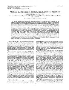
DATA Immunohistochemistry Plexin B1 in Human Brain. Plexin B1 was detected in immersion fixed paraffin embedded sections of human brain using Mouse AntiHuman Plexin B1 Monoclonal Antibody (Catalog # MAB3749) at 15 µg/mL overnight at 4 °C. Before incubation with the primary antibody, tissue was subjected to heatinduced epitope retrieval using Antigen Retrieval ReagentB a s i c ( C a t a l o g # CTS013). Tissue was stained using the Anti Mouse HRPDAB Cell & Tissue Staining Kit ( b r o w n ; C a t a l o g # CTS002) a n d counterstained with hematoxylin (blue). Specific staining was localized to neuronal cell bodies. View our protocol for Chromogenic IHC Staining of Paraffin embedded Tissue Sections.
PREPARATION AND STORAGE Reconstitution
Reconstitute at 0.5 mg/mL in sterile PBS.
Shipping
The product is shipped at ambient temperature. Upon receipt, store it immediately at the temperature recommended below.
Stability & Storage
Use a manual defrost freezer and avoid repeated freezethaw cycles. l 12 months from date of receipt, 20 to 70 °C as supplied. l 1 month, 2 to 8 °C under sterile conditions after reconstitution. l 6 months, 20 to 70 °C under sterile conditions after reconstitution.
BACKGROUND
Plexin B1 is a 300 kDa member of the B subfamily, Plexin family of Semaphorin receptors. Mature human Plexin B1 is a 2116 amino acid type I transmembrane (TM) glycoprotein that contains a 1471 aa extracellular domain (ECD) and a 612 aa cytoplasmic region. The ECD contains one Semaphorin domain, two PSI domains, and three IPT repeats. The ECD is cleaved into two subunits, a 200 kDa αchain (aa 201305) and a 100 kDa TM βchain. The subunits are nondisulfidelinked and generate a highaffinity receptor. Plexin B1 is a receptor for Semaphorin 4D/CD100. It forms a receptor complex with Neuropilins, MET, and EGF R2. Multiple splice variants are known. One shows a deletion of aa 689871. A second shows a 53 aa substitution for aa 6772135. And a third shows a 53 aa substitution for the 53 aa between aa 677729. Over aa 35150, human Plexin B1 is 90% and 97% aa identical to dog and mouse Plexin B1, respectively.
Rev. 12/2/2011 Page 1 of 1 www.RnDSystems.com For research use only. Not for use in diagnostic procedures. USA & CANADA Tel: (800) 343-7475 EUROPE Tel: +44 (0)1235 529449 CHINA Tel: +86 (21) 52380373
Journal of Neuroimmunology 252 (2012) 113–117
Contents lists available at SciVerse ScienceDirect
Journal of Neuroimmunology journal homepage: www.elsevier.com/locate/jneuroim
Short communication
Plexin-B1: A potential diagnostic biomarker for glioma and a future target for glioma immunotherapy Yun Zhang a, 1, Qi Li a, 1, Ran Zhuang a, Zhenhui Gao b, Jian Liu b, Juan Li b, Angang Yang a, Guang Cheng b,⁎, Boquan Jin a a b
Department of Immunology, the Fourth Military Medical University, Xi'an, China Department of Neurosurgery, Xijing Hospital, the Fourth Military Medical University, Xi'an, China
a r t i c l e
i n f o
Article history: Received 10 May 2012 Received in revised form 10 August 2012 Accepted 14 August 2012 Keywords: Plexin-B1 Glioma Natural killer cell
a b s t r a c t Gliomas are the most common tumors in the central nervous system. Plexin-B1 is abundantly expressed in the nervous system as an axonal guidance molecule during neuronal development. However, the correlation between its expression and the clinical characteristics of gliomas, and its therapeutic significance, remain largely unexplored. In this study, we detected the expression of Plexin-B1 in clinical glioma tissue samples. Plexin-B1 was highly expressed in the cytoplasm and on the membrane of glioma tissues, while only trace levels of Plexin-B1 were present in normal brain tissue. The expression level of Plexin-B1 in glioma tissue was associated with the pathological grade of the glioma. In addition, we used flow cytometry to analyze the expression of Plexin-B1 in glioma cell lines and its ligand, semaphorin 4D (Sema4D), in natural killer (NK) cell lines. Cytotoxicity assays showed cytolysis of the U251 glioma cell line by the NK cell line, NK92, and this was markedly downregulated when the neutralizing antibody to Plexin-B1 was added. This study demonstrates that Plexin-B1 could be used as a diagnostic biomarker, and also suggests that it may be involved in the cytotoxicity of NK cells to glioma cells. Plexin-B1 could be a useful future target for glioma immunotherapy. © 2012 Elsevier B.V. All rights reserved.
1. Introduction Gliomas are the most common tumors of the central nervous system in adults, accounting for 40% to 50% of all intracranial tumors (Iwami et al., 2011; Rainov and Heidecke, 2011). It is an aggressive form of tumor, which has a tendency to invade the surrounding brain tissue. Therefore, it is difficult to complete a surgical resection, and postoperative relapses often occur (Kros, 2011). Despite these major challenges, great progress has been made in surgical, radioactive and chemical anti-glioma treatment modalities, but their efficacy remains unsatisfactory (Strik et al., 2012). In recent years, progress has been made in the development of immunotherapy, gene therapy and other novel biological therapies against glioma (Wick et al., 2009; Hdeib and Sloan, 2011). The relationships between the biological behaviors of this disease, including the high proliferation rate of glioma cells, invasive glioma growth and relative immune response, have been increasing focuses of research. Semaphorins belong to a family of membrane-bound and secreted molecules that regulate the functional activity of axons in the nervous system (Pasterkamp and Kolodkin, 2003). Sema4A and Sema4D/CD100 were the first semaphorins that were also found to be expressed in ⁎ Corresponding author. E-mail address:
[email protected] (G. Cheng). 1 These authors contribute equally to this work. 0165-5728/$ – see front matter © 2012 Elsevier B.V. All rights reserved. http://dx.doi.org/10.1016/j.jneuroim.2012.08.005
immune cells and were therefore termed “immune semaphorins”. CD100 is abundantly expressed by T cells and NK cells, but only weakly detected in naive B cells, macrophages and DCs; Sema4A is not expressed by NK cells (Takamatsu et al., 2010). Sema4A has three functional receptors, namely Plexin-D1, Plexin-B1 and Tim-2, whereas CD100 only binds to Plexin-B1 and CD72 (Giordano et al., 2002). CD72 is also a low affinity lymphocyte receptor for CD100, which is expressed mainly on dendritic cells (DC) and B cells, and it participates in the regulation of both T and B cell responses (Kumanogoh et al., 2000). Plexin-B1, on the other hand, which is abundantly expressed in the fetal human brain and kidney, is a receptor with a high affinity for CD100 (Kd =1 × 10 −9 M) (Giordano et al., 2002). Research on CD100 and Plexin-B1 in the central nervous system showed that CD100–Plexin-B1 interactions in the central nervous system were involved in the pathogenesis of experimental autoimmune encephalomyelitis (EAE) (Okuno et al., 2010). Another study demonstrated the requirement for CD100 in the induction of antigen-specific T cells and the maturation of DCs (Kumanogoh et al., 2002). Recent study showed that semaphorins and their receptors were primarily involved in tumor formation and progression (Micucci et al., 2010). However, the expression of Plexin-B1 by gliomas and its relationship with the pathological progression of this tumor type remain unclear. In this study, we demonstrate Plexin-B1 on glioma tissue was associated with the tumor pathological grade. We further present that the treatment with mAb against Plexin-B1 was effective for cytotoxicity
114
Y. Zhang et al. / Journal of Neuroimmunology 252 (2012) 113–117
downregulation. These findings demonstrate that Plexin-B1 may be used as a diagnostic biomarker for glioma, and CD100–Plexin-B1 interactions play an important role in the cytotoxicity of NK cells against glioma cells. Plexin-B1 may provide a valuable novel therapeutic target for glioma immunotherapy. 2. Materials and methods 2.1. Patients and tissue samples This study was approved by the Research Ethics Committee of Xijing Hospital, Fourth Military Medical University, China. In total, 110 samples of surgical specimens from patients with glioma were collected from a hospital between June 2010 and December 2011 (68 males and 42 females). All patients were newly diagnosed without any history of preoperative chemotherapy, radiotherapy or any other neoadjuvant treatment. None of the patients had any inflammatory diseases. Their ages ranged from 13 to 72 years, with a mean age of 41.26 years. Tumors were histopathologically classified according to the WHO classification system for glioma (Louis et al., 2007); 57 of the 110 gliomas were classified as low grade (WHO I and WHO II), and 53 were classified as high-grade gliomas (WHO III and WHO IV). Twenty normal brain tissue biopsies were also taken from patients admitted to our hospital with either head injuries or those undergoing the insertion of decompression lines as controls. The written informed consent was obtained from all patients before their enrollment. 2.2. Immunohistochemistry Paraffin-embedded sections of glioma and normal brain tissue were conventionally stained with hematoxylin and eosin or an anti-Plexin‐B1 antibody. Briefly, slices were de-waxed, hydrated, and incubated in peroxidase inhibitor for 30 min to remove endogenous peroxidase. After blocking with diluted goat serum (Sigma, USA), all samples were incubated with a 1:50 dilution of monoclonal antibody against human Plexin-B1 (IgM, Millipore, USA) overnight at 4 °C. Non-immune IgM was used as a negative control antibody for immunohistochemical staining. After three washes in PBS, the slices were dipped into HRP-conjugated goat anti-mouse IgM (Abcam, UK) for 30 min at room temperature. Antigen–antibody complexes were incubated with DAB chromogen and observed. Sections were counterstained with Mayer's hematoxylin for 2 min, dehydrated through gradient ethanol, cleared in dimethylbenzene, and then mounted and examined using light microscopy (Olympus, Japan). An immunoreactivity score system was applied as described previously (Brown and Wahl, 1993). The scoring standard was as follows: (1) the number of positively stained cells b5% scored 0; 6–25% scored 1; 26–50% scored 2; 51–75% scored 3; > 75% scored 4; (2) intensity of stain: colorless scored 0; light brown scored 1; brown scored 2; dark brown scored 3; the scores from (1) and (2) were then multiplied. The staining score was stratified as follows: 0 score, absent; +: 1–4 score, weak; ++: 5–8 score, moderate; and +++: 9–12 score, strong according to the proportion and intensity of positively stained cells. 2.3. Flow cytometry The NK cell line, NK92, and two glioma cell lines, U251 and U87MG, were cultured in 10% FCS RPMI 1640 (Gibco, USA). Cells were collected and immunostained with PE-conjugated mouse anti-human CD100 mAb (R&D system, USA) or PE-conjugated mouse anti-human Plexin-B1 mAb (R&D system, USA). The isotype-matched mAbs (eBioscience, USA) control was used in all procedures. All staining procedures were performed according to the manufacturers' protocol. The stained cells
were then analyzed using a FACS Calibur flow cytometer and CELLQUEST software (Becton Dickinson, USA). 2.4. Cytotoxicity assays Killing assays were performed as previously reported (Markel et al., 2009). Carboxy-fluorescein diacetate succinimidyl ester (CFSE, Sigma-Aldrich, USA) pre-labeled target cells were co-incubated for 5 h with effector cells at effector-to-target ratios of 20:1, 10:1, 5:1, 2:1 and 1:1. For neutralization assay, the antibodies against Plexin-B1 or isotype-matched mAb (Santa Cruz Biotechnology, USA) were added at the time of coculture. The killing rate was quantified by PI-costaining (10 μg/ml) and assessed by the percentage of PI costaining cells out of gated CFSE-labeled cells. Background PI staining was b15% in all experiments. 2.5. Statistical analysis All statistical analyses were performed using the statistical software package, SPSS (SPSS Inc., Chicago, IL, USA). Randomized block design ANOVA was used to analyze the statistical difference among different tissue types. The associations between protein expression and different clinical parameters were evaluated by the χ 2 test. Differences were considered statistically significant when the P-value was less than 0.05. 3. Results 3.1. Immunohistochemical expression of Plexin-B1 in glioma tissues and its correlation with tumor grade Plexin-B1 expression was studied in a total of 110 glioma specimens, of which 57 were low-grade gliomas (WHO grades I and II) and 53 were high grade (grades III and IV). Twenty specimens taken from non-neoplastic brain tissues served as the control group. Based on the immunohistochemical analysis, Plexin-B1 was expressed in the cytoplasm and membrane of tumor cells in gliomas, whereas the non-neoplastic brain tissues showed only a marginal expression of Plexin-B1 (Fig. 1). Among the glioma specimens, 90 (81.8%) glioma specimens were positively stained with a significant difference of Plexin-B1 expression between glioma and non-neoplastic brain tissues (P b 0.001). Table 1 summarizes the associations between Plexin-B1 immunostaining and the clinicopathological parameters of the gliomas. Plexin-B1 expression was not significantly affected gender or age (both P > 0.05), but was closely related with tumor grade (P b 0.01). Taken together, these data indicated that Plexin-B1 may be useful as a novel biomarker for glioma. 3.2. Analysis of glioma cell lines as targets for natural killer cell-mediated cytotoxicity Human glioma cell lines U251 and U87MG were both characterized by their Plexin-B1 + phenotype. The positive frequency of Plexin-B1 in U251 cell was 78.9%, but this was relatively low in U87MG cells (Fig. 2A). Based on these data, we chose U251 cells as the target cells to study NK cell-mediated cytotoxicity. 3.3. The interaction between Plexin-B1 and CD100 played an important role in NK cell-mediated cytotoxicity against glioma cells To address the role of the interaction between Plexin-B1 and CD100 in NK cell-mediated cytotoxicity against glioma cells, the expression of CD100 on the NK cell line, NK92, was first detected (Fig. 2B). Then, NK92 and U251 cells were co-cultured at different effector/target (E:T) ratios for indicated times. The results showed that the cytotoxicity of NK cells against U251 cell increased according
Y. Zhang et al. / Journal of Neuroimmunology 252 (2012) 113–117
115
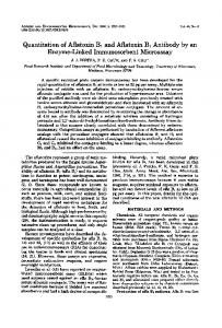
Fig. 1. Immunohistochemical staining of Plexin-B1 in non-neoplastic brain tissues (A) and glioma samples (B–E). Positive staining of Plexin-B1 is seen in the cytoplasm and cell membranes of glioma cells, whereas the non-neoplastic brain tissues showed marginal expression of Plexin-B1 (×400).
to the E:T ratio. As expected, the level of cell lysis was greatly downregulated when a neutralized mAb against Plexin-B1 was added to the culture medium (Fig. 2C). Collectively, these results demonstrate that the interaction between Plexin-B1 and CD100 participated in the NK cell-mediated killing of glioma cell lines. 4. Discussion In the current study, we examined the expression of Plexin-B1 in 110 cases of human glioma and compared its expression with tumor grade. Our data demonstrated that the Plexin-B1 protein was overexpressed in gliomas compared to non-neoplastic brain tissues. In addition, increasing expression levels of Plexin-B1 correlated with tumor grade with the trend from WHO grade I to WHO grade IV. Furthermore, the level of cytotoxicity of the NK cell line, NK92, to a glioma cell line was greatly downregulated by the addition of a neutralized mAb against Plexin-B1. These results suggest that Plexin-B1 might participate in the carcinogenesis and progression of glioma. The expression level of Plexin-B1 in gliomas is a novel
Table 1 Plexin-B1 expression in human glioma tissues with different clinical–pathological features. Clinicopathological features
Gender (M/F) Male Female Age (yr) b50 ≥50 WHO grade I II III IV
No. of cases
Plexin-B1 (n)
p
−
+
++
+++
68 42
12 8
12 6
23 14
21 14
61 49
9 11
19 10
22 16
11 12
23 34 25 28
8 7 3 2
10 15 7 4
3 7 9 10
2 5 6 12
NS
NS
b0.01
biomarker for glioma. Moreover, Plexin-B1 may be a new target for glioma immunotherapies that are based on NK cells. Plexin-B1 is a member of the plexin family of transmembrane proteins. It is a receptor that is prominently expressed in the fetal brain and kidney, and demonstrates a high binding affinity for Sema4D/ CD100 (Tamagnone et al., 1999). In the nervous system, semaphorin– plexin signaling has been shown to mediate diverse neural functions by regulating GTPase activities and cytoplasmic/receptor-type protein kinases (Kruger et al., 2005; Puschel, 2007). These plexinmediated signals are involved in integrin-mediated attachment, actomyosin contraction and microtubule destabilization (Aurandt et al., 2002). In the immune system, CD100 is expressed by T cells, activated B cells and NK cells (Delaire et al., 1998). CD100 promotes the activation of B cells and DCs to induce antibody production and antigenspecific T cells, respectively (Shi et al., 2000). In particular, CD100 is crucially involved in T-cell-mediated neurological inflammatory diseases. CD100-deficient mice were resistant to experimental autoimmune encephalomyelitis (EAE) due to impaired antigen-specific T-cell responses in the draining lymph nodes and attenuated levels of inflammation in the central nervous system (Okuno et al., 2010). In addition, T-cell-derived CD100 has been implicated in the collapse of process extension of immature oligodendrocytes and the death of immature neural cells in the spinal cords of patients with human T-cell lymphotropic virus type 1-associated myelopathy (Giraudon et al., 2004). In particular, CD100 is expressed on resting NK cells which were originally described as large granular lymphocytes with natural cytotoxicity against tumor cells (Vivier et al., 2008). Studies have shown that CD100 expressed on NK cells enhances the secretion of IFNγ and the killing of target cells that express CD72 (Mizrahi et al., 2007). Consistent with these previous reports, our group also confirmed the high expression of CD100 on the NK cell line, NK92. We further analyzed the expression of Plexin-B1 on human glioma cell lines. The results showed that the frequency of Plexin-B1 + U251 cells was much higher than that of U87MG cells. More importantly, we found that the cytotoxicity of NK cells to U251 cells was 74.8% at
116
Y. Zhang et al. / Journal of Neuroimmunology 252 (2012) 113–117
Fig. 2. Interaction between CD100 and Plexin-B1 in the NK cell cytotoxicity against glioma cell lines. (A) Expression of Plexin-B1 on glioma cell lines. U87MG (left) and U251 (right) cells were stained with PE-conjugated-mouse anti-human Plexin-B1 mAb. The percentage of Plexin-B1+ cells was analyzed by FCM. (B) Expression of CD100 on NK92 cell line. NK92 cells were stained with PE-conjugated mouse anti-human CD100 mAb. The percentage of CD100+ cells was analyzed by FCM. (C) Cytotoxicity of NK92 cells to U251 cells. U251 cells were pre-labeled with CFSE as the target cells, and cocultured with NK92 cells at different E:T ratio for 5 h. Cells were then stained with PI. The percentages of lysed cells were then calculated.
an E:T ratio of 20:1, and could be greatly downregulated to 56.8% when the neutralized mAb against Plexin-B1 was added. These results suggested that the interaction of Plexin-B1 and CD100 might play an important role in NK cell-mediated cytotoxicity against glioma cells. In summary, our data indicated that Plexin-B1 may not only be a novel biomarker for glioma, but may also constitute a new therapeutic target for glioma immunotherapy. It is hoped that a greater understanding of these molecular characteristics will lead to new insights into the immune mechanisms that are involved in glioma progression, and the development of more effective targeted therapies. Acknowledgments This study was supported by the National Natural Science Foundation of China (Grants 30972722, 30873402, 30930087). References Aurandt, J., Vikis, H.G., Gutkind, J.S., Ahn, N., Guan, K.L., 2002. The semaphorin receptor Plexin-B1 signals through a direct interaction with the Rho-specific nucleotide exchange factor, LARG. Proc. Natl. Acad. Sci. 99, 12085–12090. Brown, R.S., Wahl, R.L., 1993. Overexpression of Glut-1 glucose transporter in human breast cancer: an immunohistochemical study. Cancer 72, 2979–2985.
Delaire, S., Elhabazi, A., Bensussan, A., Boumsell, L., 1998. CD100 is a leukocyte semaphorin. Cell. Mol. Life Sci. 54, 1265–1276. Giordano, S., Corso, S., Conrotto, P., Artigiani, S., Gilestro, G., Barberis, D., Tamagnone, L., Comoglio, P.M., 2002. The semaphorin 4D receptor controls invasive growth by coupling with Met. Nat. Cell Biol. 4, 720–724. Giraudon, P., Vincent, P., Vuaillat, C., Verlaeten, O., Cartier, L., Marie-Cardine, A., Mutin, M., Bensussan, A., Belin, M.F., Boumsell, L., 2004. Semaphorin CD100 from activated T lymphocytes induces process extension collapse in oligodendrocytes and death of immature neural cells. J. Immunol. 172, 1246–1255. Hdeib, A., Sloan, A.E., 2011. Convection-enhanced delivery of (131)I-chTNT-1/B mAB for treatment of high-grade adult gliomas. Expert Opin. Biol. Ther. 11, 799–806. Iwami, K., Natsume, A., Wakabayashi, T., 2011. Cytokine networks in glioma. Neurosurg. Rev. 34, 253–263. Kros, J.M., 2011. Grading of gliomas: the road from eminence to evidence. J. Neuropathol. Exp. Neurol. 70, 101–109. Kruger, R.P., Aurandt, J., Guan, K.L., 2005. Semaphorins command cells to move. Nat. Rev. Mol. Cell Biol. 6, 789–800. Kumanogoh, A., Watanabe, C., Lee, I., Wang, X., Shi, W., Araki, H., Hirata, H., Iwahori, K., Uchida, J., Yasui, T., Matsumoto, M., Yoshida, K., Yakura, H., Pan, C., Parnes, J.R., Kikutani, H., 2000. Identification of CD72 as a lymphocyte receptor for the class IV semaphorin CD100: a novel mechanism for regulating B cell signaling. Immunity 13, 621–631. Kumanogoh, A., Suzuki, K., Ch'ng, E., Watanabe, C., Marukawa, S., Takegahara, N., Ishida, I., Sato, T., Habu, S., Yoshida, K., Shi, W., Kikutani, H., 2002. Requirement for the lymphocyte semaphorin, CD100, in the induction of antigen-specific T cells and the maturation of dendritic cells. J. Immunol. 169, 1175–1181. Louis, D.N., Ohgaki, H., Wiestler, O.D., Cavenee, W.K., Burger, P.C., Jouvet, A., Scheithauer, B.W., Kleihues, P., 2007. The 2007 WHO classification of tumours of the central nervous system. Acta Neuropathol. 114, 97–109.
Y. Zhang et al. / Journal of Neuroimmunology 252 (2012) 113–117 Markel, G., Seidman, R., Cohen, Y., Besser, M.J., Sinai, T.C., Treves, A.J., Orenstein, A., Berger, R., Schachter, J., 2009. Dynamic expression of protective CEACAM1 on melanoma cells during specific immune attack. Immunology 126, 186–200. Micucci, C., Orciari, S., Catalano, A., 2010. Semaphorins and their receptors in stem and cancer cells. Curr. Med. Chem. 17, 3462–3475. Mizrahi, S., Markel, G., Porgador, A., Bushkin, Y., Mandelboim, O., 2007. CD100 on NK cells enhance IFNgamma secretion and killing of target cells expressing CD72. PLoS One 2, e818. Okuno, T., Nakatsuji, Y., Moriya, M., Takamatsu, H., Nojima, S., Takegahara, N., Toyofuku, T., Nakagawa, Y., Kang, S., Friedel, R.H., Sakoda, S., Kikutani, H., Kumanogoh, A., 2010. Roles of Sema4D–Plexin-B1 interactions in the central nervous system for pathogenesis of experimental autoimmune encephalomyelitis. J. Immunol. 184, 1499–1506. Pasterkamp, R.J., Kolodkin, A.L., 2003. Semaphorin junction: making tracks toward neural connectivity. Curr. Opin. Neurobiol. 13, 79–89. Puschel, A.W., 2007. GTPases in semaphorin signaling. Adv. Exp. Med. Biol. 600, 12–23. Rainov, N.G., Heidecke, V., 2011. Clinical development of experimental therapies for malignant glioma. Sultan Qaboos Univ. Med. J. 11, 5–28.
117
Shi, W., Kumanogoh, A., Watanabe, C., Uchida, J., Wang, X., Yasui, T., Yukawa, K., Ikawa, M., Okabe, M., Parnes, J.R., Yoshida, K., Kikutani, H., 2000. The class IV semaphorin CD100 plays nonredundant roles in the immune system: defective B and T cell activation in CD100-deficient mice. Immunity 13, 633–642. Strik, H.M., Kolodziej, M., Oertel, W., Basecke, J., 2012. Glycobiology in malignant gliomas: expression and functions of galectins and possible therapeutic options. Curr. Pharm. Biotechnol. 13, 2299–2307. Takamatsu, H., Okuno, T., Kumanogoh, A., 2010. Regulation of immune cell responses by semaphorins and their receptors. Cell. Mol. Immunol. 7, 83–88. Tamagnone, L., Artigiani, S., Chen, H., He, Z., Ming, G.I., Song, H., Chedotal, A., Winberg, M.L., Goodman, C.S., Poo, M., Tessier-Lavigne, M., Comoglio, P.M., 1999. Plexins are a large family of receptors for trans-membrane, secreted, and GPI-anchored semaphorins in vertebrates. Cell 99, 78–80. Vivier, E., Tomasello, E., Baratin, M., Walzer, T., Ugolini, S., 2008. Functions of natural killer cells. Nat. Immunol. 9, 503–510. Wick, W., Platten, M., Weller, M., 2009. New (alternative) temozolomide regimens for the treatment of glioma. Neuro Oncol. 11, 69–79.
Anti-Plexin B1, clone 19A7.1 Monoclonal Antibody Cat. # MAB10402 Lot # 2115669
pack size: 100 μg Store at 2-8°C
FOR RESEARCH USE ONLY NOT FOR USE IN DIAGNOSTIC PROCEDURES NOT FOR HUMAN OR ANIMAL CONSUMPTION
Certificate of Analysis page 1 of 2
Applications
Species Cross-Reactivity
Antibody Isotype
Epitope/ Region
Host Species
Molecular Weight
Accession #
WB, IH
M, R, H
IgMκ
N/A
M
~230 kDa
NP_001123554
Background
Quality Control Testing
Plexin B1 is a member of the plexin family of proteins that are receptors for semaphorins, repulsive axon guidance molecules. Plexin B1 binds activated Rac and binds Rho when clustered causing stress fiber development. Plexin B1 may play a part in follicular growth and steroidogenesis in mouse ovaries. It is believed to play a role in trophoblastendometrium interactions by increasing adhesion properties. Furthermore, Plexin B is thought to effect mouse development and tumor induced angiogenesis although it is excessive and not strictly required. Plexin B1 has also been shown to inhibit ovarian cancer cell movement and invasion when silenced.
Evaluated by Western Blotting in rat brain tissue lysate.
Western Blotting Analysis: 0.5 µg/mL of this antibody detected Plexin B1 on 10 µg of rat brain tissue lysate.
Purified mouse monoclonal IgMκ in buffer containing PBS with 0.05% sodium azide.
Western Blotting Analysis: Representative lot data. Rat brain tissue lysate was probed with Anti-Plexin B1, clone 19A7.1 (0.5 µg/mL). Proteins were visualized using a Goat Anti-Mouse IgM conjugated to HRP and a chemiluminescence detection system.
Concentration
Arrow indicates Plexin B1 (~230 kDa).
Presentation
1 mg/mL Species Cross-reactivity
References 1. Driessens, M.H., et al. (2002). FEBS Lett. 529(2-3):168-172. 2. Oinuma, I., et al. (2004). Science. 305(5685): 862-865. 3. Regev, A., et al. (2005). Fertil Steril. 84(2): 1210-1219. 4. Harduf, H., et al. (2007). Fertil Steril. 87(6): 1419-1427. 5. Fazzari, P., et al. (2007). BMC Dev Biol. 7:55. 6. Ye, S., et al. (2010). BMC Cancer. 10:611.
Demonstrated to react with human, mouse, and rat. Immunogen recombinant GST-tagged corresponding to human Plexin B1.
protein
Molecular Weight ~230 kDa observed Method of Purification Ion Exchange
Stable for 1 year at 2-8°C from date of receipt. Control Rat brain tissue lysate
APPLICATION LEGEND: WB Western Blotting IP Immunoprecipitation IC Immunocytochemistry IF Immunofluorescence IH Immunohistochemistry (Tissue) IH(P) Immunohistochemistry (Paraffin) FC Flow Cytometry SPECIES LEGEND: H Human M Mouse R Rat Rb Rabbit WR Most Common Vertebrates ( ) Predicted Reactivity
Please visit www.millipore.com for additional product information, test data and references. Submit your published journal article, and earn credit toward future Millipore purchases. Visit www.millipore.com/publicationrewards to learn more!
Rev.A/2011-01-24/MAB10402CA/MK
Storage and Handling
Anti-Plexin B1, clone 19A7.1 Cat # MAB10402 Lot # 2115669
page 2 of 2
Additional Research Applications Immunohistochemistry Analysis: 1:50 dilution from a representative lot detected Plexin B1 in normal human, normal mouse, and normal rat brain tissues. Weak staining was also observed at 1:250. A dilution between 1-50 and 1-250 would be optimal. PROTOCOL Western Blotting 1. Perform SDS-polyacrylamide gel electrophoresis (SDS-PAGE) on cell lysate and transfer the proteins to a PVDF membrane. Wash the PVDF membrane twice with water. 2. Block the blotted PVDF membrane in freshly prepared 5% BSA or milk with 0.05% Tween®-20 surfactant for 1 hour at room temperature with constant agitation. 3. Incubate the PVDF with the recommended dilution of the primary antibody, diluted in freshly prepared 5% BSA or milk for 1 hour at room temperature or overnight with agitation at 2-8°C. 4. Wash the PVDF 3 times with TBST.Incubate the PVDF in the secondary reagent of choice in 5% milk for 1 hour with agitation at room temperature. 5. Wash the PVDF 3-5 times with TBST. 6. Visualize with enhanced chemiluminescence (ECL) method of choice. (Tween is a registered trademark of ICI Americas, Inc.)
Immunohistochemistry Analysis: Representative lot data. Paraffin-embedded normal human brain (Fig. 1 & 2), normal mouse brain (Fig. 3 & 4), and normal rat brain (Fig. 5 & 6) tissues were prepared using heat-induced epitope retrieval in citrate buffer, pH 6.0. Immunostaining was performed using a 1:50 dilution of Cat. No. MAB10402, Anti-Plexin B1, clone 19A7.1. Reactivity was detected using the IHC-Select® Detection Kit (Cat. No. DAB050). Staining pattern appears to be restricted to the cell membrane of all three species (Figures 1, 3 and 5). Respective, speciesspecific negative control tissue staining can be seen in Fig. 2, 4 & 6.
RELATED PRODUCTS (specific)
RELATED PRODUCTS (non-specific)
cat #
description
cat #
description
AP128P MAB10401
Goat anti-Mouse IgM, μ chain, HRP conjugate Anti-Plexin-D1, clone 19C6.1
WBAVDBASE WBAVDABTR
SNAP i.d.® Protein Detection System SNAP i.d. Antibody Collection Tray
MAB10419 MAB10424
Anti-Plexin-A1, clone 14A1.2 Anti-Plexin-B3, clone 17H10.1
WBAVDR0LL WBAVDBH03 WBAVDBH01 WBAVDBH02 IPVH00010 IPFL00010 IPVH07850 ISEQ00010
SNAP i.d. Blot Roller SNAP i.d. Triple Well Blot Holder SNAP i.d. Single Well Blot Holder SNAP i.d. Double Well Blot Holder Immobilon®-P 26.5 cm x 3.75 m Roll PVDF 0.45 μm membrane Immobilon-FL 26.5 cm x 3.75 m Roll PVDF 0.45 μm membrane Immobilon-P 7 x 8.4 cm PVDF 0.45 mm membrane (sheet) 50/pk Immobilon-P SQ 26.5 cm x 3.75 m 1 roll PVDF 0.2 μm membrane
ISEQ07850 IPFL07810 WBKLS0100 2060 2500 B2080-175GM WBLUC0500 WBLUR0500
Immobilon-P 7 x 8.4 cm PVDF 0.2 mm membrane (sheet) 50/pk Immobilon-FL 7 x 8.4 cm PVDF 0.45 mm membrane (sheet) 10/pk Immobilon Western Chemilum HRP Substrate 100 mL Re-Blot™ Western Blot Recycling Kit Re-Blot Plus Western Blot Recycling Kit Blot Quick Blocker™ Membrane Blocking Agent 175G Luminata Classico Western HRP substrate, 500 mL Luminata Crescendo Western HRP substrate, 500 mL
antibodies
Multiplex products
biotools
cell culture
enzymes
kits
proteins/peptides
siRNA/cDNA products
Please visit www.millipore.com for additional product information, test data and references 28820 Single Oak Drive • Temecula, CA 92590 Technical Support: T: 1-800-MILLIPORE (1-800-645-5476) • F: 1-800-437-7502 FOR RESEARCH USE ONLY. Not for use in diagnostic procedures. Not for human or animal consumption. Purchase of this Product does not include any right to resell or transfer, either as a stand-alone product or as a component of another product. Any use of this Product for purposes other than research is strictly prohibited.
Millipore, the M mark, Upstate, Chemicon, Linco and all other trademarks, unless specifically identified above in the text as belonging to a third party, are owned by Millipore Corporation. Copyright ©2008-2012 Millipore Corporation. All rights reserved.