Mar 14, 1996 - IDR was inhibited by 4-aminopyridine (4-AP), tetraeth- ylammonium .... fuged at 1800 rpm for 10 min at 4°C in a Sorvall RT6000 refrigerated.
J. Membrane Biol. 152, 117–130 (1996)
The Journal of
Membrane Biology © Springer-Verlag New York Inc. 1996
I. Ion Channels in Human THP-1 Monocytes S.Y. Kim,1 M.R. Silver,1 T.E. DeCoursey2 1
Department of Medicine, Pulmonary Division, Rush Presbyterian St. Luke’s Medical Center, 1653 West Congress Parkway, Chicago, IL 60612 Department of Molecular Biophysics and Physiology, Rush Presbyterian St. Luke’s Medical Center, 1653 West Congress Parkway, Chicago, IL 60612 2
Received: 19 September 1995/Revised: 14 March 1996
Abstract. The THP-1 human monocytic leukemia cell line is a useful model of macrophage differentiation. Patch clamp methods were used to identify five types of ion channels in undifferentiated THP-1 monocytes. (i) Delayed rectifier K+ current, IDR, was activated by depolarization to potentials positive to −50 mV, inactivated with a time constant of several hundred msec, and recovered from inactivation with a time constant ∼21 sec. IDR was inhibited by 4-aminopyridine (4-AP), tetraethylammonium (TEA+), and potently by charybdotoxin (ChTX). (ii) Ca-activated K+ current (ISK) dominated whole-cell currents in cells studied with 3–10 mM [Ca2+]i. ISK was at most weakly voltage-dependent, with reduced conductance at large positive potentials, and was inhibited by ChTX and weakly by TEA+, Cs+, and Ba2+, but not 4-AP or apamin. Block by Cs+ and Ba2+ was enhanced by hyperpolarization. (iii) Nonselective cation current, Icat, appeared at voltages above +20 mV. Little time-dependence was observed, and a panel of channel blockers was without effect. (iv) Chloride current, ICl, was present early in experiments, but disappeared with time. (v) Voltage-activated H+ selective current is described in detail in a companion paper (DeCoursey & Cherny, 1996. J. Membrane Biol. 152:2). The ion channels in THP-1 cells are compared with channels described in other macrophage-related cells. Profound changes in ion channel expression that occur during differentiation of THP-1 cells are described in a companion paper (DeCoursey et al., 1996. J. Membrane Biol. 152:2). Key words: THP-1 — Ion channels — Phagocytes — K+ channels — Macrophages — Monocytes — Cellular differentiation
Correspondence to: T.E. DeCoursey
Introduction THP-1 is a human monocytic leukemia cell line (Tsuchiya et al., 1980). When stimulated with phorbol ester, THP-1 cells differentiate into adherent macrophage-like cells which mimic native human macrophages with regard to morphological characteristics, expression of membrane antigens, receptors, and oncogenes, and production of secretory products (Auwerx, 1991). Compared to other human myeloid cell lines such as HL-60, U937, KG-1 or HEL, differentiated THP-1 cells more closely resemble native monocyte-derived macrophages (Auwerx, 1991), and therefore are a widely studied and useful model of cellular regulation and differentiation in the monocytic lineage. The expression of ion channels in monocytes, macrophages, and related cell lines changes, sometimes dramatically, during the course of cell maturation, differentiation, or activation (reviewed by Gallin, 1991). This suggests that ion channels play important roles in cellular regulation of differentiation and of function. Although several studies have explored the effects of ion channel blockers on functional properties of THP-1 cells (Pelassy, Cattan & Aussel, 1992; Gallin, Mason & Moran, 1994; Crutchley, Conanan & Que, 1995), at present there is little information on what types of channels are present and their pharmacological sensitivity. Two types of K+ channels, probably corresponding with IDR and ISK, have been reported in THP-1 cells (Grygorczyk & Rodger, 1993), and we have presented preliminary accounts of this work (Kim, DeCoursey & Silver, 1994; Kim et al., 1995). Here we identify seven main types of ion channels in either undifferentiated or differentiated THP-1 cells. Four are K+ selective, one each is H+ or Cl− selective, and one is nonselective among small cations. Five types of ion channels are expressed by undifferentiated THP-1 cells, of which four are described in this
118
paper. We compare the properties of these channels with those described previously in other macrophages and related cells. The properties of H+ currents and changes which occur during differentiation are described in the following paper (DeCoursey & Cherny, 1996). The final paper in the series describes two additional K+ channels present only in differentiated THP-1 cells and delineates changes in ion channel expression and corresponding ion channel mRNA levels that occur during differentiation (DeCoursey et al., 1996). The proposed functions of ion channels in THP-1 and other macrophage-related cells are discussed in the next two papers. Materials and Methods CELL PREPARATION THP-1 cells were obtained from American Type Culture Collection (Rockville, MD). Cells were cultured in suspension in RPMI medium supplemented with 0.29 mg/ml glutamine, 10% fetal bovine serum (Gibco Laboratories, Grand Island, NY), 100 U/ml of penicillin, 100 mg/ml streptomycin, and 0.25 mg/ml Fungizone (Amphotericin B, Gibco). Cells were incubated at 37°C in a humidified atmosphere of 5% CO2 in air. Every 2–3 days about half of the media was replaced with fresh media, and once per week the cells were removed, centrifuged at 1800 rpm for 10 min at 4°C in a Sorvall RT6000 refrigerated centrifuge with a Sorvall H1000B rotor. The cell pellet was resuspended in fresh media at 1–2 × 10⁶ cells/ml. In early experiments, the cells and media were simply diluted with fresh media without centrifugation. Using this procedure THP-1 cells expressed either IDR or IIR, in contrast with the studies reported here in which control cells expressed only IDR. We interpreted this behavior to indicate that the media has become contaminated by the accumulation of some substance (e.g., endotoxin from occasional bacteria) which inadvertently activated the cells. After we adopted the more stringent procedure of passaging the cells by centrifugation and removal of all media each week, the behavior of the cells became quite consistent. The ∼200 cells studied before adopting the new procedure have been excluded from this study.
S.Y. Kim et al.: Ion Channels in THP-1 Monocytes positive pressure, the pipette was placed on or near a cell and then suction was initiated.
SOLUTIONS Solutions were varied according to the ion channel studied and are listed in the Table. Most salts and buffers and apamin were purchased from Sigma Chemical (St. Louis, MO). Methanesulfonate (MeSO−3) salts were prepared by titrating methanesulfonic acid (Aldrich Chemical, Milwaukee, WI) with the appropriate cation hydroxide to make a stock solution at 1 M, from which the solutions were prepared. Measurements of the relative permeability of Tl+ were made in TlNO3, bracketed between and compared with KNO3 solution, because TlCl is insoluble. Charybdotoxin was purchased from Bachem (King of Prussia, PA). Ca2+-activated K+ currents were examined using solutions buffered to various free Ca2+ concentrations, [Ca2+], by adding various amounts of CaCl2 to 10 mM EGTA, assuming apparent binding constants of 0.216 M for Mg2+ and 3.76 × 10⁻⁷ M for Ca2+ (derived from Martell & Smith, 1974). The [Ca2+] of these solutions measured with a Ca2+-sensitive electrode (F2002 Ca2+ Selectrode, Radiometer, Copenhagen, Denmark) using the Radiometer Ion 83 Ion Meter agreed very roughly with the calculated value except at high [Ca2+]. In the text we give the nominal values for [Ca2+] because it is not clear whether the measured values were necessarily more accurate. In any case, the solutions with higher [Ca2+] should be considered quite approximately determined because they are above the optimal range of Ca2+ buffering by EGTA. Liquid junction potentials between the pipette solution and the bath, and between the bath and the bath electrode were calculated or measured and were corrected in derived data (e.g., current-voltage plots) but not in raw current records.
DATA ANALYSIS Fitting digitized currents, I(t), with a single exponential was done by adjusting by eye the amplitude, A, time constant, t, and steady-state current, ISS, of a curve drawn according to: I(t) 4 A(exp(−t/t)) + ISS, which was superimposed on the data points. Boltzmann functions were fitted to conductance data by nonlinear least squares: g/gmax 4 {1 + exp[(V − V1/2)/Vslope]}−1, allowing gmax, V1/2, and Vslope to vary. Doseresponse curves were fitted by eye by superimposing on the data points a curve drawn according to: I/Imax 4 (1 + [drug]/Ki)−1, where [drug] is the concentration of blocker, and Ki is the dissociation constant, or half-blocking concentration.
ELECTROPHYSIOLOGY Micropipettes were pulled in several stages using a Flaming Brown automatic pipette puller (Sutter Instruments, San Rafael, CA) from EG-6 glass obtained from Garner Glass (Claremont, CA). Pipettes were coated with Sylgard 184 (Dow Corning, Midland, MI) and heatpolished to a tip resistance measured in Ringer’s solution of typically 1-5 MV. Pipette and the initial bath solutions were filtered at 0.1 mm (Millipore, Bedford, MA). The current signal from the patch clamp (Axopatch-1A, Axon Instruments, Burlingame, CA, or List EPC-7, Darmstadt, Germany) was digitized and stored in computer files for offline analysis using Indec Laboratory Data Acquisition and Display Systems (Indec Corporation, Sunnyvale, CA). Experiments were done using the whole-cell, on-cell, and excised patch configurations (Hamill et al., 1981). Inside-out patches were formed by lifting the pipette into the air briefly and returning it to a bath solution with low free Ca2+. Experiments are done at room temperature (20–23°C). THP-1 cells are nonadherent. An aliquot of cells was added to the recording chamber and allowed to settle. With maintained weak
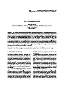
Results DELAYED RECTIFIER K+ CURRENT (IDR) Figure 1 illustrates whole-cell currents in THP-1 monocytes dominated by delayed rectifier currents, IDR. A family of voltage-clamp currents in Ringer’s solution is shown in Fig. 1A. The corresponding peak and steadystate current-voltage relations in Fig. 1B show that K+ currents in this cell were activated by depolarizing pulses positive to −40 mV. Activation was half maximal at −20 ± 4.4 mV (mean ± SD, 7 cells), determined by fitting a Boltzmann function (Materials and Methods) to the chord conductance-voltage data generated assuming a reversal potential of −86 mV. In cells studied with iso-
S.Y. Kim et al.: Ion Channels in THP-1 Monocytes
119
Fig. 1. Properties of IDR. (A) A family of superimposed delayed rectifier K+ currents in a THP-1 cell in Ringer’s solution, with KCl solution in the pipette. Depolarizing pulses 2 sec in duration were applied from a holding potential, Vhold, of −80 mV, with an interval of 23 sec between pulses. Illustrated pulses are from −60 to +60 mV in 10 mV increments (before correction for liquid junction potentials). Filter 2 kHz. (B) Current-voltage relationship for the cell in part A. Peak current (l) and current at the end of the 2-sec pulses (L) are shown, with the points connected by straight lines. (C) A family of voltage-clamp currents in a cell bathed in isotonic K+ Ringer’s solution. From Vhold 4 −80 mV, 4-sec pulses were applied in 10 mV increments at 47-sec intervals from −70 to +70 mV. Filter 2 kHz. (D) Peak current-voltage relationship for the experiment shown in C. (E) State dependent block of IDR by TEA+. Currents recorded during identical 4-sec pulses to +20 mV are superimposed. The indicated concentration of TEA+ (in mM) was added to the Ringer’s solution, with KMeSO3 in the pipette. The cell was ‘‘rested’’ for >60 sec before each pulse. Note that TEA+ slowed the decay of current, and that the control current (arrow) crosses over the others. (F) Block of IDR by ChTX. Superimposed are currents during 4-sec test pulses to +20 mV, with >60 sec at Vhold 4 −80 mV before each pulse.
tonic K+ Ringer’s in the bath (Fig. 1C), large inward K+ currents were observed between −50 mV and 0 mV and outward currents at more positive potentials. The peak current-voltage relationship in Fig. 1D for this experiment reversed near 0 mV in symmetrical K+ solutions, consistent with this conductance being K+ selective. The average reversal potential, Vrev, in 4.5 mM [K+]o was -85.3 ± 3.9 mV (mean ± SD, n 4 6 cells), measured using tail currents, and in 150 mM [K+]o was +1.6 ± 0.9 mV (n 4 5). The observed shift of 86.9 mV is close to the 88.6 mV calculated from the Nernst equation for a perfectly K+ selective channel.
tions. This slowing of inactivation has been interpreted to mean that open channel block by TEA+ interferes with channel inactivation (Grissmer & Cahalan, 1989a). A similar phenomenon was observed in alveolar epithelial K+ currents (Jacobs & DeCoursey, 1990). Bath application of 100 mM 4-AP abolished IDR (data not shown). Figure 1F shows inhibition by charybdotoxin, ChTX, a toxin extracted from Leiurus quinquestriatus scorpion venom. ChTX appeared to scale down the currents without affecting the kinetics, with a mean Ki of 1.7 ± 0.5 nM (n 4 3). Inactivation of IDR
Blockers +
Several K channel inhibitors were tested to compare the sensitivity of the delayed rectifier in THP-1 monocytes with that of K+ channels in other cells. Figure 1E shows inhibition by external TEA+. The peak current was reduced with Ki 4 12.5 ± 2.5 mM (mean ± SD, n 4 5). TEA+ slowed the inactivation of IDR with the result that the currents cross over with increasing TEA+ concentra-
Delayed rectifier K+ currents inactivated during long depolarizing pulses (Fig. 1A). Inactivation was substantial but not complete in Ringer’s solution during 2-sec pulses (open symbols in Fig. 1B), and appeared to be slower and less complete in K+ Ringer’s solution (Fig. 1C), consistent with effects of [K+]o on delayed rectifier K+ channels in other cells (Ruben & Thompson, 1984; Kostyuk & Martynyuk, 1988; Grissmer & Cahalan, 1989b; De-
120
S.Y. Kim et al.: Ion Channels in THP-1 Monocytes
Fig. 2. Inactivation of IDR. (A) Voltage dependence of prepulse inactivation. From Vhold 4 −80 mV, 4-sec prepulses to the potential on the abscissae were followed immediately by a test pulse to +60 mV. The inactivating current (i.e., the difference between peak and steady-state current) during this test pulse is plotted, normalized to the fitted maximum test current, 770 pA in this cell. The curve shows the fit to a Boltzmann (Materials and Methods) with midpoint V1/2 −24.1 mV and slope factor Vslope 4.15 mV. The cell was rested for 60 sec at Vhold before each pulse. Ringer’s solution in the bath, KCl in the pipette. (B) Accumulation of inactivation of delayed rectifier during repeated voltage ramps. After ‘‘resting’’ the membrane at −80 mV for at least 60 sec, the membrane potential was ramped from −120 to +100 mV at a rate of 716 mV/sec, and was held for 50 msec at Vhold 4 −80 mV and then 60 msec at −120 mV between successive ramps. The outward current was largest during the first ramp, and decreased progressively with repeated ramps. The obvious accumulation of inactivation was characteristic of IDR. (C) Voltage dependence of the time constant of inactivation in a typical cell in Ringer’s solution, with KCl in the pipette. The decay of K+ current during 4-sec depolarizing pulses was fitted with a single exponential. (D) Time course of recovery from inactivation in Ringer’s solution, with KCl in the pipette. From Vhold 4 −80 mV, pairs of 4-sec pulses to 0 mV were applied with various intervals between pulses. The amplitude of the peak current during the second pulse is plotted, after normalization to the peak current during the first pulse. The data were fitted by nonlinear least-squares to a single exponential to obtain the time constant of recovery, 20.5 sec in this experiment.
Coursey, 1990; Demo & Yellen, 1991). The apparent voltage dependence of inactivation of IDR was examined using 3–4 sec prepulses to various voltages followed by a test pulse to +40 or +60 mV. Figure 2A shows the test current amplitude in one cell, fitted with a Boltzmann function (Materials and Methods). The average midpoint was −35.7 ± 4.7 mV (mean ± SD, n 4 4). In experiments in which we wanted to eliminate IDR to observe other currents, we set Vhold at −20 mV. Inactivation accumulated during rapidly repeated depolarizing pulses or voltage ramps, suggesting that the rate of recovery from inactivation was slow. The superimposed ramp records in Fig. 2B illustrate the accumu-
lation of inactivation of IDR. This behavior was one criterion used to identify IDR. The time course of IDR inactivation during depolarizing pulses in Ringer’s solution (Fig. 1A) was fit by a single exponential with time constant, ti. Inactivation was faster at more positive voltages and ti was virtually independent of voltage at potentials above −10 mV (Fig. 2C). The average ti at +20 mV in 4.5 mM [K+]o was 334 ± 139 msec (mean ± SD, n 4 4), with KMeSO3 in the pipette and 579 ± 148 msec (n 4 9) with KCl. Effects of intracellular anions on ti of IDR in human T lymphocytes have been described previously (Cahalan et al., 1985). To examine the time course of recovery from inactivation, we used a
S.Y. Kim et al.: Ion Channels in THP-1 Monocytes
121
Fig. 3. Single-channel IDR current-voltage relationship in a cellattached patch. The pipette contained Mg2+-free KMeSO3. Currents elicited by voltage ramps were sorted according to whether a channel was open, and the average closed-channel current was subtracted from the average open-channel current. Channels like this one were identified as IDR by their voltage-dependent gating, inactivation (periods of many seconds in which no channels opened), rectification, and conductance consistent with IDR channels in other cells. In addition, cells in which IDR channels were identified in cell-attached patch configuration had macroscopic IDR in whole-cell configuration. The patch was held at RP −60 mV, and the voltage was ramped from RP −60 to RP + 160 mV at 0.44 V/sec. The bath contained Ringer’s solution, filter 1 kHz.
pulse protocol consisting of a pair of identical 4-sec voltage steps to 0 mV from a holding potential of −80 mV with variable intervals between pulses. The normalized amplitude of the second pulse is plotted in Fig. 2D as a function of the interval between pulses. The time constant of recovery from inactivation fit by first-order kinetics was t 4 21.2 ± 3.2 sec (mean ± SD, n 4 3). This much slower recovery than onset of inactivation is the cause of the characteristic accumulation of inactivation in cells with IDR. Single delayed rectifier channel currents were often observed in cell-attached patches of THP-1 monocytes. The net (leak-subtracted) single channel current-voltage relationship is plotted in Fig. 3. The unitary current rectifies inwardly, and actually decreases at large positive potentials, possibly due to block by an intracellular cation. The slope conductance at Vrev was roughly 20 pS in experiments with high [K+] pipette solutions. SMALL CONDUCTANCE Ca-ACTIVATED K+ CURRENT (ISK) Figure 4A illustrates whole-cell ramp currents in a THP-1 monocyte studied with a pipette solution in which Ca2+ was weakly buffered, with 0.1 mM EGTA and no added Ca2+. The only current evident in Ringer’s solution was IDR. Addition of ionomycin to the bath increased the conductance at all potentials. This calciumactivated conductance was evidently K+ selective be-
Fig. 4. (A) Ionomycin elicits ISK. The whole-cell current during a voltage ramp in Ringer’s solution in a cell containing KCl with no added Ca2+ and 0.1 mM EGTA shows only prominent IDR (which inactivated progressively during subsequent ramps). After addition of 2 mM ionomycin to the bath, a large conductance appeared which reversed near EK and exhibited little time dependence during voltage pulses, ISK. (B) Single-channel ISK currents in a cell-attached patch. The pipette contained KMeSO3, the bath K+ Ringer’s solution, with 10 mM A23187 added. The resting potential of the cell was presumably ‘‘clamped’’ near 0 mV by this solution. The holding potential was −40 mV (relative to the resting potential), the broken line indicates the current level when no channels were open. No channel openings were detected before adding A23187 to the bath.
cause it reversed near the K+ equilibrium potential, EK. To study the properties of this conductance more systematically, we used pipette solutions with [Ca2+] buffered to various levels. We use the term ISK advisedly, recognizing that small conductance Ca2+-activated K+ channels vary widely in their properties (Latorre et al., 1989); the term ‘‘intermediate’’ or IK has also been applied to ChTX-sensitive, apamin-insensitive channels (Varnai et al., 1993). In some cells, delayed rectifier currents could be seen in the presence of the ISK, but in others ISK was so large that it swamped out other currents which might have been present. Single-channel currents attributable to ISK channels are illustrated in Fig. 4B. In cell-attached patch configuration, there was initially no channel activity. When A23187 was added to the bathing solution, two channels appeared. The unitary conductance was roughly 20–25
122
S.Y. Kim et al.: Ion Channels in THP-1 Monocytes
potentials, but Vrev was near −80 mV, suggesting that a K+ selective conductance was active, rather than a simple leak. When the bath was changed to 150 mM K+ Ringer’s solution, Vrev was 0 mV, confirming the K+ selectivity of this conductance. In other experiments with 3–10 mM Ca2+ in the pipette solution, whole-cell currents usually reversed between −70 and −80 mV in Ringer’s solution and near 0 mV in K+ Ringer’s, both close to EK. Any nonselective leak conductance will bring Vrev in Ringer’s solution toward more positive values, and in a given cell a shift of Vrev towards 0 mV in Ringer’s solution was taken as an indication that the membrane was becoming damaged and nonselectively leaky. We did not pursue the alternative possibility that high [Ca2+]i also activates nonselective channels. Figure 5B and C illustrate ISK currents during voltage pulses. In general, there was no obvious or consistent time dependence. In the cell illustrated, there was a suggestion of turn-on during pulses to positive potentials, but in other cells this was not observed and sometimes partial decay occurred. Because it was not possible to be sure in any given cell that the entire conductance was due to ISK alone, we are reluctant to ascribe much significance to the weak and variable evidence of time dependence, and therefore consider ISK to be practically time independent. A consistent feature of ISK however, was the reduced conductance at large positive potentials, apparent both during ramps (Fig. 5A) and during pulses in both Ringer’s and K+ Ringer’s solution (Fig. 5B and C, respectively). This behavior is strongly reminiscent of voltage-dependent ionic blockade by intracellular cations. The only cation other than K+ in the pipette solution was 2 mM Mg2+, which produces similarly weak block of outward current through a number of K+ channels. Whether Mg2+ block causes the observed rectification was not explored.
Fig. 5. (A) ISK during voltage ramps in the same cell in Ringer’s solution (4.5 K+) and K+ Ringer’s (150 K+), with 10 mM [Ca2+] KCl in the pipette. Note the pronounced inward rectification at depolarized potentials. The time-independence of ISK can be seen in families of currents during voltage pulses in Ringer’s solution (B) and in K+ Ringer’s solution (C) in another cell studied with the same pipette solution. Voltages were incremented in 20-mV steps between the indicated extremes. Vhold was −80 mV in Ringer’s and 0 mV in K+ Ringer’s solution; arrows indicate zero current.
pS. Similar currents were seen in inside-out patches exposed to elevated [Ca2+]. ISK at Different [K+]o In the whole-cell experiment illustrated in Fig. 5A, the pipette contained 10 mM free Ca2+. In Ringer’s solution (4.5 mM K+) the membrane conductance was high at all
[Ca2+]i Dependence of ISK ISK was obvious in cells studied at 10 mM [Ca2+]i, and was also seen in many cells at lower nominal [Ca2+]i. The average slope conductance in Ringer’s solution at Vrev was 2.3 ± 1.8 nS (mean ± SD, n 4 21) at 10 mM [Ca2+]i, 1.8 ± 1.3 nS (n 4 11) at 3 mM [Ca2+]i, and 0.5 ± 0.5 nS (n 4 3) at 1 mM [Ca2+]i. With nominally 38 nM [Ca2+]i (Table, KCl pipette solution) small inward currents resembling ISK were observed in only 7 of 37 cells and had an average amplitude of 0.24 nS in those cells. With pipette solutions of 1 or 3 mM [Ca2+], the amplitude of ISK appeared to be somewhat variable with time in some cells, presumably related to changes in the access resistance, i.e., the patency of the pipette tip. Accurate determination of the [Ca2+]i dependence of ISK would require a more effective Ca2+ buffer than EGTA or an independent estimate of [Ca2+]i. Nevertheless, it is ap-
S.Y. Kim et al.: Ion Channels in THP-1 Monocytes
123
Table. Composition of solutions Name
K+
Ca2+
Mg2+
Cl−
−
MeSO3
EGTA
pH
11 10 11 10a
7.2 7.2 7.2 7.2
HEPES
Pipette solutions (mM) KCl KCl, 10mM Ca2+ KMeSO3 KMeSO3, Mg2+ free
167 162 161 167
1 9.65 1 2
2 2 2 0
146 143.3 6 4
0 0 133 120
10 10 10 10
External solutions (mM) Name
K+
Ringer’s K+ Ringer’s KMeSO3 d + X Ringer’s
4.5 150 150 164
Na+ 149 0 0 0
Ca2+ 2 2 2 2
Mg2+ 1 1 0 1
Cl− 150.5 151 4 166
MeSO−3 0 0 150 0
pH 7.4 7.4 7.0 7.4
buffer 5b 10b 20c 10b
Includes also 10 mM EDTA. The final K+ concentration after titrating the pH with KOH is given. CsCl, NaCl, and TMACl pipette solutions were identical to KCl except all of the K+ was replaced with the appropriate cation. b HEPES, c BES, d X+ means any cation (Rb+, TMA+, Na+, Li+, Cs+, NH+4, TEA+). See Materials and Methods for a description of the Tl+ Ringer’s solution.
a
parent that ISK is activated by elevated [Ca2+]i. The data are generally consistent with [Ca2+]i sensitivity of ISK channels in human T lymphocytes or HL-60 cells, which are maximally activated at ∼1 mM [Ca2+]i (Grissmer, Nguyen & Cahalan, 1993; Varnai et al., 1993). Selectivity of ISK The selectivity of ISK was explored by applying voltage ramps and changing the cation in the bath solution, with a 10 mM [Ca2+]i K+ solution in the pipette (data not shown). We assumed that the main conductance under these conditions was ISK. For K+, Rb+, and Cs+ measurements Vhold was set at 0 mV to inactivate IDR, and for less permeant ions with quite negative Vrev, IDR would not be active near Vrev anyway. The permeability to a specific cation, relative to K+, PX/PK, was calculated for these bi-ionic conditions from the Goldman-HodgkinKatz voltage equation (Hille, 1992). Best estimates for the relative permeabilities are: Tl+ . K+ . Rb+ . NH4+ . Cs+ . Li+ ù Na+ . TMA+, TEA+. 1.77 1.0 0.89 0.22 0.094 ø0.005 ø0.004 ,0.001 ,0.001
The measured value for Vrev, particularly for the weakly permeant ions, may be erroneously positive due to any leak current which is present. The PX values given are based on 1 to 6 measurements, but each represents the average of the two values with the lowest PX, because we did not correct for leak current. Leak current would lead to an overestimate of PX. The sequence should be correct, unless the leak itself is selective, because it is based on comparisons within individual cells. For example,
studied in the same cell Vrev was distinctly more negative in TMA+ or TEA+ solutions than in Na+ or Li+. Pharmacological Sensitivity of ISK The traditional K+ channel blocker tetraethylammonium, TEA+, was moderately effective for ISK. Fig. 6A illustrates a family of ISK currents in Ringer’s solution. Addition of 30 mM TEA+ (Fig. 6B) appeared to scale down the current uniformly at all potentials. In the presence of 30 mM TEA+ ISK was evidently not completely blocked, because Vrev was still near EK. This is more apparent in Fig. 6C, which shows the dose-response relationship for TEA+ using voltage ramps. Without correcting for leak current, Ki was estimated to be 10–25 mM in Ringer’s solution in two cells. Figure 6D and E illustrate that ChTX inhibits ISK, like TEA+, without obvious voltage dependence. The dose-response relationship for ChTX using voltage ramps (Fig. 6F) indicates that this toxin is a potent inhibitor of ISK, although it is a more potent blocker of IDR (Fig. 1F). The Ki estimated for ChTX block of ISK was 4–12 nM in Ringer’s solution (n 4 2) and 10–30 nM in K+ Ringer’s solution (n 4 2). Figure 7 illustrates block of ISK in K+ Ringer’s solution by TEA+ (A), Cs+ (B), and Ba2+ (C). No timedependence was detected during voltage pulses for any blockers of ISK, so the voltage dependence seen in ramps is a reasonable indication of steady-state behavior. As in Ringer’s solution, TEA+ added to K+ Ringer’s solution produced practically voltage-independent block (Fig. 7A). Half block of ISK in K+ Ringer’s solution by TEA+ was estimated in two cells at several potentials to be 15–25 mM. Onset and removal of block occurred as rap-
124
S.Y. Kim et al.: Ion Channels in THP-1 Monocytes
Fig. 6. Block of ISK by TEA+ and ChTX in Ringer’s solution. (A) ISK elicited by voltage pulses, in 20 mV increments from −120 to +100 mV from Vhold 4 −80 mV. The zero current potential was near Vhold here and in B. Pipette solution 10 mM [Ca2+] KCl, Vhold 4 −80 mV. (B) The identical pulse family in the presence of 30 mM TEA+. (C) Currents during voltage ramps from −120 to +100 mV in the same cell in the presence of the indicated concentrations of TEA+, substituted for Na+. Vhold 4 −80 mV. (D) A family of currents during voltage pulses in a different cell studied with 10 mM [Ca2+]i KCl in the pipette. Pulses were applied in 20 mV increments between −120 and +20 mV. Arrow indicates zero current level, which was near Vhold in E. (E) Currents during pulses identical with D applied in the presence of 30 nM ChTX. (F) Block of ISK currents by ChTX in the same cell. Ramps from −120 to +100 mV from Vhold 4 −80 mV.
idly as the solutions were changed (on the order of 10 sec). For Cs+ and Ba2+, block was much greater at more negative potentials. The outward current was reduced in an apparently voltage-independent manner, but this inhibition appeared qualitatively different from the voltage-dependent block of inward currents. Voltagedependent block of inward current occurred immediately when Cs+ or Ba2+ was added, whereas outward currents were reduced gradually and progressively over tens of seconds after the bath change was complete. As the outward current was reduced, there was also a comparable scaling down of inward currents. The illustrated ramp currents were recorded 1–2 min after each solution change. Upon washout, the voltage-dependent block was immediately removed, but the scaling down of the conductance was reversed more slowly, and not always completely. The impression received was that some
slow process modulated the overall level of activation of the ISK conductance, in addition to the specific reversible voltage-dependent block by Cs+ or Ba2+ of the open channel. The voltage-dependent ionic blockade of ISK by Cs+ or Ba2+ was rather weak, being distinctly incomplete at moderate negative voltages even at 150 mM Cs+ or 100 mM Ba2+. External 4-AP at 1 mM and apamin at 1 mM had no effect on ISK. NONSELECTIVE CATION CURRENT (Icat) A time-independent outward current (Icat) was observed in addition to the inactivating outward K+ current in some cells. Figure 8A shows both Icat and IDR elicited by depolarizing voltage steps from a holding potential of −80
S.Y. Kim et al.: Ion Channels in THP-1 Monocytes
125
Fig. 7. Block of ISK by TEA+, Cs+, and Ba2+ in a cell studied in K+ Ringer’s solution, with 10 mM Ca2+ KCl in the pipette. All blockers were added from 1 M stock solutions to K+ Ringer’s solution, resulting in some dilution of its constituents (e.g., by 10% for 100 mM blocker), as well as hypertonicity. (A) ISK during voltage ramps from +80 to −140 mV in the indicated concentrations of TEACl added to K+ Ringer’s solution. Block and recovery from block of both inward and outward currents occurred rapidly during solution changes. (B) ISK in the presence of the indicated concentrations of Cs+. Both block and unblock were rapid for the voltage-dependent component of block, but slower for the voltage-independent scaling down of both inward and outward currents. (C) ISK in the indicated concentrations of Ba2+. The ISK current recovered only half its original amplitude after washout of high Ba2+ concentrations. The ramps in Ba2+ were done before those in TEA+ (A) or Cs+ (B) thus their control currents are smaller. Illustrated ramps currents were recorded 1–2 min after each solution change. Note the pronounced enhancement of block at more negative voltages.
mV. When Vhold was −30 mV (Fig. 8B) IDR was inactivated and only the time-independent Icat remained. Icat was evidently responsible for most of the current at the end of long depolarizing pulses. In some cells (e.g., Fig. 1A) little current remained after IDR had inactivated, but in others (e.g., Fig. 8A and B) Icat was prominent. Icat was small immediately after establishing wholecell configuration, becoming larger over several minutes. Figure 8C illustrates the development of Icat in a THP-1 monocyte studied with NaCl in the pipette. Whole-cell currents during voltage ramps are superimposed, measured at several times after establishing whole-cell configuration. At 2 min there was only a small linear leak current. By 10 min there was pronounced Icat, with its characteristic steep voltage dependence, activating above +40 mV. The amplitude increased further over the next 8 min. When voltage pulses were applied in this cell (not shown), the currents exhibited little time-dependence and the same voltage dependence as in the ramps. Large Icat was observed in cells studied with K+ (Fig. 8A and B), Cs+ (Fig. 9A-C), or Na+ (Fig. 9D-F) in the pipette solution, but not TMA+ or TEA+ (not shown). The identity of the extracellular cation (Na+, K+, Cs+, TEA+, TMA+) had no obvious effect on these currents. Replacing external Cl− with MeSO−3 did not reduce Icat, which thus appears to be selective for small cations. The following ion channel inhibitors, applied externally, did not inhibit Icat: 1 mM Zn2+, >100 mM TEA+, 1 mM 4-AP, 100 nM ChTX, 5 mM Ba2+, 1 mM Gd2+, 1 mM SITS, 100 mM amiloride. No inhibitor of Icat has been found.
CHLORIDE CURRENTS (ICl) Chloride currents, ICl, were often present in THP-1 cells shortly after whole-cell configuration was established, but ran down progressively over a few minutes or tens of minutes. The typical appearance of ICl is illustrated in Fig. 10A. Whole-cell currents during voltage ramps are plotted in normal Ringer’s solution, and in low Cl− Ringer’s solution. Replacing the external Cl− with MeSO−3 greatly reduced the outward current and shifted the zero current potential to more positive potentials. These changes were reversible. Some outward rectification persisted in symmetrical MeSO−3 solutions, suggesting that MeSO−3 carries some current through Cl− channels. No time- or voltage-dependent gating was evident at moderate potentials in some cells, but in other cells the outward current decayed at large positive potentials. Block Figure 10B illustrates block of ICl by 1 mM SITS (4acetamido-48-isothiocyanostilbene-2,28-disulfonic acid). In this experiment the pipette contained TMACl, thus the outward rectification cannot be attributed to asymmetrical Cl− concentrations and must reflect a property of the gCl. Addition of SITS decreased both inward and outward currents. Block by SITS was not reversible. Because of the temporal changes in gCl and the difficulty of distinguishing it from leak current, we did not attempt to quantify the amplitude of ICl.
126
S.Y. Kim et al.: Ion Channels in THP-1 Monocytes
was not obviously voltage dependent at moderate potentials. In one inside-out patch, outward single-channel currents persisted when Na+ replaced K+ in the bath, but substituting MeSO−3 for Cl− reduced the conductance from 350 pS to 210 pS. PROTON CURRENT (IH) Voltage-activated H+ selective currents were observed in THP-1 cells before and after differentiation. The properties and expression of IH are described in the next paper (DeCoursey & Cherny, 1996). Discussion DELAYED RECTIFIER K+ CURRENT (IDR)
Fig. 8. Family of currents in a cell containing KCl pipette solution and bathed in Ringer’s solution, exhibiting both IDR and nonselective cation current, Icat. In A the holding potential was −80 mV and both currents are seen, IDR inactivating and Icat time-independent. When Vhold was set at −30 mV (B) the same pulses elicited only Icat. In both the pulses were in 20 mV increments from −60 to +100 mV. (C) Development of Icat with time in a different cell. Currents during voltage ramps are superimposed for the indicated times (min) after establishing whole-cell configuration. Several consecutive ramp currents were averaged for each record. The bath contained Ringer’s solution, and the pipette NaCl.
The delayed rectifier in THP-1 monocytes, IDR, activated with depolarization above −50 mV, inactivated during sustained depolarizations with a ti ∼300–600 msec (faster with MeSO−3 than Cl− pipette solutions), recovered slowly from inactivation with t ∼21 sec, and was inhibited by external TEA+ (Ki 12.5 mM) and ChTX (Ki 1.7 nM). Single IDR channels had a conductance of ∼20 pS at Vrev in symmetrical high [K+]. IDR in THP-1 monocytes thus resembles that in other human and mouse macrophages (Ypey & Clapham, 1984; Gallin & Sheehy, 1985; Randriamampita & Trautmann, 1987; Nelson, Jow & Popovich, 1990b), although to our knowledge ChTX block of IDR has not been previously demonstrated in macrophages, and only recently in microglia (Eder et al., 1995). The properties, especially the ChTX sensitivity and inactivation kinetics, of the delayed rectifier K+ channel in THP-1 cells are virtually identical with those of type ‘n’ K+ channels in rat alveolar epithelial cells (DeCoursey, Jacobs & Silver, 1988; Jacobs & DeCoursey, 1990; DeCoursey, 1990) and in human and murine T lymphocytes (Cahalan et al., 1985; Deutsch, Krause & Lee, 1986; DeCoursey et al., 1987; Sands, Lewis & Cahalan, 1989). In a companion study we use the polymerase chain reaction (PCR) to show that Kv1.3 mRNA is present in THP-1 cells and suggest that it codes for IDR (DeCoursey et al., 1996).
Large Anion Channels
SMALL CONDUCTANCE Ca-ACTIVATED K+ CURRENT (ISK)
In rare instances we observed large unitary currents which behaved differently than IBK, the high-conductance voltage- and [Ca2+]i-activated K+ channels described in the third paper in this series (DeCoursey et al., 1996). These channels were observed too infrequently to allow complete characterization. Their open probability
Selectivity The selectivity sequence found for ISK is similar to that of many types of K+ channels (Hille, 1992), and is consistent with the relative permeability of ISK channels in human erythrocytes (Christophersen, 1991) and in hu-
S.Y. Kim et al.: Ion Channels in THP-1 Monocytes
127
Fig. 9. Nonselective cation currents in cells perfused with CsCl (A–C) or NaCl (D–F) pipette solutions, all in Ringer’s solution. Illustrated for each cell is a family of currents during voltage pulses between −80 and +100 mV (A and D), the corresponding current-voltage relationship (B and E), and the current during a voltage ramp (C and F). Vhold was 0 mV in both experiments; in A and D the holding current was near zero.
man T lymphocytes (Grissmer et al., 1993). Cs+ was clearly permeant through ISK in THP-1 cells. The biionic reversal potentials in both Li+ and Na+ were clearly positive to those in TMA+ or TEA+ when measured in the same cell, thus Li+ and Na+ are detectably permeant as well. Block ISK was blocked by ChTX and by TEA+, in both cases somewhat less potently than was IDR. Block of ISK by both drugs appeared to be voltage independent. The slightly more potent block of IDR than ISK by both TEA+ and ChTX is quite similar to the pattern observed in activated human B lymphocytes (Partiseti et al., 1992); in activated T lymphocytes IDR was more sensitive than ISK to TEA+ but was equipotently blocked by ChTX (Grissmer et al., 1993). Both Cs+ and Ba2+ produced distinctly voltage-dependent block of ISK, but were rela-
tively weak in comparison with their potency for blocking inward rectifier K+ channels in THP-1 cells (DeCoursey et al., 1996). Thus, block of ISK was not quite complete at 100 mM Ba2+, and was clearly incomplete at 30 mM Ba2+. In 150 mM Cs+ there was still substantial inward and outward current. Similarly, in human macrophages ISK was blocked in a voltage-dependent manner by Ba2+ and more weakly than was IIR (Gallin, 1989). Gating ISK appeared to be largely time- and voltage-independent. Sometimes there was a suggestion that the current turned on or off at certain potentials, but this behavior was neither consistent from cell-to-cell nor was it always convincingly distinguishable from poor capacity compensation or contamination from other ion channels which may have been present. However, the ISK current consistently was reduced at large positive potentials.This
128
S.Y. Kim et al.: Ion Channels in THP-1 Monocytes
other types of channels contaminate the data in some cells, or that ISK is influenced by additional, unidentified factors. A voltage- and time-independent K+ conductance induced by G protein activators in murine macrophages and J774 cells generally resembles ISK but is not blocked by ChTX (McKinney & Gallin, 1992). This conductance however, has been shown to appear spontaneously at high [Ca2+]i in the absence of receptor stimulation (Fan & McCloskey, 1994) and therefore might conceivably have contaminated ISK measurements in some cells in our study, because ChTX block was not tested in every cell. This G protein-related K+ conductance rectifies outwardly, whereas ISK usually rectified inwardly. Occasional cells in which K+ selective currents at high [Ca2+]i that rectified outwardly were observed may have expressed both types of K+ channels. The G protein-related K+ channel was described in murine macrophages and J774 cells (McKinney & Gallin, 1992; Fan & McCloskey, 1994), but to our knowledge has not been reported previously in human macrophages. NONSELECTIVE CATION CURRENT (Icat)
Fig. 10. (A) Whole-cell chloride currents during voltage ramps. The record labeled ‘Cl−’ was recorded in Ringer’s solution, the smaller trace is the average of several currents recorded before and after in ‘‘Cl-free’’ Ringer’s solution, identical to Ringer’s but with all but 6 mM of the Cl− replaced by MeSO−3. Recorded 6 min after whole-cell configuration was established. Pipette solution KMeSO3. (B) Block of ICl by SITS. Whole-cell ICl during a voltage ramp in the absence and presence of 1 mM SITS. Pipette solution TMACl, bath Ringer’s solution. Ramps were applied from −120 to +100 mV with 5 sec at Vhold 4 0 mV between successive ramps.
reduction produced a region of negative slope conductance in some cells. Presence in Other Monocytes A similar ISK channel has been described in human macrophages (Ince et al., 1987; Kakuta et al., 1988; Gallin, 1989), murine macrophages (Randriamampita & Trautmann, 1987; Hara et al., 1990), HL-60 cells (Wieland et al., 1992; Varnai et al., 1993), human B lymphocytes and rat thymocytes (Mahaut-Smith & Schlichter, 1989), and activated human T lymphocytes (Grissmer et al., 1993). The extent of inward rectification of the ISK currentvoltage relationship is somewhat variable from one study to another; however, we observed substantial cell-to-cell variability in this property. This variability may indicate that multiple types of ISK channels are involved, that
A nonselective cation current, Icat, was present in some THP-1 cells. This current was time-independent and became pronounced at potentials above +20 mV. Icat was not affected by holding the cell at depolarized potentials which inactivated IDR. No blockers were found. An apparently similar nonselective cation current has been described in corneal endothelial cells (Watsky, 1995). Although a variety of nonselective currents have been reported at the single-channel level (reviewed by Gallin, 1991), to our knowledge no comparable conductance has been described in macrophages. CHLORIDE CURRENT (ICl) We observed outwardly-rectifying Cl− currents early in whole-cell experiments, which disappeared over several minutes. Generally similar macroscopic Cl− currents have been reported in human monocyte-derived macrophages (Nelson, Jow & Jow, 1990a). In human neutrophils, an outwardly-rectifying Cl− conductance is activated by cell swelling (Stoddard, Steinbach & Simchowitz, 1993). Although we did not explore its sensitivity to osmotic stress, it is possible that ICl in THP-1 cells disappeared with time as a transient osmotic imbalance equilibrated. Large conductance voltage-dependent Cl− channels have been described in murine macrophages (Schwarze & Kolb, 1984; Randriamampita & Trautmann, 1987) and in other cells. Generally similar large anion channels were observed infrequently in THP-1 cells. The macroscopic ICl described here exhibited neither voltage-dependence nor discrete current levels of comparable size, and thus must be due to a different
S.Y. Kim et al.: Ion Channels in THP-1 Monocytes
channel. Smaller conductance single Cl− channels have been reported in human U937 monocytes (Kanno & Takishima, 1990). A SITS-sensitive anion conductance contributes to regulatory volume decrease in THP-1 cells challenged with hypotonic solutions (Gallin et al., 1994). The authors gratefully acknowledge a critique of the manuscript by Leslie C. McKinney, and the technical assistance of Donald R. Anderson. This project was supported at various stages by the Division of Pulmonary Medicine (SK), a Parker B. Francis Award (MS), a Grantin-Aid from the American Heart Association (TD), and by National Institutes of Health grants R01-HL37500, R01-HL52671, and Research Career Development Award KO4-1928 (TD).
References Auwerx, J. 1991. The human leukemia cell line, THP-1: a multifaceted model for the study of monocyte-macrophage differentiation. Experientia 47:22–31 Cahalan, M.D., Chandy, K.G., DeCoursey, T.E., Gupta, S. 1985. A voltage-gated potassium channel in human T lymphocytes. J. Physiol. 358:197–237 Christophersen, P. 1991. Ca2+-activated K+ channel from human erythrocyte membranes: single channel rectification and selectivity. J. Membrane Biol. 119:75–83 Crutchley, D.J., Conanan, L.B., Que, B.G. 1995. K+ channel blockers inhibit tissue factor expression by human monocytic cells. Circ. Res. 76:16–20 DeCoursey, T.E. 1990. State-dependent inactivation of K+ currents in rat type II alveolar epithelial cells. J. Gen. Physiol. 95:617–646 DeCoursey, T.E., Chandy, K.G., Gupta, S., Cahalan, M.D. 1987. Two types of potassium channels in murine T lymphocytes. J. Gen. Physiol. 89:379–404 DeCoursey, T.E., Cherny, V.V. 1996. Voltage-activated proton currents in human THP-1 monocytes. J. Membrane Biol. 152:2 DeCoursey, T.E., Jacobs, E.R., Silver, M.R. 1988. Potassium currents in rat type II pulmonary alveolar epithelial cells. J. Physiol. 395:487–505 DeCoursey, T.E., Kim, S.Y., Silver, M.R., Quandt, F.N. 1996. Ion channel expression in PMA-differentiated human THP-1 macrophages. J. Membrane Biol. 152:2 Demo, S.D., Yellen, G. 1991. The inactivation gate of the Shaker K+ channel behaves like an open-channel blocker. Neuron 7:743–753 Deutsch, C., Krause, D., Lee, S.C. 1986. Voltage-gated potassium conductance in human T lymphocytes stimulated with phorbol ester. J. Physiol. 372:405–423 Eder, C., Fischer, H.-G., Hadding, U., Heinemann, U. 1995. Properties of voltage-gated currents of microglia developed using macrophage colony-stimulating factor. Pfluegers Arch. 430:526–533 Fan, Y., McCloskey, M.A. 1994. Dual pathways for GTP-dependent regulation of chemoattractant-activated K+ conductance in murine J774 macrophages. J. Biol. Chem. 269:31533–31543 Gallin, E.K. 1989. Evidence for a Ca-activated inwardly rectifying K channel in human macrophages. Am. J. Physiol. 257:C77–C85 Gallin, E.K. 1991. Ion channels in leukocytes. Physiol. Rev. 71:775– 811 Gallin, E.K., Mason, T.M., Moran, A. 1994. Characterization of regulatory volume decrease in the THP-1 and HL-60 human myelocytic cell lines. J. Cell. Physiol. 159:573–581 Gallin, E.K., Sheehy, P.A. 1985. Differential expression of inward and outward potassium currents in the macrophage-like cell line J774.1. J. Physiol. 369:475–499
129 Grissmer, S., Cahalan, M.D. 1989a. TEA prevents inactivation while blocking open K+ channels in human T lymphocytes. Biophys. J. 55:203–206 Grissmer, S., Cahalan, M.D. 1989b. Divalent ion trapping inside potassium channels of human T lymphocytes. J. Gen. Physiol. 93:609–630 Grissmer, S., Nguyen, A.N., Cahalan, M.D. 1993. Calcium-activated potassium channels in resting and activated human T lymphocytes: expression levels, calcium dependence, ion selectivity, and pharmacology. J. Gen. Physiol. 102:601–630 Grygorczyk, R., Rodger, I.W. 1993. Potassium channels in THP-1 human monocytes. Biophys. J. 64:A200 (Abstr.) Hamill, O.P., Marty, A., Neher, E., Sakmann, B., Sigworth, F.J. 1981. Improved patch-clamp techniques for high-resolution current recording from cells and cell-free membrane patches. Pfluegers Arch. 391:85–100 Hara, N., Ichinose, M., Sawada, M., Imai, K., Maeno, T. 1990. Activation of single Ca2+-dependent K+ channel by external ATP in mouse macrophages. FEBS Lett. 267:281–284 Hille, B. 1992. Ionic Channels of Excitable Membranes. Sinauer Associates, Sunderland, MA Ince, C., Van Duijn, B., Ypey, D.L., Van Bavel, E., Weidema, F., Leijh, P.C.J. 1987. Ionic channels and membrane hyperpolarization in human macrophages. J. Membrane Biol. 97:251–258 Jacobs, E.R., DeCoursey, T.E. 1990. Mechanisms of potassium channel block in rat alveolar epithelial cells. J. Pharmacol. Exp. Ther. 255:459–472 Kakuta, Y., Okayama, H., Aikawa, T., Kanno, T., Ohyama, T. Sasaki, H.T., Kato, T., Takishima, T. 1988. K channels of human alveolar macrophages. J. Allergy Clin. Immunol. 81:460–468 Kanno, T., Takishima, T. 1990. Chloride and potassium channels in U937 human monocytes. J. Membrane Biol. 116:149–161 Kim, S.Y., DeCoursey, T.E., Silver, M.R. 1994. Ion channels in the human macrophage cell line THP-1. Biophys. J. 66:A328. (Abstr.) Kim, S.Y., DeCoursey, T.E., Cherny, V.V., Silver, M.R. 1995. Altered ion channel expression during PMA-induced differentiation of THP-1 monocytes. Biophys. J. 68:A44. (Abstr.) Kostyuk, P.G., Martynyuk, A.E. 1988. Potassium outward current dependent on extracellular calcium in snail neuronal membrane. Neurosci. 24:1081–1087 Latorre, R., Oberhauser, A., Labarca, P., Alvarez, O. 1989. Varieties of calcium-activated potassium channels. Ann. Review Physiol. 51:385–399 Mahaut-Smith, M.P., Schlichter, L.C. 1989. Ca2+-activated K+ channels in human B lymphocytes and rat thymocytes. J. Physiol. 415:69–83 Martell, A.E., Smith, R.M. 1974. Critical Stability Constants. Volume 1: Amino Acids. Plenum Press, New York McKinney, L.C., Gallin, E.K. 1992. G-protein activators induce a potassium conductance in murine macrophages. J. Membrane Biol. 130:265–276 Nelson, D.J., Jow, B., Jow, F. 1990a. Whole cell currents in macrophages: I. Human monocyte-derived macrophages. J. Membrane Biol. 117:29–44 Nelson, D.J., Jow, B., Popovich, K.J. 1990b. Whole cell currents in macrophages: II. Alveolar macrophages. J. Membrane Biol. 117:45–55 Partiseti, M., Choquet, D., Diu, A., Korn, H. 1992. Differential regulation of voltage- and calcium-activated potassium channels in human B lymphocytes. J. Immunol. 148:3361–3368 Pelassy, C., Cattan, N., Aussel, C. 1992. Changes in phospholipid metabolism induced by quinine, 4-aminopyridine and tetraethylammonium in the monocytic cell line THP1. Biochem. J. 282:443–446
130 Ramdriamampita, C., Trautmann, A. 1987. Ionic channels in murine macrophages. J. Cell Biol. 105:761–769 Ruben, P., Thompson, S. 1984. Rapid recovery from K current inactivation on membrane hyperpolarization in molluscan neurons. J. Gen. Physiol. 84:861–875 Sands, S.B., Lewis, R.S., Cahalan, M.D. 1989. Charybdotoxin blocks voltage-gated K+ channels in human and murine T lymphocytes. J. Gen. Physiol. 93:1061–1074 Schwarze, W., Kolb, H.A. 1984. Voltage dependent kinetics of an anionic channel of large unit conductance in macrophages and myotube membranes. Pfluegers Arch. 402:281–291 Stoddard, J.S., Steinbach, J.H., Simchowitz, L. 1993. Whole cell Cl− currents in human neutrophils induced by cell swelling. Am. J. Physiol. 265:C156–C165 Tsuchiya, S., Yamabe, M., Yamaguchi, Y., Kobayashi, Y., Konno, T., Tada, K. 1980. Establishment and characterization of a human
S.Y. Kim et al.: Ion Channels in THP-1 Monocytes acute monocytic leukemia cell line (THP-1). Int. J. Cancer 26:171– 176 Varnai, P., Demaurex, N., Jaconi, M., Schlegel, W., Lew, D.P., Krause, K.H. 1993. Highly co-operative Ca2+ activation of intermediateconductance K+ channels in granulocytes from a human cell line. J. Physiol 472:373–390 Watsky, M.A. 1995. Nonselective cation channel activation during wound healing in the corneal endothelium. Am. J. Physiol. 268:C1179–C1185 Wieland, S.J., Gong, Q-H., Chou, R.H., Brent, L.H. 1992. A lineagespecific Ca2+-activated K+ conductance in HL-60 cells. J. Biol. Chem. 267:15426–15431 Ypey, D.L., Clapham, D.E. 1984. Development of a delayed outwardrectifying K+ conductance in cultured mouse peritoneal macrophages. Proc. Natl. Acad. Sci. USA 81:3080–3087