JOURNAL OF BACTERIOLOGY, Aug. 2007, p. 5566–5573 0021-9193/07/$08.00⫹0 doi:10.1128/JB.00483-07 Copyright © 2007, American Society for Microbiology. All Rights Reserved.
Vol. 189, No. 15
IcsA Surface Presentation in Shigella flexneri Requires the Periplasmic Chaperones DegP, Skp, and SurA䌤 Georgiana E. Purdy,† Carolyn R. Fisher, and Shelley M. Payne* Institute for Cellular and Molecular Biology and Section of Molecular Genetics and Microbiology, The University of Texas at Austin, Austin, Texas 78712 Received 30 March 2007/Accepted 10 May 2007
A Shigella flexneri degP mutant, which was defective for plaque formation in Henle cell monolayers, had a reduced amount of IcsA detectable on the bacterial surface with antibody. However, the mutant secreted IcsA to the outer membrane at wild-type levels. This suggests that IcsA adopts an altered conformation in the outer membrane of the degP mutant with reduced exposure on the cell surface. IcsA is, therefore, unlikely to be accessible to actin-nucleating proteins within the eukaryotic cell cytoplasm, which is required for bacterial movement within the host cell and cell-to-cell spread. The degP mutant was somewhat more sensitive to detergents, antibiotics, and the antimicrobial peptide magainin, indicating that the degP phenotype was not limited to IcsA surface presentation. The plaque defect of the degP mutant, which is independent of DegP protease activity, was suppressed by overexpression of the periplasmic chaperone Skp but not by SurA. S. flexneri skp and surA mutants failed to form plaques in Henle cell monolayers and were defective in cell surface presentation and polar localization of IcsA. Therefore, the three periplasmic folding factors DegP, Skp, and SurA were all required for IcsA localization and plaque formation by S. flexneri. Shigella flexneri is a gram-negative facultative intracellular pathogen that causes bacillary dysentery. Important aspects of Shigella pathogenesis are the ability of the bacteria to invade colonic epithelial cells by bacterial-induced phagocytosis, lyse the phagocytic vesicle, multiply within the cytosol, and subsequently spread to adjacent cells by polymerizing the eukaryotic cell actin (38). The ability of Shigella to spread from cell to cell requires the expression and polar surface localization of IcsA (VirG), a 110-kDa outer membrane protein (OMP) encoded on the 220-kb virulence plasmid. Inside the host cell, the amino-terminal domain of IcsA is exposed on the bacterial surface and interacts with the eukaryotic proteins vinculin and neural Wiskott-Aldrich syndrome protein to nucleate assembly of F-actin tails that propel the bacterium through the host cytosol and into adjacent cells (44, 45). icsA mutants do not form plaques in Henle cell monolayers, and defects in polar IcsA localization also result in either reduced plaque size or an inability to form plaques in cultured cell monolayers (3, 24, 25). IcsA is a member of the autotransporter family of outer membrane proteins, which includes several other bacterial virulence factors (16). Autotransporters are believed to mediate their own translocation to the outer membrane without periplasmic chaperones. The carboxy-terminal domain of the protein forms a -barrel in the outer membrane, through which the amino-terminal portion, or “passenger domain,” is transported and exposed on the bacterial surface (10, 11). IcsA is secreted across the inner membrane by the Sec secretion apparatus; it transits the periplasm and inserts itself into the
outer membrane (5). In actively dividing bacteria, IcsA is localized to the old pole of the bacillus (11, 12). Although the mechanism for this localization is unclear, most evidence indicates that IcsA inserts directly at the pole (10, 11, 42). In an investigation of S. flexneri virulence factors, a number of mutations were identified that affect proper IcsA localization and/or intercellular spread (18, 19). Some mutations that affect the lipopolysaccharide (LPS) biosynthesis pathway result in mislocalization of IcsA and in the inability to form wild-type plaques in Henle cell monolayers (19, 36, 37). Smooth LPS consists of three regions: lipid A, core oligosaccharide, and the serotype-specific O-antigen repeating molecule. The S. flexneri rfaL mutant lacks O-antigen side chains and has a rough LPS phenotype. In an rfaL mutant, IcsA is distributed over the entire bacterial surface, and the mutant forms either pinpoint plaques or no plaques in tissue culture monolayers (19, 36). Some S. flexneri 2a strains have two modal lengths of O-antigen repeats: the 11- to 17-repeat mode, determined by rol or wzz (27), and the ⬎90-repeat mode determined by cld (43). The very long O-antigen LPS side chains increase serum resistance but also mask IcsA, and the ratio of short and very long chains is important (26). We reported previously that an S. flexneri degP mutant also has a defect in the surface expression of IcsA, resulting in a smallplaque phenotype (32). DegP is a member of the HtrA family of proteases, which are highly conserved among bacteria and higher organisms, including Saccharomyces cerevisiae and humans (7, 29). Escherichia coli DegP has both protease and chaperone activities in vitro toward purified, denatured MalS and citrate synthase (41). The switch between the two activities is temperature dependent, with chaperone activity predominant at low temperatures (28°C) and protease function active at and above 37°C. This switch may involve a conformational change that makes the active-site serine more accessible. Site-directed mutagenesis of the protease active-site Ser-210 or His-105 results in proteolyti-
* Corresponding author. Mailing address: University of Texas, 1 University Station, A5000, Austin, TX 78712-1095. Phone: (512) 4719258. Fax: (512) 471-7088. E-mail:
[email protected]. † Present address: Cornell University, College of Veterinary Medicine, Department of Microbiology and Immunology, Ithaca, NY 14853. 䌤 Published ahead of print on 25 May 2007. 5566
SHIGELLA IcsA LOCALIZATION REQUIRES DegP, Skp, AND SurA
VOL. 189, 2007
cally inactive DegP mutants (40). We showed that S. flexneri expressing catalytically inactive DegP (DegP with the mutation Ser210Ala) formed wild-type-size plaques in Henle cell monolayers. This suggests a direct or indirect role for DegP as a chaperone in IcsA localization and intercellular spread (32). Periplasmic chaperones are involved in the folding and targeting of proteins to the outer membrane. SurA, a member of the parvulin family of peptidyl-prolyl cis/trans-isomerases (PPIases), functions as a chaperone independently of its PPIase activity (23, 34). A surA mutant had reduced levels of the major OMPs and had general outer membrane defects such as increased sensitivity to detergents (34). Both phenotypes were complemented by a SurA mutant lacking PPIase activity, suggesting that SurA chaperone function, not PPIase activity, aids in the folding and assembly of OMPs (2). Skp (OmpH/HlpA) was identified as a periplasmic chaperone that maintains the solubility of the periplasmic intermediates of OMPs, and skp mutants have a reduced concentration of proteins in the outer membrane (6, 39). Recent genetic studies have shown that double mutations in degP and surA and in skp and surA result in a lethal phenotype. It has therefore been suggested that there are two overlapping, periplasmic chaperone pathways for delivery of proteins to the outer membrane; the first uses DegP and Skp, and the other uses SurA, and at least one of these pathways must be functional for viability (33). In this study we investigate further the role of these periplasmic chaperones in IcsA surface presentation. Our results indicate that all three chaperones are required for proper IcsA presentation and for plaque formation in S. flexneri. MATERIALS AND METHODS Bacterial strains, plasmids, and growth conditions. Bacterial strains and plasmids are listed in Table 1. S. flexneri strains were plated on tryptic soy broth (BBL) agar plates containing 0.01% Congo red. When necessary, antibiotics were added at the following concentrations: carbenicillin, 125 g/ml; chloramphenicol, 30 g/ml; and kanamycin, 50 g/ml. Mutations in the 2457T background were constructed by P1 transduction of kanamycin insertion mutations from the Keio collection and were confirmed by PCR (1, 35). IcsA deletion strain. To obtain icsA mutants, virulence plasmid deletion mutants of SM100 and SM1100 were identified as Crb⫺ isolates on Congo red agar. These were then screened by PCR to select those that lacked icsA and sopA but retained the remainder of the virulence plasmid. These strains were designated GP100 (icsA sopA) and GP1100 (degP::Cm icsA sopA). Plasmid construction. For inducible icsA expression, icsA was amplified by PCR from SM100 genomic DNA using a 10:1 ratio of Taq and Pfu DNA polymerases and primers icsASDRI (5⬘-GGGAATTCCTGATAATATAGTGC ATGAATCAAATTCAC-3⬘) and icsARIstop (5⬘-GCGAATTCTCAGAAGGT ATATTTCAC-3⬘); the product was digested with EcoRI and inserted into the EcoRI site of pBAD30, generating pGP58.2. To construct pGP25.7, S. flexneri degP was removed as a NotI fragment from plasmid pGP25.5 (32) and ligated into the NotI site of pBluescript SK(⫺). For cloning of skp and surA, the genes were amplified from S. flexneri SA100 by PCR using a 10:1 ratio of Taq and Pfu DNA polymerases. To construct pGP56.4, skp was amplified using primers RIskp0366 (5⬘-CGGAATTCCGAAAGCAGTTTA CTTC-3⬘) and BamHIskp1881 (5⬘-GCGGATCCTGGTTACGTTCGCCCAGA G-3⬘). The PCR product was digested with BamHI and EcoRI and cloned into the BamHI-EcoRI sites of pBluescript. To construct pGP60.1, surA was amplified using primers surA3073RI (5⬘-GGAATTCCCGTTGAGTTTCATCCC-3⬘) and surA5355RI (5⬘-GGAATTCAATGCGAACAAGCAAGC-3⬘). The PCR product was digested with EcoRI and cloned into the EcoRI site of pBluescript. The clones were verified by sequence analysis. Indirect immunofluorescence labeling. Bacteria were grown to late logarithmic phase and then fixed in 4% (vol/vol) paraformaldehyde. For inducible expression of IcsA, GP100/pGP58.2 and GP1100/pGP58.2 strains were grown until the A600 value was 0.6; then arabinose was added to a final concentration of 0.1 mM, and growth was continued for 30 or 60 min. The cells were fixed in 4%
5567
TABLE 1. Strains and plasmids Strain or plasmid
Characteristic(s)
Source or reference
Strains S. flexneri SA100 SM100 SM1100 SA514 SA555-38 GP100 GP1100 2457T 2457T degP 2457T skp 2457T surA E. coli JWK0157_1 JWK0173_1 JWK0052_1
S. flexneri serotype 2a SA100 Strr SM100 degP::Cm SA100 eld SA100 rfaL::TnphoA SM100 icsA sopA Crb⫺ SM1100 icsA sopA Crb⫺ S. flexneri serotype 2a degP::kan skp::kan surA::kan
30 32 32 19 19 This This 9 This This This
degP::kan skp::kan surA::kan
1 1 1
Plasmids pBAD30 pGP25.2 pGP25.7 pGP38.1 pGP56.4 pGP58.2 pGP60.1
Arabinose-inducible expression vector S. flexneri degP in pWKS30 S. flexneri degP in pBluescript SK(⫺) pWKS30 carrying icsA S. flexneri skp in pBluescript SK(⫺) pBAD30 carrying icsA S. flexneri surA in pBluescript SK(⫺)
14 32 This 32 This This This
work work work work work
work work work work
(vol/vol)paraformaldehyde and labeled by indirect immunofluorescence as described previously (32), using rabbit polyclonal antibody against IcsA (Rabbit 35), provided by Edwin Oaks (Walter Reed Army Institute of Research), and a fluorescein isothiocyanate-conjugated goat anti-rabbit secondary antibody. SDS-PAGE and Western blotting. Whole-cell and subcellular fraction proteins were normalized to the number of bacterial cells and analyzed by sodium dodecyl sulfate—12% polyacrylamide gel electrophoresis (SDS–12% PAGE). Proteins were transferred to nitrocellulose for Western analysis. Immunoblotting was performed using primary antibodies against IcsA and goat anti-rabbit horseradish peroxidase (HRP)-conjugated secondary antibody. Quantitative detection of IcsA. To determine the relative amounts of IcsA on intact bacteria, S. flexneri strains were grown to late logarithmic phase, centrifuged, washed two times with phosphate-buffered saline (PBS), and then fixed in PBS containing 4% (vol/vol)paraformaldehyde. The bacteria were washed twice with PBS, resuspended in a 1:100 dilution of S. flexneri Poly Group B antisera (Difco) or a 1:200 dilution of anti-IcsA antiserum and incubated for 1 h. After three washes in PBS, the bacteria were resuspended in a 1:100 dilution of HRP-conjugated goat anti-rabbit secondary antibody (Bio-Rad) and incubated for 1 h. After washing, the bacteria were resuspended in a final volume of 100 l of peroxidase substrate (Roche Molecular Biology). After a 10-min incubation, the bacteria were removed by centrifugation, and the supernatant was transferred to a 96-well plate, where the absorbance was measured at 405 nm using a BioTek Instruments EL311 microplate reader. Membrane fractionation and isolation of periplasmic proteins by spheroplasting. To isolate outer membrane proteins, a 50-ml culture of S. flexneri was grown to an A595 of 1.0 and harvested by centrifugation at 8,000 ⫻ g for 15 min at 4°C. The bacterial pellet was resuspended in 5 ml of 10 mM HEPES buffer, pH 7.4, and a Complete EDTA-free protease inhibitor cocktail pellet (Boehringer Mannheim) was added. The bacteria were lysed by sonication with six 30-s pulses. Unbroken cells were removed by centrifugation at 8,000 ⫻ g for 20 min at 4°C. The supernatant was then centrifuged at 100,000 ⫻ g for 45 min at 12°C to collect the total membranes (20). To isolate the Sarkosyl-insoluble outer membrane fraction, the method of Filip et al. was used (8). Periplasmic proteins from late-logarithmic-phase bacteria were isolated by lysozyme-EDTA treatment as described by Kaback (21). OMPs and periplasmic proteins were resolved by SDS–12% PAGE and transferred to nitrocellulose for Western analysis. LPS extraction and analysis. LPS was extracted from S. flexneri by the method of Hitchcock and Brown (17). The samples were subsequently analyzed by SDS–11% PAGE and silver stained or transferred to nitrocellulose for Western
5568
PURDY ET AL.
J. BACTERIOL.
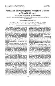
FIG. 1. Localization of IcsA expressed from the native promoter or an inducible promoter in the DegP⫹ and DegP⫺ strains. IcsA localization was determined by indirect immunofluorescence in strains GP100 (DegP⫹) and GP1100 (degP::Cm) with icsA expressed from its own promoter on pGP38.1 (A) or from an arabinose-inducible promoter on the plasmid pGP58.2 (B). Arrows with large arrowheads point to IcsA localized at the pole of GP100/pGP38.1 and GP1100/pGP38.1. The arrow with a small arrowhead points to a bacterium with IcsA localized at the pole but showing less intense staining. In panel B, immunofluorescence was performed 30 and 60 min following induction of icsA expression with 0.1 mM arabinose. Duplicate images with exposure times of 300 ms and 600 ms are shown. The increased exposure allows visualization of IcsA on the degP mutant cells. Arrows point to the same bacterium in each set of images.
analysis using S. flexneri Poly Group B antisera (Difco). Serum sensitivity was determined as described previously (19). Magainin 2 and antibiotic sensitivity. Sensitivity to the antimicrobial peptide magainin 2 (GIGKFLHSAKKFGKAFVGEIMNS) (Advanced ChemTech, Louisville, KY) was determined as previously described by Groisman et al. (13). Briefly, bacterial cultures were grown to an A595 value of ⬇0.6, and then 5 ⫻ 104 CFU in 50 l of PBS were incubated with 50 g/ml magainin 2 for 1 h with shaking. Serial dilutions of the cell suspension were then plated, and the percent survival was calculated as follows: (number of bacterial CFU at t ⫽ 2 h)/(number of bacterial CFU at t ⫽ 0), where t is time. Antibacterial MICs were determined by diluting stationary phase cultures 1:100 into medium containing twofold dilutions of polymyxin B or carbenicillin and incubating them overnight at 37°C with shaking. The MIC was the highest dilution at which no growth was observed. Tissue culture and plaque assays. Henle cells (Intestinal 407; American Type Culture Collection) were routinely cultured in Earle’s minimal essential medium (Invitrogen) containing 10% tryptose phosphate broth (Difco), 2 mM glutamine, 0.1 mM nonessential amino acids, and 10% fetal bovine serum (Invitrogen). The ability of S. flexneri to invade Henle cells was determined by the procedure of Hale and Formal (15). Plaque assays in Henle cell monolayers were performed as described by Oaks et al. (28).
RESULTS Localization defect in S. flexneri degP mutant upon induction of icsA expression. Our previous results showed that sur-
face presentation of IcsA was impaired in an S. flexneri degP mutant, SM1100 (32). The amount of IcsA synthesized in the degP mutant was the same as in the wild-type strain, suggesting that the reduced level of detectable IcsA on the surface could reflect differences in IcsA secretion or in its conformation. To assess the localization and kinetics of IcsA surface presentation, icsA was expressed under the control of an inducible promoter (Fig. 1), as described by Steinhauer et al. (42), who showed that, following induction, IcsA is first detected by immunofluorescence at the bacterial pole. icsA was expressed either from its native promoter or from an arabinose-inducible promoter in an icsA deletion background (Fig. 1). When the cloned icsA was expressed from its native promoter, IcsA was localized predominantly to the pole in both the DegP⫹ and DegP⫺ strains, with a small amount of IcsA along the lateral surface of the bacteria (Fig. 1A). However, the degP mutant showed an overall reduction in staining for IcsA compared to the DegP⫹ parent, as had been observed previously (32). The strains carrying icsA fused to an inducible promoter had no detectable IcsA on the surface prior to induction (data not shown), but 30 and 60 min following arabi-
VOL. 189, 2007
SHIGELLA IcsA LOCALIZATION REQUIRES DegP, Skp, AND SurA
5569
FIG. 2. Quantitative surface immunodetection of IcsA. (A) Western analysis was performed with anti-IcsA antiserum on whole-cell lysates or on outer membranes (om) isolated from equivalent numbers of wild-type SM100 and the S. flexneri degP mutant SM1100 cells. (B) Whole bacteria were treated with anti-Shigella or anti-IcsA antibodies. The amount of bound antibody was determined by labeling with HRP-conjugated secondary antibody and measuring the HRP enzymatic activity (A405). ␣, anti.
nose induction, IcsA was localized to the pole of cells of both strains (Fig. 1B). The staining of IcsA expressed from the inducible promoter at 60 min following induction resembles the IcsA localization seen when icsA was expressed from its native promoter in the DegP⫹ strain (Fig. 1A). While the degP mutant had IcsA localized at the pole, a smaller proportion of the bacteria had detectable IcsA on the surface, and the bacteria stained less intensely than when icsA was induced in the DegP⫹ bacteria, especially 60 min after arabinose induction (Fig. 1B). These data indicate that while IcsA is targeted to the pole in the degP mutant, the level of IcsA in the outer membrane or the exposure of IcsA in its normal conformation on the bacterial surface is reduced. Quantitative detection of antibody-accessible IcsA on the bacterial surface. To quantitate the difference between the amount of IcsA detected by immunofluorescence on the surface of the wild type and degP mutant, a modified immunodetection procedure was performed. An HRP-conjugated secondary antibody was used, and the amount of HRP signal generated from the degP mutant and its wild-type parent strain was determined by quantitating the HRP enzymatic activity. To normalize the IcsA-HRP signal to the number of bacteria, immunodetection was also performed with anti-Shigella LPS antisera, and the ratio of anti-IcsA to anti-LPS signals was determined for each strain. While Western analysis of wildtype and the degP mutant whole-cell lysates indicated that the same amount of IcsA was present (Fig. 2A, upper panel), the HRP immunodetection assay indicated that IcsA levels on the surface of the degP mutant SM1100 were approximately 60% that of wild-type bacteria (Fig. 2B). These data indicate that there is less IcsA accessible to antibody on the surface of the degP bacteria, consistent with the reduced staining in the immunofluorescence experiments. IcsA localizes to the outer membrane in the S. flexneri degP mutant. Because the total amount of IcsA was the same in wild-type and degP mutant cells but less was detected on the surface, the location of the IcsA within the cell was analyzed. The bacterial cells were fractionated to determine whether IcsA was present in the outer membrane of the degP mutant at the same level as in wild-type bacteria or whether it was sequestered elsewhere in the cell. Western analysis showed that the same amount of IcsA was present in the outer membrane of the degP mutant as in wild-type S. flexneri (Fig. 2A, lower
FIG. 3. LPS profiles of S. flexneri strains. Shigella LPS was extracted, resolved by SDS–11%PAGE and silver stained (A) or analyzed by Western immunoblotting with S. flexneri Poly Group B antisera (B). The position of the LPS core is indicated. Bands labeled as 1 unit or 2 units are LPS molecules with only one or two O-antigen repeating units attached. The brackets indicate the location of LPS molecules with either 11 to 17 O-antigen repeating units or ⬎90 repeating units.
panel). The other cell fractions were also examined, and no differences were detected between the wild type and degP mutant in the amount of IcsA in the cytoplasm, cytoplasmic membrane, or periplasmic fraction (data not shown). These data indicate that IcsA was delivered to the outer membrane and did not accumulate in another compartment in the cell. LPS and outer membrane phenotypes of the S. flexneri degP mutant. The conformation of IcsA in the outer membrane could be directly or indirectly affected by the loss of DegP chaperone function. IcsA has not been reported to require a periplasmic chaperone, and we were unable to detect a direct interaction between DegP and IcsA by coimmunoprecipitation (data not shown). An interaction between DegP and IcsA could be transient, but it is also possible that DegP influences IcsA accessibility on the outer membrane surface by affecting LPS or other outer membrane components. Mutants that are defective in the synthesis of LPS O-antigen side chains or that overproduce very long O-antigen side chains affect IcsA surface exposure and the ability of the strain to form wild-type plaques (19, 26, 37). To determine whether the degP mutant had altered LPS, the LPS from the degP mutant was extracted and analyzed by SDS-PAGE and Western immunoblotting (Fig. 3). LPS from the degP mutant was compared with LPS from wild type, an rfaL mutant whose LPS lacked O-antigen side chains, and a cld mutant whose LPS lacks the O-antigen modal length of ⬎90 repeats. The LPS from the degP mutant had a wild-type LPS profile, including both the 11- to 17-repeat mode (Fig. 3A) and the very long (⬎90) repeats of O-antigen (Fig. 3B). Wild-type LPS of Shigella confers resistance to serum killing (19); therefore, a
5570
PURDY ET AL.
J. BACTERIOL.
TABLE 2. Serum sensitivity of S. flexneri DegP⫹ and DegP⫺ strains Strain
Genotype
Serum sensitivity (log10 kill)a
SM100 SM1100 SA555-38
Wild type degP::Cm rfaL::TnphoA
0.31 0.38 4.2
a Bacteria were incubated in Luria broth or in Luria broth containing 10% rabbit serum for 2 h at 37°C. Serum killing is calculated as follows: log10 kill ⫽ (log10 CFU/ml after 2 h in broth) ⫺ (log10 CFU/ml after 2 h in broth containing 10% serum). Higher numbers indicate increased killing by serum.
serum sensitivity assay was performed to confirm that there were no other alterations in LPS composition. Like wild-type S. flexneri, the degP mutant survived in Luria broth containing 10% serum, conditions under which an rfaL mutant, SA555-38, was killed (Table 2). Because the degP mutant LPS exhibits a wild-type profile and the degP mutant had no increase in serum sensitivity, it is unlikely that the degP mutant intercellular spread defect or the altered IcsA localization is a result of altered LPS composition. Identification of a membrane defect in the degP mutant. Defects in IcsA localization could result from an altered outer membrane structure. Although no differences in the OMP profiles were detected following SDS-PAGE (data not shown), changes in the levels of minor membrane protein constituents might not have been detected by this procedure. To obtain additional measures of potential membrane defects in the degP mutant, sensitivities to SDS and antibiotics that target the cell envelope were determined. In SDS sensitivity assays, the degP mutant was slightly more sensitive than the wild type (data not shown), and there was a twofold decrease in the MICs of polymyxin B (0.25 g/ml compared to 0.5 g/ml for the wild type) and carbenicillin (2.5 g/ml compared to 5 g/ml for the wild type) for the degP mutant. Further, there was a marked reduction in survival of the degP mutant upon exposure to the antimicrobial peptide magainin 2 relative to the wild-type strain (Fig. 4). Sensitivity to SDS, antibiotics, and antimicrobial peptides are indicative of a minor outer membrane defect that could influence the proper insertion of IcsA into the outer membrane.
FIG. 4. Magainin 2 sensitivity of the degP mutant. Bacteria were incubated with 50 g/ml of magainin, and samples were taken at time zero and at 1 h. Dilutions were plated, and the number of CFU at each time point was determined to measure the percent survival. The mean and standard deviations of three independent experiments are shown.
FIG. 5. Multicopy suppression of the degP plaque formation defect by skp but not surA. Plaque formation in confluent monolayers of Henle cells was determined for the degP mutant overexpressing skp (SM1100/pGP56.4; lower left) or surA (SM1100/pGP60.1; lower right). The wild-type SA100 and the degP mutant SM1100 carrying the vector (degP/vector) are shown for comparison (top).
Multicopy suppression of the S. flexneri degP mutant by the periplasmic chaperone Skp. Because it is the chaperone rather than protease activity of DegP that appears to be required for plaque formation (32) and for proper insertion of IcsA into the outer membrane, we determined whether other known periplasmic chaperones, specifically Skp and SurA, play a role in this process. Single mutations in degP, skp, or surA are viable, but skp surA and degP surA double mutants are lethal in E. coli, suggesting functional redundancy of these periplasmic folding factors (33). To further understand whether the chaperone activity required for IcsA localization is specific to DegP, both skp and surA were examined for the ability to suppress the Shigella degP intercellular spread phenotype. S. flexneri skp and surA were expressed from the high-copynumber plasmid pBluescript SK(⫺) in the degP mutant SM1100. The plaque size of SM1100 carrying the skp plasmid, pGP56.4, was restored to that of wild type, indicating that the intercellular spread defect caused by the degP mutation was suppressed by Skp (Fig. 5). Analysis of IcsA on the bacterial cell surface showed that the presence of skp on a multicopy plasmid, like complementation with degP, restored the normal surface localization of IcsA in the degP mutant (Fig. 6). Therefore, it is likely that Skp and DegP have overlapping functions in S. flexneri, and increased amounts of Skp compensate for loss of DegP. In contrast, the degP mutant expressing surA from the same high-copy-number vector (SM1100/pGP60.1) did not form wild-type plaques (Fig. 5), indicating that SurA could not suppress the degP phenotype in the plaque assay. Functional overlap of Skp and DegP has been shown by previous genetic studies in E. coli, but this is the first report of multicopy suppression of a degP mutant phenotype by another chaperone (2, 33). Loss of plaque formation by S. flexneri skp and surA mutants. The plaque size phenotype of the degP mutant was rescued by overexpression of the periplasmic chaperone skp, which suggests that intercellular spread requires one or more periplasmic chaperones. To determine whether the presence of Skp or SurA was also necessary for wild-type plaque formation by S. flexneri, we generated mutants defective in skp and in surA. Because we could not obtain a defined surA mutation that could be complemented by the cloned gene in strain SM100, the surA and skp mutants and an additional degP mutant were constructed in the closely related, sequenced S.
VOL. 189, 2007
SHIGELLA IcsA LOCALIZATION REQUIRES DegP, Skp, AND SurA
5571
mented by a plasmid carrying the wild-type gene, indicating that each gene was expressed from the high-copy-number vector, but neither the skp mutant nor the surA mutant plaque defect could be suppressed by the other gene or by degP expressed from this vector (Table 3). The surA mutant produced smaller colonies than the wild type on agar plates, and this growth defect may contribute to the inability of this mutant to form wild-type plaques. DISCUSSION
FIG. 6. Effect of mutations in degP, skp, and surA on the presence of IcsA on the surface of S. flexneri. (A) IcsA localization was determined by indirect immunofluorescence in strain 2457T (wild type) and in the 2457T degP, 2457T skp, and 2457T surA strains. (B) IcsA localization was determined by indirect immunofluorescence in the degP mutant carrying degP (SM1100/pGP25.2) or skp (SM1100/pGP56.4) on a multicopy plasmid.
flexneri 2a strain 2457T. We confirmed the degP phenotype in the 2457T background (Table 3). As in the SM100 degP mutant, the 2457T degP mutant was able to invade cultured epithelial cells (Table 3). The percentage of Henle cells that were invaded by the degP mutant was somewhat lower than the wild type, but the invasion was enhanced by growth in deoxycholate (DOC) (Table 3), and the percentage of cells invaded was within the range observed for invasive strains (31). Like the SM100 degP mutant, 2457T degP was defective for plaque formation (Table 3). In the 2547T background, however, the degP mutation caused a complete loss of plaque formation and thus had a stronger phenotype than the SM100 mutant, which produced small plaques. Plaque formation by 2457T degP was restored to wild-type levels by expression of degP from a plasmid vector and was suppressed by expression of skp from a high-copy-number vector (Table 3), in agreement with the results obtained with the SM100 degP mutant. The skp and the surA mutants were also tested in Henle cell invasion and plaque assays. Both mutants invaded at wild-type levels but were defective for plaque formation, suggesting defects in intercellular spread (Table 3). As noted with the degP mutant, the presence of IcsA on the surface of the skp and surA mutants was greatly reduced (Fig. 6), although there was no difference in the amount of IcsA in the cells as determined by Western analysis (data not shown). Each mutant was comple-
The ability of Shigella to invade intestinal epithelial cells, multiply within the host cell cytoplasm, and spread to adjacent cells is key to Shigella virulence. Intercellular spread is mediated by IcsA-dependent actin polymerization at the bacterial pole, and in a previous study we had shown that the periplasmic chaperone DegP was required for proper IcsA surface localization (32). IcsA was detected on the mutant bacteria by immunofluorescence, but the amount of immunofluorescence appeared less than wild-type levels, suggesting that degP mutant bacteria have less IcsA on the surface. In the present study, we determined that the mutant had wild-type levels of IcsA in the outer membrane; however, a quantitative surface immunoassay indicated that IcsA in the degP mutant was less accessible to antibody labeling on the cell surface. The most likely explanation is that IcsA is secreted to the outer membrane, but the protein is inserted in such a way that the epitope is inaccessible to antibody. In the host cell cytoplasm, the N-terminal domain of IcsA on the bacterial surface interacts with the host proteins vinculin and neural Wiskott-Aldrich syndrome protein to nucleate F-actin polymerization. Actin polymerization at the bacterial pole propels the bacteria into the adjacent cell. If there is reduced detection of IcsA with antibody, it is likely that there is less IcsA accessible for interactions with the actin-nucleating proteins in the host cell cytoplasm. Altered actin polymerization would result in inefficient intercellular spread of the degP mutant. The IcsA protein is an autotransporter and consists of an N-terminal passenger domain and a C-terminal -domain, which is predicted to form a -barrel of amphipathic antiparallel sheets. The steps to its proper insertion in the outer membrane have been described. IcsA is secreted across the
TABLE 3. Invasion assays and plaque formation by S. flexneri degP, skp, and surA mutants Invasion (%)a Strain
2457T 2457T degP 2457T skp 2457T surA
⫹DOC
⫺DOC
100 81 100 NDc
63 ⫾ 1.1 23 ⫾ 1.6 53 ⫾ 4.8 67 ⫾ 2.7
Plaque formationb pBluescript pGP25.7 pGP56.4 pGP60.1 vector (DegP⫹) (Skp⫹) (SurA⫹)
⫹ ⫺ ⫺ ⫺
ND ⫹ ⫺ ⫺
ND ⫹ ⫹ ⫺
ND ⫺ ⫺ ⫹
a Percent of the Henle cells that contained 3 or more intracellular bacteria. Bacteria were grown in Luria broth alone or with 2.5 mM DOC. Values are means of three or more independent experiments with 1 standard deviation (⫺DOC) or two experiments (⫹DOC). b Plaque formation in Henle cell monolayers by mutant strains carrying the indicated plasmid. ⫹, wild-type size and number of plaques; ⫺, no plaques; ND, not determined. c ND, not determined. Growth of 2457T surA was inhibited by DOC.
5572
PURDY ET AL.
inner membrane by the Sec secretion system, is present transiently in the periplasm as a soluble protease-resistant intermediate, and is then inserted into the outer membrane, where the IcsA passenger domain is threaded through the channel formed by the -domain (4, 5). The rate-limiting step appears to be the translocation of the IcsA passenger domain to the surface (4). Since wild-type levels of IcsA fractionate with the outer membrane of the degP mutant, the IcsA localization defect in the mutant may be in the translocation of the IcsA passenger domain to the bacterial surface. The mechanism by which DegP affects IcsA surface localization appears to be through its chaperone function (32). The autotransporters have not been shown to require periplasmic chaperones (22, 46), suggesting that the role of DegP in IcsA presentation may be in processing another cellular factor that interacts with IcsA. LPS composition, which is known to affect IcsA localization and intercellular spread, was examined in the S. flexneri degP mutant but was found to be the same as wild type. Another possibility was that the outer membrane of the degP mutant was altered in some way that would affect protein insertion or translocation to the bacterial surface. The degP mutant was found to be slightly more sensitive than the wild type to detergent and antibiotics. Analysis of the OMP composition by SDS-PAGE revealed no obvious differences (data not shown), but it is possible that even a minor change in OMP composition in the degP mutant could affect IcsA insertion into the outer membrane in its proper conformation. Three periplasmic chaperones, DegP, Skp, and SurA, have a role in the proper folding and insertion of proteins into the outer membrane of E. coli. Two overlapping chaperone pathways for OMPs have been proposed in E. coli: one consisting of DegP and Skp and the other of SurA (2, 33). DegP chaperone activity was important for IcsA localization in S. flexneri, and we examined strains lacking Skp and SurA to assess their role in intercellular spread and the ability of each chaperone to compensate for the lack of another. It is interesting that single mutations in each of these chaperones resulted in a defect in intercellular spread, which implies that outer membrane factors that affect proper IcsA localization are folded and delivered to the outer membrane by DegP, Skp, and SurA. Suppression by skp of the degP mutant phenotype and a requirement for both SurA and DegP or Skp support the two-pathway model proposed by Rizzitello et al. (33). In this model, proteins may be chaperoned across the periplasm to the outer membrane by either the DegP/Skp route or by SurA, with some proteins showing a preference for one of the two pathways. As overexpression of degP was insufficient to restore wild-type levels of intercellular spread to the skp mutant, the role of Skp in the DegP/Skp pathway may be greater than that of DegP for surface presentation of IcsA. The defect in plaque formation exhibited by the surA mutant suggests that SurA also plays a role, which this model predicts to be functionally distinct from that of DegP and Skp. However, the mutation in surA appears to have pleiotropic effects on bacterial growth and physiology that may also impact IcsA or other factors required for virulence. In E. coli, surA mutation compromises proper folding of OMPs (23), and a comparison of protein composition of the wild type and surA mutant of S. flexneri by SDS-PAGE indicated that the mutant had slightly reduced amounts of the major OMPs (data not shown). This and other
J. BACTERIOL.
membrane defects may have affected the localization of virulence-associated proteins including IcsA, but the total amount of IcsA in the mutant was the same as the wild type. This indicates that IcsA was not degraded to a greater extent in this mutant. Further investigation of the surA mutant is required to define more clearly the role of SurA in outer membrane biogenesis of S. flexneri and in intercellular spread. ACKNOWLEDGMENTS We are grateful to Edwin Oaks (Walter Reed Army Institute of Research) for generously providing antiserum, Nicola Davies for strain construction, Elizabeth Wyckoff for DNA sequence analysis and critical reading of the manuscript, Emily Helton and Matthew Ramsey for expert technical assistance, and Tom Silhavy for strains. This work was supported by grant AI16935 from the National Institutes of Health. REFERENCES 1. Baba, T., T. Ara, M. Hasegawa, Y. Takai, Y. Okumura, M. Baba, K. A. Datsenko, M. Tomita, B. L. Wanner, and H. Mori. 2006. Construction of Escherichia coli K-12 in-frame, single-gene knockout mutants: the Keio collection. Mol. Syst. Biol. 2:2006 0008. 2. Behrens, S., R. Maier, H. de Cock, F. X. Schmid, and C. A. Gross. 2001. The SurA periplasmic PPIase lacking its parvulin domains functions in vivo and has chaperone activity. EMBO J. 20:285–294. 3. Bernardini, M. L., J. Mounier, H. d’Hauteville, M. Coquis-Rondon, and P. J. Sansonetti. 1989. Identification of icsA, a plasmid locus of Shigella flexneri that governs bacterial intra- and intercellular spread through interaction with F-actin. Proc. Natl. Acad. Sci. USA 86:3867–3871. 4. Brandon, L. D., N. Goehring, A. Janakiraman, A. W. Yan, T. Wu, J. Beckwith, and M. B. Goldberg. 2003. IcsA, a polarly localized autotransporter with an atypical signal peptide, uses the Sec apparatus for secretion, although the Sec apparatus is circumferentially distributed. Mol. Microbiol. 50:45–60. 5. Brandon, L. D., and M. B. Goldberg. 2001. Periplasmic transit and disulfide bond formation of the autotransported Shigella protein IcsA. J. Bacteriol. 183:951–958. 6. Chen, R., and U. Henning. 1996. A periplasmic protein (Skp) of Escherichia coli selectively binds a class of outer membrane proteins. Mol. Microbiol. 19:1287–1294. 7. Clausen, T., C. Southan, and M. Ehrmann. 2002. The HtrA family of proteases: implications for protein composition and cell fate. Mol. Cell 10:443– 455. 8. Filip, C., G. Fletcher, J. L. Wulff, and C. F. Earhart. 1973. Solubilization of the cytoplasmic membrane of Escherichia coli by the ionic detergent sodiumlauryl sarcosinate. J. Bacteriol. 115:717–722. 9. Formal, S. B., G. J. Dammin, E. H. Labrec, and H. Schneider. 1958. Experimental Shigella infections: characteristics of a fatal infection produced in guinea pigs. J. Bacteriol. 75:604–610. 10. Fukuda, I., T. Suzuki, H. Munakata, N. Hayashi, E. Katayama, M. Yoshikawa, and C. Sasakawa. 1995. Cleavage of Shigella surface protein VirG occurs at a specific site, but the secretion is not essential for intracellular spreading. J. Bacteriol. 177:1719–1726. 11. Goldberg, M. B., O. Barzu, C. Parsot, and P. J. Sansonetti. 1993. Unipolar localization and ATPase activity of IcsA, a Shigella flexneri protein involved in intracellular movement. Infect. Agents Dis. 2:210–211. 12. Goldberg, M. B., J. A. Theriot, and P. J. Sansonetti. 1994. Regulation of surface presentation of IcsA, a Shigella protein essential to intracellular movement and spread, is growth phase dependent. Infect. Immun. 62:5664– 5668. 13. Groisman, E. A., F. Heffron, and F. Solomon. 1992. Molecular genetic analysis of the Escherichia coli phoP locus. J. Bacteriol. 174:486–491. 14. Guzman, L. M., D. Belin, M. J. Carson, and J. Beckwith. 1995. Tight regulation, modulation, and high-level expression by vectors containing the arabinose PBAD promoter. J. Bacteriol. 177:4121–4130. 15. Hale, T. L., and S. B. Formal. 1981. Protein synthesis in HeLa or Henle 407 cells infected with Shigella dysenteriae 1, Shigella flexneri 2a, or Salmonella typhimurium W118. Infect. Immun. 32:137–144. 16. Henderson, I. R., and J. P. Nataro. 2001. Virulence functions of autotransporter proteins. Infect. Immun. 69:1231–1243. 17. Hitchcock, P. J., and T. M. Brown. 1983. Morphological heterogeneity among Salmonella lipopolysaccharide chemotypes in silver-stained polyacrylamide gels. J. Bacteriol. 154:269–277. 18. Hong, M., Y. Gleason, E. E. Wyckoff, and S. M. Payne. 1998. Identification of two Shigella flexneri chromosomal loci involved in intercellular spreading. Infect. Immun. 66:4700–4710. 19. Hong, M., and S. M. Payne. 1997. Effect of mutations in Shigella flexneri chromosomal and plasmid-encoded lipopolysaccharide genes on invasion and serum resistance. Mol. Microbiol. 24:779–791.
VOL. 189, 2007
SHIGELLA IcsA LOCALIZATION REQUIRES DegP, Skp, AND SurA
20. Inouye, M., and J. P. Guthrie. 1969. A mutation which changes a membrane protein of E. coli. Proc. Natl. Acad. Sci. USA 64:957–961. 21. Kaback, H. R. 1970. Bacterial membranes. Methods Enzymol. 22:99–120. 22. Kostakioti, M., C. L. Newman, D. G. Thanassi, and C. Stathopoulos. 2005. Mechanisms of protein export across the bacterial outer membrane. J. Bacteriol. 187:4306–4314. 23. Lazar, S. W., and R. Kolter. 1996. SurA assists the folding of Escherichia coli outer membrane proteins. J. Bacteriol. 178:1770–1773. 24. Lett, M. C., C. Sasakawa, N. Okada, T. Sakai, S. Makino, M. Yamada, K. Komatsu, and M. Yoshikawa. 1989. virG, a plasmid-coded virulence gene of Shigella flexneri: identification of the virG protein and determination of the complete coding sequence. J. Bacteriol. 171:353–359. 25. Makino, S., C. Sasakawa, K. Kamata, T. Kurata, and M. Yoshikawa. 1986. A genetic determinant required for continuous reinfection of adjacent cells on large plasmid in S. flexneri 2a. Cell 46:551–555. 26. Morona, R., and L. Van Den Bosch. 2003. Lipopolysaccharide O antigen chains mask IcsA (VirG) in Shigella flexneri. FEMS Microbiol. Lett. 221: 173–180. 27. Morona, R., L. van den Bosch, and P. A. Manning. 1995. Molecular, genetic, and topological characterization of O-antigen chain length regulation in Shigella flexneri. J. Bacteriol. 177:1059–1068. 28. Oaks, E. V., M. E. Wingfield, and S. B. Formal. 1985. Plaque formation by virulent Shigella flexneri. Infect. Immun. 48:124–129. 29. Pallen, M. J., and B. W. Wren. 1997. The HtrA family of serine proteases. Mol. Microbiol. 26:209–221. 30. Payne, S. M., D. W. Niesel, S. S. Peixotto, and K. M. Lawlor. 1983. Expression of hydroxamate and phenolate siderophores by Shigella flexneri. J. Bacteriol. 155:949–955. 31. Pope, L. M., K. E. Reed, and S. M. Payne. 1995. Increased protein secretion and adherence to HeLa cells by Shigella spp. following growth in the presence of bile salts. Infect. Immun. 63:3642–3648. 32. Purdy, G. E., M. Hong, and S. M. Payne. 2002. Shigella flexneri DegP facilitates IcsA surface expression and is required for efficient intercellular spread. Infect. Immun. 70:6355–6364. 33. Rizzitello, A. E., J. R. Harper, and T. J. Silhavy. 2001. Genetic evidence for parallel pathways of chaperone activity in the periplasm of Escherichia coli. J. Bacteriol. 183:6794–6800. 34. Rouviere, P. E., and C. A. Gross. 1996. SurA, a periplasmic protein with peptidyl-prolyl isomerase activity, participates in the assembly of outer membrane porins. Genes Dev. 10:3170–3182.
5573
35. Sambrook, J., and D. W. Russell. 2001. Molecular cloning: a laboratory manual, 3rd ed. Cold Spring Harbor Laboratory Press, Cold Spring Harbor, NY. 36. Sandlin, R. C., M. B. Goldberg, and A. T. Maurelli. 1996. Effect of O side-chain length and composition on the virulence of Shigella flexneri 2a. Mol. Microbiol. 22:63–73. 37. Sandlin, R. C., K. A. Lampel, S. P. Keasler, M. B. Goldberg, A. L. Stolzer, and A. T. Maurelli. 1995. Avirulence of rough mutants of Shigella flexneri: requirement of O antigen for correct unipolar localization of IcsA in the bacterial outer membrane. Infect. Immun. 63:229–237. 38. Sansonetti, P., C. Egile, and C. Wenneras. 2001. Shigellosis: from disease symptoms to molecular and cellular pathogenesis, p. 335–385. In E. A. Groisman (ed.), Principles of bacterial pathogenesis. Academic Press, Inc., New York, NY. 39. Schafer, U., K. Beck, and M. Muller. 1999. Skp, a molecular chaperone of gram-negative bacteria, is required for the formation of soluble periplasmic intermediates of outer membrane proteins. J. Biol. Chem. 274:24567–24574. 40. Skorko-Glonek, J., A. Wawrzynow, K. Krzewski, K. Kurpierz, and B. Lipinska. 1995. Site-directed mutagenesis of the HtrA (DegP) serine protease, whose proteolytic activity is indispensable for Escherichia coli survival at elevated temperatures. Gene 163:47–52. 41. Spiess, C., A. Beil, and M. Ehrmann. 1999. A temperature-dependent switch from chaperone to protease in a widely conserved heat shock protein. Cell 97:339–347. 42. Steinhauer, J., R. Agha, T. Pham, A. W. Varga, and M. B. Goldberg. 1999. The unipolar Shigella surface protein IcsA is targeted directly to the bacterial old pole: IcsP cleavage of IcsA occurs over the entire bacterial surface. Mol. Microbiol. 32:367–377. 43. Stevenson, G., A. Kessler, and P. R. Reeves. 1995. A plasmid-borne Oantigen chain length determinant and its relationship to other chain length determinants. FEMS Microbiol. Lett. 125:23–30. 44. Suzuki, T., H. Miki, T. Takenawa, and C. Sasakawa. 1998. Neural WiskottAldrich syndrome protein is implicated in the actin-based motility of Shigella flexneri. EMBO J. 17:2767–2776. 45. Suzuki, T., S. Saga, and C. Sasakawa. 1996. Functional analysis of Shigella VirG domains essential for interaction with vinculin and actin-based motility. J. Biol. Chem. 271:21878–21885. 46. Thanassi, D. G., C. Stathopoulos, A. Karkal, and H. Li. 2005. Protein secretion in the absence of ATP: the autotransporter, two-partner secretion and chaperone/usher pathways of gram-negative bacteria (review). Mol. Membr. Biol. 22:63–72.