Identification and Characterization of Endoplasmic Reticulum-Associated Degradation Proteins Differentially Affected by Endoplasmic Reticulum Stress1 Mariana E. Kirst, David J. Meyer, Bryan C. Gibbon, Rudolf Jung, and Rebecca S. Boston* Department of Botany, North Carolina State University, Raleigh, North Carolina 27695–7612 (M.E.K., R.S.B.); Pioneer Hi-Bred International, Incorporated, a DuPont Company, Johnston, Iowa 50131 (D.J.M., R.J.); and Department of Plant Sciences, University of Arizona, Tucson, Arizona 85721 (B.C.G.)
The disposal of misfolded proteins from the lumen of the endoplasmic reticulum (ER) is one of the quality control mechanisms present in the protein secretory pathway. Through ER-associated degradation, misfolded substrates are targeted to the cytosol where they are degraded by the proteasome. We have identified four maize (Zea mays) Der1-like genes (Zm Derlins) that encode homologs of Der1p, a yeast (Saccharomyces cerevisiae) protein implicated in ER-associated degradation. Zm Derlins are capable of functionally complementing a yeast Der1 deletion mutant. Such complementation indicates that the Der1p function is conserved among species. Zm Derlin genes are expressed at low levels throughout the plant, but appear prevalent in tissues with high activity of secretory protein accumulation, including developing endosperm cells. Expression of three of the four Zm Derlin genes increases during ER stress, with Zm Derlin1-1 showing the strongest induction. Subcellular fractionation experiments localized Zm Derlin proteins to the membrane fraction of microsomes. In maize endosperm, Zm Derlin proteins were found primarily associated with ER-derived protein bodies regardless of the presence of an ER stress response.
The endoplasmic reticulum (ER) serves as a versatile gatekeeper of the secretory pathway. It is not only the entry point for translocation of newly synthesized proteins, but also the site of quality control processes that discriminate between conformationally correct proteins in a native state and those that are terminally misfolded. In the latter case, continued protein accumulation could be toxic to the organism, possibly impairing its development. To cope with the accumulation of misfolded proteins and restore homeostasis, the cell initiates an ER stress response that has been linked to development of ER-rich tissues, upregulation of molecular chaperones, selective translational attenuation, and activation of an ER-associated degradation (ERAD) process (Zhang and Kaufman, 2004). ERAD ensures that aberrant proteins do not transit through the secretory pathway. Instead, misfolded proteins are targeted for removal from this pathway by retrotranslocation through the ER membrane to the cytosol where they undergo ubiquitination and degradation by the proteasome (Brodsky and
1
This work was supported by the U.S. Department of Energy (grant no. DE–FG02–00ER150065 to R.S.B.), by Pioneer Hi-Bred International, and by the North Carolina Agricultural Research Service (to R.S.B.). * Corresponding author; e-mail
[email protected]; fax 919– 515–3436. Article, publication date, and citation information can be found at www.plantphysiol.org/cgi/doi/10.1104/pp.105.060087. 218
McCracken, 1999; Plemper and Wolf, 1999; McCracken and Brodsky, 2003). Attempts to characterize the ERAD process have allowed identification of several components of the ERAD machinery, some of which are co-opted from their classical roles in importing proteins to additional functions in protein export. Genetic studies are consistent with a model whereby molecular chaperones could present ERAD substrates to the Sec61 complex (Nishikawa et al., 2001; Molinari et al., 2002). Upon association of ERAD components, the Sec61 complex would form a channel for retrotranslocation of the misfolded proteins (Plemper et al., 1999). Proteins emerging from the ER would be ubiquitinated by the combined action of ubiquitin-conjugating enzymes (ubc6, ubc1, and/or ubc7) and the ubiquitin-ligase complex Hrd1/Der3. The ubiquitinated substrates could then be recognized by the Cdc48 AAA-ATPase complex and removed from the ER membrane for degradation by the proteasome (Ye et al., 2001; Jarosh et al., 2002; Muller et al., 2005). In plants, little is known about the degradation mechanisms of the ER. A link between ER stress and ERAD was observed in a genome-wide analysis of Arabidopsis (Arabidopsis thaliana) cells treated with pharmacological agents to induce ER stress (Martinez and Chrispeels, 2003). Use of ricin A as a reporter for degradation in transformed tobacco (Nicotiana tabacum) protoplasts revealed cytosolic accumulation after treatment with a proteasome inhibitor (di Cola et al., 2001). Expression of a dominant-negative form of the
Plant Physiology, May 2005, Vol. 138, pp. 218–231, www.plantphysiol.org Ó 2005 American Society of Plant Biologists
Maize Derlin Proteins
AAA-ATPase Cdc48 in Arabidopsis plants stabilized the misfolded wheat protein, MLO-1, a finding consistent with this protein being a substrate for ERAD (Muller et al., 2005). In an effort to identify changes in gene expression that were associated with the ER stress response in seeds, we undertook an open-ended RNA-profiling study. This study took advantage of three maize (Zea mays) endosperm mutants that provide easily assayed models. The three mutants, floury-2 (fl2), Mucronate (Mc), and defective endosperm B30 (De*-B30) exhibit ER stress responses that are specific to the endosperm and coincident with the deposition of defective storage proteins in the seed (Boston et al., 1991; Coleman et al., 1995; Kim et al., 2004). The endosperm mutants exhibit different degrees of ER stress, depending on the deposition of defective storage proteins. The strongest phenotype is observed in the fl2 mutant, which produces a 24-kD a-zein that is blocked for cleavage of the signal peptide. As a result, the defective storage protein accumulates as a membrane-anchored protein in the ER and in ERderived protein bodies (Coleman et al., 1995; Gillikin et al., 1997). The accumulation of this terminally misfolded protein causes an ER stress that leads to an increase in the accumulation of molecular chaperones such as binding protein (BiP). Mc and De*-B30 also have defective storage proteins (Kim et al., 2004). Mc is caused by a frameshift mutation (R. Jung and R.S. Boston, unpublished data), while De*-B30 is another zein signal peptide-processing mutant. Each of the mutations also leads to a decrease in the amount of the mutant protein along with other storage proteins in the seed (Soave and Salamini, 1984). This decrease, together with the presence of ER stress, is consistent with an attenuation of protein synthesis as a result of the ER stress response and degradation of the defective proteins by the ERAD pathway. We hypothesized that the profiling of maize endosperm mutants would uncover changes not only associated with molecular chaperone gene expression but also associated with vesicle trafficking and ERAD. This was indeed the case as numerous genes showed changes in mRNA accumulation. Two of these were particularly interesting to us as they both showed homology to Der1p, a protein linked to ERAD almost 10 years ago but still lacking a functional characterization (Knop et al., 1996). In yeast (Saccharomyces cerevisiae), Der1p has been implicated in ERAD by mutant analysis in which a Der1 deletion mutant accumulated a misfolded reporter protein in the lumen of the ER (Knop et al., 1996; Hill and Cooper, 2000; Taxis et al., 2003). A much more dramatic effect was seen in double mutants lacking both Der1p and Ire1p, the protein kinase/endonuclease responsible for signaling and transducing ER stress, as the double mutation was lethal at restrictive temperatures (Travers et al., 2000). Recently, homologs for the yeast Der1 protein have been identified in other organisms, including humans (Hitt and Wolf, 2004; Lilley and Ploegh, 2004; Ye et al., 2004). These proteins Plant Physiol. Vol. 138, 2005
have been designated as Derlins (for Der1-like). Attempts to identify Der1p and Derlin activities to date have failed to provide a uniform picture with reports of involvement in degradation of soluble proteins in yeast and membrane reporter proteins in mammalian cells (Knop et al., 1996; Taxis et al., 2002, 2003; Lilley and Ploegh, 2004). Derlins have been linked to other ERAD-associated proteins by protein-protein interaction studies, but the mechanisms by which they contribute to the ERAD function remain unknown (Ito et al., 2001; Giot et al., 2003; Li et al., 2004; Ye et al., 2004). Here, we report the discovery of four Derlin genes in maize and provide a functional characterization of the gene family. RESULTS Discovery of Maize Derlin Genes by RNA Profiling
To identify genes associated with the ER stress response, we performed a high-throughput, openarchitecture transcript-profiling assay (GeneCalling; Crasta and Folkerts, 2003) with immature endosperm samples from the corn inbred line W64A (1) and its near-isogenic conversions Mc, fl2, and De*-B30 (Hunter et al., 2002). GeneCalling analysis measures, in a highly reproducible manner, differences in the abundance of individual cDNA fragments. In general, these differences are directly related to differences in the level of expression of specific transcripts (Shimkets et al., 1999; Bruce et al., 2000). Using 64 restriction enzyme pairs, we detected about 20,000 cDNA fragments from this analysis. From a comparison of electrophoretically obtained traces corresponding to cDNA fragments, we identified about 200 cDNA fragments that consistently showed differences between samples from the normal and mutant lines. The majority of these fragments were identified by either (1) competitive PCR confirmations (trace poisoning) based on their match to the Pioneer-DuPont corn gene database; or (2) cloning and sequencing. A subset of the identified differentially expressed genes matched genes that have previously been implicated in ER stress in other organisms (the comprehensive analysis is still in progress; B.C. Gibbon, R. Jung, and R.S. Boston, unpublished data). Two of the putatively up-regulated genes (Fig. 1) were of particular interest because they were members of the same gene family. One was nearly identical to, and the other a potential homolog of, a corn gene (GenBank accession no. CAB97005) that is annotated in public databases as NADH oxidoreductase. We found this annotation to be an error that probably originated because of its match to a Caenorhabditis elegans six-gene operon that, along with a homolog CAB97005, contained the coding sequence of an NADH oxidoreductase (Hough et al., 1999). BLAST searches with the deduced amino acid sequences from the two up-regulated transcripts indicated a clear homology to the yeast Der1 protein, and we therefore refer to them as Zm Derlins. 219
Kirst et al.
hypothesize that the function, although unknown, of the Der1 and Zm Derlin proteins would also be conserved. This hypothesis was further supported by reports that appeared during the course of our study describing three mammalian Derlin proteins (Lilley and Ploegh, 2004; Ye et al., 2004). Plant Derlin Genes Are Grouped into Two Subfamilies
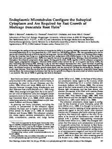
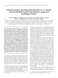
BLAST searches of the Pioneer-DuPont gene databases identified two additional Derlin genes in corn and a search of the Arabidopsis and rice wholegenome databases identified three Arabidopsis Derlin genes and two rice Derlin genes (Table I). The plant Derlin genes fell into two groups, which we call Derlin1 and Derlin2, with deduced amino acid sequences having approximately 30% overall identity between subfamilies and .90% overall identity within each subfamily. The maize Derlin genes were mapped by RFLP mapping and each pair of paralogous sequences mapped to the same map position (Table I). Zm Derlin1-1 and Zm Derlin1-2 mapped to chromosome 8 (approximately 120 cM on the IBM map) and Zm Derlin2-1 and Zm Derlin2-2 both mapped to chromosome 1 (approximately 7 cM on the IBM map). Because of the indistinguishable map positions and sequence similarity between paralogs, we searched the maize genome survey sequences (GSS) section of GenBank and assembled sequences with identical nucleotide (nt) overlaps into GSS contigs (data not shown). GSS contigs that originated from the same maize inbred line B73 were identified with nearperfect matches to each of the four Zm Derlin cDNAs, thus verifying that they originated from distinct genes and not from alleles (Table I). Figure 1. Differences in expression levels of Zm cDNA fragments detected by GeneCalling. The sections show average gel traces (red) based on nine replicate gels of digested cDNA fragments from endosperm samples of inbred W64A1 (A and B, top), De*-B30 (A, bottom), and fl2 (B, bottom) harvested 23 DAP. A, Trace section that displays a 351-bp fragment of Zm Derlin1-1 (vertical red line). B, Trace section that displays a 240-bp fragment of Zm Derlin2-1 (vertical red line). Competitive PCR reactions for the samples were performed to confirm the identity of corresponding bands (green traces) using control primers and Zm Derlin1-1-specific primers (A) or control primers and Zm Derlin2-1-specific primers (B). The x axis is in base pairs and the y axis is in relative fluorescence units.
Figure 1 shows differences in expression levels of the Zm Derlin cDNA fragments detected by GeneCalling. The relative expression of Zm Derlin1-1 (Fig. 1A) showed a more pronounced induction during ER stress (shown for De*-B30) than Zm Derlin2-1 (shown for fl2 in Fig. 1B). Similar differences for both genes were observed in all three mutants, which exhibit endosperm-specific ER stress responses (data not shown). The link to ER stress along with sequence conservation between yeast and plants led us to 220
Table I. Putative Derlin homologs from maize, rice, and Arabidopsis Species
Maize
Designation
Zm Derlin1-1 Zm Derlin1-2 Zm Derlin2-1 Zm Derlin2-2 Rice Os Derlin1-1 Os Derlin2-1 Arabidopsis At Derlin1-1 At Derlin2-1 At Derlin2-2
Predicted ORF Size
243 243 249 249 242 249 266 244 244
Accession No.
Chromosome/ Locus
AY854013a 8d AY854014 8d AY854015 1d AY854016 1d b N/A 5 AAO20072 3 NM_119078c At4g29330 NM_118301c At4g21810 NM_116724c At4g04860
a
Full-length cDNA; related sequence reported in GenBank (accession no. CAB97005) is likely a chimera between Zm Derlin1-1 and Zm b Derlin1-2. No full-length cDNA sequence available; predicted coding sequence (accession no. GRMP00000036919) for the corresponding gene in Gramene (http://www.gramene.org) is probably c incorrect. Full-length cDNA; other predicted coding sequences for At4g29330, At4g21810, and At4g04860 in GenBank are probably d incorrect. GenBank accession numbers for Zm Derlin GSS contigs are Zm Derlin1-1, AY854017; Zm Derlin1-2, AY854018; Zm Derlin2-1, AY854019; and Zm Derlin2-2, AY854020. Plant Physiol. Vol. 138, 2005
Maize Derlin Proteins
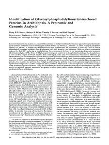
Figure 2. Protein sequence alignment of Derlin homologs. Deduced amino acid sequences of the maize Derlin homologs Zm Derlin1-1(ZmD1-1, accession no. AY854013) and Zm Derlin2-1 (ZmD2-1, accession no. AY854015), human Derlins (HsD1, accession no. NP_077271 and HsD2, accession no. NP_057125), C. elegans Derlins (CeD1, accession no. NM_066189 and CeD2, accession no. NP_49272), and the S. cerevisiae Der1 protein (ScD1, accession no. CAA63165) were aligned with ClustalW. Amino acids that were identical in at least six sequences are shown in bold. Wavy lines above ZmD1-1 and ZmD2-1 sequences indicate peptides used as antigens for antibody production. Solid lines mark transmembrane domains reported for the yeast Der1 protein (Hitt and Wolf, 2004).
Derlin Proteins Are Conserved across Kingdoms
Zm Derlin Proteins Are Integral Membrane Proteins
Figure 2 shows an alignment of the deduced amino acid sequences of Zm Derlin1-1 and Zm Derlin2-1 as prototypes of the plant Derlins, along with Der1p from yeast, and Derlin-1 and Derlin-2 from human and C. elegans. The maize proteins were similar in size to those of the other organisms, with the exception of Der1p, which had only 211 amino acids. The plant Derlin1 and Derlin2 sequences each showed about 25% to 30% identity to Derlin proteins of the other species. The plant Derlin2 sequences, however, were distinguished by an 11-amino acid region that failed to align with sequences from other organisms. Despite the low overall identity observed among the aligned proteins, there was a clear overall homology with some amino acids being conserved across the entire group (Fig. 2). Analysis of other protein features in silico revealed similarities that are perhaps more important than the sequence conservation. For example, all Derlins, including the plant proteins, contained a domain that showed distant similarity to the Rhomboid domain of intramembrane proteases (pfam01694), and hydrophobicity analysis with the program TMHMM (Krogh et al., 2001) predicted four and five transmembrane domains for Zm Derlin1 and Zm Derlin2, respectively (data not shown). Multiple transmembrane domains were also predicted for yeast Der1p (underlined in Fig. 2) and for the mammalian Derlin proteins (Knop et al., 1996; Taxis et al., 2003; Hitt and Wolf, 2004; Lilley and Ploegh, 2004).
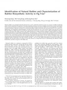
We tested the predicted membrane association of the maize Zm Derlin proteins by immunoblot analysis of subcellular fractions from the fl2 endosperm mutant. Antibodies were raised against peptides from the deduced amino acid sequence of Zm Derlin1-1 in regions outside the predicted membrane-spanning regions (wavy lines in Fig. 2). Because of the high sequence homology between Zm Derlin1-1 and Zm Derlin1-2, the antibodies cross-reacted with both; however, they did not cross-react with Zm Derlin2 proteins. To evaluate the predicted integral membrane localization of Zm Derlin1 proteins, we performed alkaline fractionation and evaluated detergent extraction of microsomal samples from immature endosperm. The microsomal fraction did not include the protein bodies whose high content of insoluble zeins blocks efficient removal of luminal contents after alkaline lysis (J. Gillikin and R.S. Boston, unpublished data; in this work, we use the terms cisternal ER and protein bodies as operational designations for the endosperm fractions containing light and heavy ER subdomains, respectively). Figure 3A shows fractions probed by immunoblot analysis for Zm Derlin1 proteins, the ER luminal molecular chaperone calreticulin, and the related membrane protein calnexin. A Zm Derlin1 signal was detected in the microsomal membrane fraction but was undetectable in the soluble fraction. Alkaline treatment of microsomes followed by centrifugation to separate membrane and soluble luminal fractions showed little or no Zm Derlin1 in the soluble fraction,
Plant Physiol. Vol. 138, 2005
221
Kirst et al.
Zm Derlin1 signals on immunoblots, but had no apparent effect on the calreticulin or calnexin signals. To verify that the association of Zm Derlins with fractions of endomembranes reflected their localization in living cells, we examined their transient expression. Green fluorescent protein fusion constructs of Zm Derlin1-1 and Zm Derlin2-1 directed by a ubiquitin promoter were introduced into maize callus tissues by microprojectile bombardment. Fluorescence from both constructs was detected in a perinuclear and reticulate pattern around cells and was absent from the nucleus or plasma membrane (D.J. Meyer and W.J. GordonKamm, unpublished data). This pattern was indicative of ER localization and was consistent with the pattern of Derlin-1 localization observed in animal cells (Lilley and Ploegh, 2004). Zm Derlin1-1 and Zm Derlin2-1 Genes Complement the Yeast Der1 Phenotype
Figure 3. Immunoblots showing association of Zm Derlin1 with microsomal membranes. Approximately equal fresh weight equivalents of endosperm (3 mg) were separated by SDS-PAGE (15%) prior to transfer and probing with antibodies against Zm Derlin1 (ZmD1) or calreticulin/calnexin (CRT, CNX). A, Localization after alkaline lysis. A microsomal fraction from fl2 endosperm (SM) was further separated into membrane (P) and luminal (S) fractions by alkaline lysis and differential centrifugation. B, Localization after detergent treatments. A fl2 microsomal fraction (SM) was treated with the detergents shown above lanes and separated by centrifugation into soluble (S) and insoluble fractions (P). DOC, deoxycholate; NP-40, Nonidet P-40.
as expected for an integral membrane localization (Fujiki et al., 1982; Shatters and Miernyk, 1991). We further characterized the membrane association of Zm Derlin1 by subjecting microsomal samples to centrifugation after treatment with various detergents. Figure 3B shows an immunoblot analysis of the solubilization of Zm Derlin1 in comparison to calreticulin and calnexin controls. Deoxycholate, Nonidet P-40, and Triton X-100 all promoted nearly complete release of the calreticulin and calnexin marker proteins from the membrane fraction. In contrast, a portion of Zm Derlin1 remained in the pellet fraction after treatment with lower concentrations of the detergents. At high detergent concentrations, little Zm Derlin1 was visible by immunodetection in either fraction. Digitonin treatment produced a different result, with calreticulin and calnexin being detected in both fractions. Zm Derlin1 partitioned with the soluble fraction at low digitonin concentrations, but was found in both fractions when the detergent concentration was raised. In general, higher detergent concentrations led to weaker 222
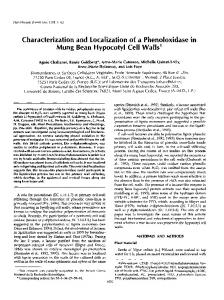
To determine whether or not the Zm Derlin1-1 and Zm Derlin2-1 proteins have a conserved function between species, we performed a yeast complementation assay. The recipient strain harbored a double mutation for Der1 and Ire1, the kinase/endonuclease sensor responsible for initiating signal transduction of ER stress (Mori et al., 1993; Travers et al., 2000). The double mutant offers an easily-scored selection as it is viable at 25°C, but has a temperature-sensitive phenotype, rendering it unable to grow at 37°C (mutants of Der1 alone, in contrast, have no growth phenotype and must be assayed biochemically). If the Zm Derlin genes complemented the phenotype, we would expect to see growth at 37°C. Strains were constructed by transformation with the open reading frames (ORFs) of maize Zm Derlin1-1, Zm Derlin2-1, and Sc Der1 in expression plasmids with selectable markers. Individual transformants were grown overnight, inoculated onto complete media plates in 10-fold serial dilutions, and incubated at either 25°C or 37°C for 4 d. Strains containing either Zm Derlin1-1 or Zm Derlin2-1 sequences grew well even under the restrictive condi-
Figure 4. Complementation of a yeast Ire1/Der1 double mutant with Zm Derlin1-1 (ZmD1-1) and Zm Derlin2-1 (ZmD2-1). Cells were grown to approximately OD600 5 2 (approximately 1 3 105 cells 5 1 OD600) serially diluted in 10-fold increments (as shown from left to right) and inoculated onto solid media. Replica plates were incubated for 4 d at 25°C (permissive temperature) or 37°C (restrictive temperature). ScD1 shows complementation of the double mutant by the yeast Der1 gene. Plant Physiol. Vol. 138, 2005
Maize Derlin Proteins
tions (Fig. 4), whereas cells containing an empty plasmid showed only minimal growth. Cells containing the yeast Der1 sequence (Sc Der1) grew well at 37°C as expected for complementation by the endogenous gene. Zm Derlin mRNAs Are Expressed throughout the Plant
The functional data provided by the yeast complementation experiment led us to predict that the Zm Derlin proteins were involved in ERAD in maize not only during ER stress, but also under nonstress conditions. Because ERAD would be needed for eliminating misfolded proteins in most, if not all, cell types, we surveyed the transcription profiles of the four Zm Derlin genes from a variety of organs, tissues, and developmental stages of the maize inbred B73 by multiple parallel signature sequencing (MPSS; Brenner et al., 2000). During MPSS, more than 1 million 17-nt-long expressed sequence tags (ESTs) from each tissue are obtained, and this allows direct quantitative comparisons of mRNA abundance within and among different tissue samples (Meyers et al., 2004b). Table II shows the abundance of MPSS ESTs in representative tissue samples from a comprehensive gene expression data set of several hundred libraries. Zm Derlin1-1, Zm Derlin2-1, and Zm Derlin2-2 genes were expressed throughout the plant, although their relative levels differed with the tissue analyzed. Zm Derlin1-1 mRNA was more abundant than transcripts of the other three Zm Derlins. Zm Derlin1-2 transcripts were detected only in the endosperm and root tissues and generally were the least prevalent of the Zm Derlin genes. At 12 days after pollination (DAP), abundance of Zm Derlin1-1 and 1-2 transcripts was reduced compared to other developmental stages. However, this combined decrease in expression was not observed at the protein level (data not shown). Expression of Zm Derlin Genes Is Differentially Induced during ER Stress
We expanded our expression analysis of the Zm Derlin genes to compare their expression during ER Table II. Relative abundance of Zm Derlin MPSS ESTsa
a
Tissue
Derlin1-1
Derlin1-2
Derlin2-1
Derlin2-2
Immature ear Root Leaf Embryo Stalk Endosperm 8 DAP Endosperm 12 DAP Endosperm 21 DAP Endosperm 30 DAP Endosperm 35 DAP Endosperm 40 DAP
118 333 128 101 119 122 95 190 203 202 80
0 13 0 0 0 96 3 51 20 16 30
7 45 33 36 48 49 74 90 61 59 73
92 33 18 22 59 19 65 110 54 58 76
Abundance shown in parts per million (ppm) MPSS ESTs.
Plant Physiol. Vol. 138, 2005
stress. Semiquantitative reverse transcription (RT)PCR and quantitative RT-PCR were carried out under nonsaturating conditions, with RNA isolated from developing endosperm tissue. Tissue was collected from the inbred line W64A(1), the near-isogenic ERstress mutants fl2, Mc, and De*-B30, and the endosperm opacity mutant opaque-2 (o2), which lacks a zein transcription factor but does not show an ER stress response (Schmidt et al., 1987; Boston et al., 1991). As shown in Figure 5A, the signal for the Zm Derlin1-1 gene was dramatically increased in the maize mutants that exhibited an ER stress response compared to the normal line and was not increased in the o2 mutant. The signals of the other three Zm Derlin genes were weaker and showed only marginal changes across samples under our assay conditions. As an amplification and loading control, we used a maize catalase gene that is uniformly expressed during endosperm development (Redinbaugh et al., 1988; Bass et al., 1994). To confirm that induction of Zm Derlin1-1 expression was specific for ER stress, we compared endosperm and embryo tissues from normal and fl2 lines. Because embryos do not synthesize zeins, they do not make mutant (or normal) zein storage proteins and thus do not exhibit the ER stress response seen in mutant endosperm. As shown in Figure 5B, only the endosperm sample from the fl2 mutant showed an obvious increase in Zm Derlin1 expression. The signal difference for the Zm Derlin2 amplification product appeared insignificant between embryo and endosperm tissues of the fl2 mutant, although perhaps increased compared to the normal endosperm sample. To gain better resolution of the expression profiles of the Zm Derlin genes during ER stress, we performed real-time quantitative PCR (qPCR) assays of RNA from endosperm (Fig. 5C). qPCR allows accurate and real-time measurement of an amplified PCR product in independent samples (for review, see Gachon et al., 2004). Figure 5C shows a comparison of mRNA levels for the Zm Derlin genes normalized to 28S rRNA and calibrated against the normalized wild-type control. Zm Derlin1-1 showed strong induction and high expression levels in the three mutants associated with ER stress and a slight induction in the o2 mutant. Zm Derlin1-2 showed a qualitatively similar profile, except that ER stress induction was less pronounced. Expression of the Zm Derlin2 genes did not correlate strictly with ER stress. Zm Derlin2-1 expression was higher in all the mutants than in the normal control, while Zm Derlin 2-2 expression was relatively constant across all of the samples. The differences in Zm Derlin expression profiles led us to search maize GSS contigs that extended into the 5# region of the Zm Derlin cDNAs (Table I) for upstream elements associated with ER stress (Roy and Lee, 1999; Kokame et al., 2001; Okada et al., 2002; Martinez and Chrispeels, 2003). This examination revealed two overlapping cis-acting elements in Zm Derlin1-1 (CCACGTtA and [CCa]CCtcatcacccagC223
Kirst et al.
Figure 5. Comparison of Zm Derlin gene expression by semiquantitative (A and B) and quantitative (C) RT-PCR. First-strand cDNA was synthesized from total RNA isolated from dissected endosperm or embryo tissues of wild-type or mutant maize lines harvested 18 DAP. A, Amplification of endosperm transcripts. Ethidium bromide-stained gels are shown for amplification products from 30 cycles with primers specific for the four Zm Derlins and a catalase 1 (CAT1) loading control. A duplicate reaction for Zm Derlin1-1 (ZmD1-1) was terminated after 20 cycles (top). B, Amplification of Zm Derlin1 (ZmD1) and Zm Derlin2 (ZmD2) transcripts in embryo and endosperm tissues from wild-type and fl2 maize. The primer sets used for amplification did not discriminate between the sequences within the two Zm Derlin families. C, Amplification of Zm Derlin transcripts under real-time qPCR conditions. Histograms show quantitative transcript data for Zm Derlin genes in wild-type and mutant endosperm. Values for each were calibrated to the normalized wild-type sample. Bars 5 mean 6 SD (n 5 3–4). Different scales are used in graphs.
CACG) that had strong homology to motifs 1 (CCACGTNA) and 2 (CCN12CCACG) previously identified on the coding strand beginning 263 nt upstream of the ORF in some Arabidopsis genes induced by ER stress (Martinez and Chrispeels, 2003). These elements, as well as one in the single Arabidopsis Derlin1 gene (Table I), share the common CCACG core sequence present in the ERSE-I and ERSE-II elements of induced mammalian genes (Roy 224
and Lee, 1999; Kokame et al., 2001). The Arabidopsis sequence, however, is present in the reverse orientation 190 nt upstream of the ORF of At Derlin1-1. A sequence (CCaaattttcaatCCACG) similar to the Arabidopsis motif 2 was present 1,910 nt upstream of the Zm Derlin2-1 ORF, but we were unable to find sequences resembling any of the ER stress elements upstream of the remaining two Zm Derlin genes (M.E. Kirst, unpublished data). Similarly, we found only Plant Physiol. Vol. 138, 2005
Maize Derlin Proteins
weak homology to a mammalian element 648 nt from the ORF of the At Derlin2-1 gene and no cis-acting element in the At Derlin2-2 sequence. The RNA expression data and virtual promoter analysis suggested that the Zm Derlin1 genes were linked specifically to a function in the ER stress response. We extended our analysis to include protein accumulation for the Zm Derlins as judged by immunoblotting. We obtained antiserum against a Zm Derlin2-1 peptide (shared with Zm Derlin2-2) and located within a domain predicted to be in the lumen of the ER. The peptide had 52% identity to the corresponding peptide in the Zm Derlin1 proteins (Fig. 2, wavy line above Zm Derlin2-1) and the antibody did not cross-react with Zm Derlin1. Figure 6 shows immunoblots probed for the Zm Derlins. Accumulation of Zm Derlin1 proteins reflected the pattern observed for mRNA with a strong up-regulation in the mutants associated with ER stress. The o2 mutant showed a slight increase of the immunoblot signal over the normal line even though both were much lower than in the mutants linked to ER stress. For Zm Derlin2, the relative signal strengths were reversed, with o2 being strongest, followed by the normal line, and then the mutants associated with ER stress having the weakest signals. We detected a strong induction of the well-characterized molecular chaperone, BiP, in endosperm of the ER stress mutants (Fig. 6), as shown in previous studies (Boston et al., 1991; Fontes et al., 1991; Marocco et al., 1991). Thus, on the protein level, Zm Derlin1 accumulation appears induced by the ER stress response, whereas Zm Derlin2 (at least the portion or polypeptide conformation that can be detected with the antibody) appears to decrease. Zm Derlin1 and Zm Derlin2 Proteins Associate with Protein Bodies
In the mutants associated with ER stress, molecular chaperones have been found in association with pro-
Figure 6. Accumulation of Zm Derlin proteins in normal and mutant endosperm tissue. Equal amounts of protein from total membrane preparations (5 mg of protein representing approximately 250 mg of endosperm) were separated through 15% SDS-polyacrylamide gels and probed by immunoblotting. Duplicate membranes probed with antiBiP or anti-aATPase antibodies were used as ER stress and protein loading controls, respectively. Plant Physiol. Vol. 138, 2005
Figure 7. Distribution of Zm Derlin proteins in subcellular fractions of normal and fl2 endosperm tissue. Cisternal ER and protein body (PB) fractions from discontinuous Suc gradients were analyzed by immunoblotting based on equal fresh weight equivalents (1.25 mg). Blots were probed for Zm Derlin1 (ZmD1), Zm Derlin2 (ZmD2), or calnexin (CNX) and calreticulin (CRT).
tein bodies, which are the predominant location of the mutant zeins (Galante et al., 1983; Zhang and Boston, 1992). To determine whether Zm Derlins similarly localized preferentially to protein bodies in endosperm of ER stress mutants, we separated ER into cisternal and protein body fractions (Gillikin et al., 1995). Figure 7 shows proteins from W64A1 and W64Afl2 endosperm loaded by equivalent fresh weights and probed for Zm Derlin1 or Zm Derlin2 by immunoblot analysis. Both Zm Derlin1 and Zm Derlin2 were detected primarily in the protein body fractions from the normal or fl2 samples. Consistent with the data in Figure 6, we detected more Zm Derlin1 in the mutant, whereas Zm Derlin2 protein appeared lower in the mutant sample when compared to the protein body fraction isolated from normal endosperm. Calnexin and calreticulin controls are shown as references for the distribution of known molecular chaperones between membrane and luminal fractions of the two organelles, respectively. In the experiment shown in Figure 7, the Zm Derlin1 signal was detected as a doublet. We frequently detected the migration of Zm Derlin1 bands as a doublet that can occur in both normal and mutant endosperm (e.g. sample from the normal line in Fig. 6). The doublet is unlikely the result of proteolytic processing of the protein as the smaller of the two bands is detected by antibodies against the NH2-terminal region shared between Zm Derlin1-1 and Zm Derlin1-2 (Fig. 2, wavy lines), as well as by an antibody developed against a COOH-terminal peptide (PPANGNSGSGVFRGRSYRLN) of Zm Derlin1-1 (M.E. Kirst and R.S. Boston, unpublished data).
DISCUSSION
The data presented here indicate that proteins associated with the ERAD machinery in yeast and animals are conserved in plants. Among putatively up-regulated genes in endosperm of maize ER stress 225
Kirst et al.
mutants, we detected two homologs of a yeast Der1 gene. This finding is of interest because, although very little is known about the ERAD pathway in plants, mammalian Derlins and yeast Der1p have been directly implicated in the retrotranslocation of misfolded proteins from the ER lumen into the cytosol (Lilley and Ploegh, 2004). Searches of gene databases identified two additional maize Derlin genes as well as multiple Derlin homologs in other plant species (Table I). The four maize genes are members of two Derlin gene subfamilies, Derlin1 and Derlin2, each represented by two nearly identical maize paralogs. Based on RFLP mapping, we placed both Zm Derlin1 paralogs at the same locus on chromosome 8. Similarly, both Zm Derlin2 paralogs mapped to one locus on chromosome 1. The emergence of these two pairs of paralogs is therefore consistent with recent tandem gene duplications rather than duplications resulting from the allotetraploidy event that occurred 5 to 10 million years ago in the progenitor of maize (Helentjaris et al., 1988; Gaut and Doebley, 1997; Swigonova et al., 2004). The putative Derlin proteins from other plant species, including mosses, gymnosperms, and eudicots, group in phylogenetic alignments in deeply rooted clades either with the Zm Derlin1 proteins or with the Zm Derlin2 proteins (R. Jung, unpublished data). Interestingly, Derlin proteins from animals also fall into two deeply rooted protein subfamilies. Moreover, this clustering extends beyond the plant kingdom, although the posterior probability values are much lower. In alignments of plant Derlins with animal Derlins (Lilley and Ploegh, 2004), one group of animal Derlins (e.g. Hs Derlin-1) appears to cluster with the plant Derlin1 subfamily, and the other group of animal Derlins (e.g. Hs Derlin-2 and Hs Derlin-3) appears to group with the plant Derlin2 proteins. In contrast, the single yeast Derlin protein, Der1p, has similar overall identity (approximately 15%) to both plant Derlin subfamilies. Together, these data are indicative of the emergence of the two Derlin gene subfamilies prior to the development of vascular plants and perhaps even earlier, during eukaryotic evolution, predating the split of plants and animals. The maintenance of two diverged Derlin protein subfamilies during evolution strongly suggests that, in addition to a core Derlin function, each subfamily has a nonredundant and important, if not essential, biological activity. Zm Derlin1-1 and Zm Derlin2-1 were able to suppress the mutant phenotype of a yeast strain lacking Der1 and the capacity to sense ER stress (Dire1). This complementation confirmed that the heterologous maize proteins possess characteristics essential for the core function of a Derlin protein in yeast. At 37°C, the yeast growth was less robust than at 25°C. This difference, however, was observed for both the yeast and Zm Derlin transformants. A similar result was observed for the C. elegans Derlin gene (GenBank accession no. NM_066189), which reestablished growth of the yeast mutant in a complementation experiment (Hitt and Wolf, 2004). The C. elegans Derlin also partially restored 226
the capacity of the yeast mutant to degrade a soluble misfolded substrate, thus confirming conservation of the ERAD function across widely diverged species. The plant Derlin proteins have diverged considerably from the yeast Der1 protein (approximately 15% amino acid identity) during evolution. However, both Zm Derlin1-1 and Zm Derlin2-1 have a conserved Ser residue, suggested by Hitt and Wolf (2004) to be essential for Der1p function. Whether this residue is, in fact, important in preserving Der1 activity among species remains to be investigated. Because the maize proteins have little sequence homology to the yeast Der1p and members of both subfamilies complement the yeast mutant, members of both plant Derlin families may be considered orthologs of yeast Der1p. The induction of Zm Derlin1-1 and Zm Derlin1-2 mRNAs in the fl2, Mc, and De*-B30 endosperm mutants (Fig. 5) was consistent with previous reports of expression of putative ERAD genes being upregulated during pharmacologically induced ER stress (Travers et al., 2000; Martinez and Chrispeels, 2003). ER-stress induction has been linked to the presence of sequence motifs that can be found in either orientation upstream of many, but not all, target genes (Roy and Lee, 1999; Kokame et al., 2001; Martinez and Chrispeels, 2003). Zm Derlin1-1, which shows a strong ER-stress response, has multiple such cis-acting elements located in close proximity to the coding region. In contrast, Zm Derlin1-2 had no recognizable ER stress elements, even though it shows a similar, albeit attenuated, induction as Zm Derlin1-1. Zm Derlin2-1, which showed weak up-regulation during ER stress as well as in the o2 mutant (Figs. 1 and 5), has a single cis-acting element located 1,910 nt upstream of the protein coding region, while Zm Derlin 2-2 lacks any recognizable ERstress motifs. The upstream regions of Zm Derlin homologs in Arabidopsis show similar features, with the apparent ortholog of Zm Derlin1 having a more proximal element than the apparent orthologs of Zm Derlin2 (only one of which has an element). These putative regulatory features, along with the transcript and protein accumulation data, suggest that Zm Derlin1-1 and Zm Derlin1-2 have biological roles in maintaining and restoring ER homeostasis after perturbation of the protein secretory pathway. Zm Derlin2-1 may also participate in the ER stress response; however, its role would appear to be more general as it was also induced in the o2 mutant (Fig. 5C). Such a situation has precedent in the well-studied stress 70 family of molecular chaperones whose members show differential responses to stress. Hsp70 proteins are induced by heat stress, whereas BiPs are induced by ER stress, and some of the other Hsc70 members appear to be needed during normal growth and development (Guy and Li, 1998; Lin et al., 2001). Hsp70 expression is regulated by the heat stress transcription factors that bind to cis-acting elements, whereas elements upstream of BiP are recognized by different factors (Yoshida et al., 1998; Foti et al., 1999; Pirkkala et al., 2001). Plant Physiol. Vol. 138, 2005
Maize Derlin Proteins
The induction of the Zm Derlin1-1 gene by ER stress led to increases in both RNA and protein. The defective zeins in the ER stress mutants are clearly responsible for initiating induction of the ER stress response and they may also be the targets of Derlinmediated degradation. Normal zeins have no Lys residues and thus lack the epsilon amino groups that serve as ubiquitin conjugation sites. Each of the mutations, however, leaves the affected zein with a Lys residue. The new Lys in the fl2 and De*-B30 mutants lies in the uncleaved signal peptide at the NH2 terminus, while the one in the Mc mutant lies very near the COOH terminus (J. Gillikin, R. Jung, and R.S. Boston, unpublished data). Mc has a dramatically lower accumulation of the 16-kD g-zein and fl2 has less of the 22-kD a-zein when compared to a normal maize line or other endosperm mutants (Hunter et al., 2002). The possible decrease of the 19-kD a-zein in the De*-B30 mutant cannot easily be assessed because the corresponding gene accounts for only 4% of the transcripts encoding members of the large 19-kD a-zein gene family. As a result, the protein encoded by the normal allele would be masked by the more abundant protein family members. In animals, ER stress can lead to an inhibition of translation mediated through the transmembrane kinase, PERK (for review, see Liu and Kaufman, 2003). Thus, it may be possible that the lower amounts of defective zeins are due to selective translational attenuation. Nevertheless, we think removal by ERAD is most likely because the lower accumulation appears rather specific for the defective zein in each respective mutant. In none of the other mutants associated with ER stress did we see a specific decrease in the accumulation of proteins encoded by the corresponding nondefective alleles. Through subcellular fractionation, we observed that the Zm Derlin proteins predominantly associated with the protein bodies that are protein storage compartments derived from the ER. Although both types of Zm Derlins were detected in the protein body fraction, Zm Derlin1 was more abundant in the fl2 mutant compared to normal maize, whereas the converse pattern was found for Zm Derlin2, which accumulated to higher levels in the normal line. At present, we cannot explain this difference. It is plausible that the apparent decrease of Zm Derlin2 is the result of translational attenuation of mRNAs not needed during an ER stress response (Harding et al., 2002). Alternatively, it is also possible that, in response to the ER stress, the immunoepitope has undergone a posttranslational modification, rendering it no longer recognizable by the antipeptide antibody. Our attempts to raise specific antibodies against a different Zm Derlin2 epitope so far have been unsuccessful (D.J. Meyer, M.E. Kirst, and R. Jung, unpublished data), and we have not been able to test this hypothesis. In this context, it is noteworthy that Zm Derlin1 migrated electrophoretically as a doublet of approximately 23.5 and 25 kD, from which the faster migrating band was detected equally well with peptide antibodies regardPlant Physiol. Vol. 138, 2005
less of whether they were made against the NH2 or COOH terminus of the protein (Figs. 6 and 7). This observation rules out a simple proteolytic cleavage as an explanation for the size difference and leaves us favoring the possibility of another posttranslational modification of the Zm Derlin1 polypeptides. It is further remarkable that the prevalence of one polypeptide of the doublet over the other seemed to change in response to the presence of ER stress (Fig. 7). The observations that Zm Derlin1-1 and Zm Derlin2-1 complemented the yeast Der1p function and associated with protein bodies, but that primarily Zm Derlin1 was strongly induced during ER stress, suggest that the two proteins have undergone some functional divergence in maize. One possibility is that both proteins serve a function in normal cellular metabolism, and possibly protein degradation, while only Zm Derlin1 is part of the ERAD pathway operating in response to ER stress. The Zm Derlins contain GxxxG motifs that have been linked to interactions between transmembrane segments and oligomerization of transmembrane helices (Ubarretxena-Belandia and Engelman, 2001; Langosch et al., 2002). They also contain weak homology to some elements of the Rhomboid family, which are multimembranespanning proteins with intramembrane Ser protease activity (Koonin et al., 2003). Even though the exact function of Derlins remains unknown in any system, studies in mammalian cells have shown its requirement for dislocation of misfolded proteins from the ER (Lilley and Ploegh, 2004). In preliminary yeast two-hybrid experiments, Zm Derlin1-1 interacted with a putative ubiquitin-binding protein containing UBQ and UBA domains suggested to recruit ubiquitinated substrates for release to the proteasome (M.E. Kirst and R. Jung, unpublished data; Buchberger, 2002). Several putative interacting partners for Der1 have been identified in high-throughput yeast two-hybrid screens (Ito et al., 2001; Giot et al., 2003; Li et al., 2004). For example, Drosophila and C. elegans putative Derlin1 homologs interacted with proteins encoded by genes induced during ER stress. The Derlin2 homolog in Drosophila also showed a possible link to ERAD, as both it and the Cdc48 AAA ATPase that is associated with proteasomal targeting, bound to the same phosphatase. However, other interacting partners identified in the large-scale screens lacked an obvious link to ERAD or ER stress (Ito et al., 2001; Giot et al., 2003; Li et al., 2004). Clearly, further confirmation is needed to understand the protein-protein interactions that are functionally important for the Derlins during ERAD. A step toward this goal was made in an animal cell culture system in which immunoprecipitation assays placed Derlin-1 in a complex with the Cdc48 AAA-ATPase (Ye et al., 2004). Further investigation into the interacting partners of the various Zm Derlins will likely provide much-needed insight into the role of these proteins in ERAD or other cellular processes. 227
Kirst et al.
MATERIALS AND METHODS Plant Material The normal maize (Zea mays) inbred W64A(1) and its near-isogenic mutants, fl2, Mc, o2, and De*-B30, were grown and self-pollinated at the Central Crops Research Station, Clayton, North Carolina, during summer field seasons for all experiments, except the GeneCalling experiment where plants were grown in the summer of 1998 in field plots at the Pioneer Hi-Bred International genetic nursery in Johnston, Iowa (Hunter et al., 2002). Wellfilled ears of each line were harvested 18 and 23 d after self-pollination between 7:30 and 9:30 AM, immediately frozen in liquid nitrogen, and stored at 280°C until use.
GeneCalling Analysis Endosperm was dissected from frozen seed from the middle portions of the ears. To minimize the effect of biological variation between ears on the gene expression analysis, equal numbers of endosperms from three ears were pooled and treated as one sample. Total RNA was isolated from ground endosperm tissues using the PUREscript kit (Gentra Systems, Minneapolis), according to the manufacturer’s instructions, and mRNA profiling was performed at CuraGen (New Haven, CT) by GeneCalling, essentially as described by Shimkets et al. (1999). In brief, cDNA was synthesized from three independently pooled endosperm samples per genotype (biological repeats). Each of the resulting 12 cDNA preparations was further divided into three aliquots (technical repeats) to provide together nine repeats per genotype for the profiling analysis. Each of these cDNA aliquots was digested in parallel reactions with 64 different combinations of restriction enzyme pairs. Fragments from each digest were ligated to adapters; the fragments were amplified with primers that have unique tags (biotin on one end, fluorescent marker at the other). Labeled fragments were purified using streptavidin beads and were resolved by high-resolution gel electrophoresis to generate traces showing peaks whose position and height represented Mr and abundance of cDNA fragment(s), respectively. Trace data were used for qualitative (Mr) and quantitative (abundance) comparisons between the W64A1 and mutant samples. GeneCalling software compiled a list of differentially abundant fragments and assigned a ranking (significance) to each detected difference. The software further searched a nucleic acid database for fragments with the same length and end sequences and predicted likely gene candidates. The identity of predicted fragments was confirmed by competitive amplification with an unlabeled gene-specific primer (poisoning) or by cloning and sequencing the fragment (Shimkets et al., 1999).
Sequence Alignment Alignment of deduced amino acid sequences of two Zm Derlin proteins with putative homologs from other organisms was performed by ClustalW analysis in the software package Vector NTI suite 8 (Informax, Bethesda, MD).
Subcellular Fractionation Endosperm was removed from kernels and ground (1:2, w/v) in buffer B (10 mM Tris-HCl, pH 8.5, at 25°C, 10 mM KCl, 5 mM MgCl2, and 7.2% [w/v] Suc) by mortar and pestle (Shank et al., 2001). Homogenates were incubated on ice to allow the starch and cell debris to settle, and the remaining material was used as crude extracts. All extraction and cellular fractionation steps were carried out at 0°C to 4°C. Microsomal membrane isolation (Fig. 3; Shank et al., 2001): Crude extracts were subjected to centrifugation at 5,000g for 10 min to remove protein bodies (pellet fraction), leaving a microsomal fraction containing cisternal ER and other membranes. The supernatant was subjected to centrifugation at 100,000g for 30 min in a fixed-angle rotor to collect the microsomes. For separation of the microsomal fraction into membrane and luminal fractions (Fujiki et al., 1982), samples were prepared as described by Shatters and Miernyk (1991), with minor additional modifications. The microsomal pellet was resuspended in 1 mL of 100 mM Na2CO3, pH 11.5, and incubated under constant agitation at 4°C for 30 min before being placed on a Suc pad (100 mM NaHCO3, pH 8.3, 500 mM Suc) and subjected to centrifugation at 100,000g for 30 min. This fractionation step leaves the luminal contents in the supernatant while
228
membranes form a pellet below the Suc pad. The membrane fraction was resuspended in buffer B. For detergent solubilization, microsomal pellets were resuspended in 50 mM Tris-HCl, pH 6.8, containing the nonionic detergents digitonin, Nonidet P-40, or Triton X-100, or the ionic detergent deoxycholate at a final concentration of either 0.4% or 1.0%, and incubated for 30 min at 4°C under constant agitation prior to centrifugation at 100,000g for 30 min. Digitonin was further purified according to the method of Gorlich and Rapoport (1993) prior to use. For total membrane isolation (Fig. 6), crude extracts were subjected to centrifugation at 100,000g to yield a single pellet containing protein bodies, cisternal ER, and other membranes. Protein was quantified with a bicinchoninic acid protein assay kit and bovine serum albumin standard (Pierce, Rockford, IL). For fractionation by Suc density gradient centrifugation (Fig. 7), crude extract was filtered through two layers of miracloth, overlayed on a 2.0 M Suc solution in buffer B, and subjected to centrifugation for 10 min at 164g in a swinging-bucket rotor. The supernatant fraction from the low-speed centrifugation was recovered and applied to a discontinuous Suc gradient prepared as 2-mL steps of 2.0 M, 1.5 M, 1.0 M, and 0.5 M Suc in buffer B. Gradients were subjected to centrifugation at 80,000g in a TFT-41.14 swinging-bucket rotor for 30 min at 4°C. The cisternal ER was collected from the 1.0/ 1.5 M Suc interface and the protein bodies were collected from the 1.5/2.0 M Suc interface (Gillikin et al., 1995).
Immunoblot Analysis Samples were adjusted to 23 SDS-PAGE sample buffer (Laemmli, 1970) and boiled for 5 min prior to fractionation through 15% SDS-polyacrylamide gels. Proteins were transferred by semidry blotting to Immobilon-P membranes (Millipore, Billerica, MA) in the presence of transfer buffer (48 mM Tris base, 39 mM Gly, pH 9.2) for 90 min at 1 mA/cm2 of gel area. Membranes were blocked with 5% (w/v) nonfat dry milk for 1 h prior to incubation with primary antibody. For detection of the Zm Derlin proteins, immunoblots were probed with polyclonal antibodies raised in rabbits against peptide SPAEYYKSLPPISKAYG from the Zm Derlin1 proteins at a 1:10,000 dilution in Tris-buffered saline plus Tween (TBST; 20 mM Tris-HCl, pH 7.5, at 25°C, 0.14 M NaCl, 0.1% [v/v] Tween 20) or antibodies against peptide RYCKLLEENSFRGRTAD from the Zm Derlin2 proteins at a 1:15,000 dilution in TBST. Antibodies against BiP (ID9; Stressgen, Victoria, British Columbia, Canada), calnexin and calreticulin (Pagny et al., 2000), and 19- or 27-kD zeins (Hunter et al., 2002), were used at 1:10,000 dilution in TBST. Antiserum against the mitochondrial aATPase (Luethy et al., 1993) was used at a 1:1,000 dilution in TBST. Following incubation with primary antibodies, immunoblots were incubated with the appropriate secondary antibody against rabbit or mouse coupled to horseradish peroxidase (Bio-Rad Laboratories, Hercules, CA) at a 1:10,000 dilution in TBST. Visualization of proteins was made with chemiluminescent substrates (Pierce) by exposure to x-ray film (X-OMAT; Eastman-Kodak, Rochester, NY).
Quantitative Expression Analysis of Derlin Genes by MPSS The mRNA from a variety of maize tissue samples (inbred line B73) was previously isolated and MPSS was performed by Lynx Therapeutics (Hayward, CA) as described (Brenner et al., 2000; Meyers et al., 2004a). The resulting MPSS ESTs, here defined as the first 17 bp, including and following downstream of the most 3# Sau3A site (GATC) of a gene transcript, are quantified and reported on a ppm basis (1–2 million sequencing reactions performed per sample) in a searchable database. The quantity of Derlin MPSS ESTs in each tissue sample were then obtained by queries of this database with the exact string of the conceptual MPSS ESTs identified for Zm Derlin1-1 (5#-GATCCATGGTCGGAGTG-3#); for Zm Derlin1-2 (5#-GATCTAAATGTTTTTTC-3#); for Zm Derlin2-1 (5#-GATCTGAGATGCGGCAG-3#); and for Zm Derlin2-2 (5#-GATCGTGATGGGCAAAC-3#).
RT-PCR Analysis Total RNA was isolated from embryo (18 DAP) or endosperm (18 DAP) tissues (200 mg) using the TRIzol reagent (Invitrogen, Carlsbad, CA) by
Plant Physiol. Vol. 138, 2005
Maize Derlin Proteins
a modification of the manufacturer’s protocol in which the RNA precipitation step was carried out by addition of 0.5 volume of a 1.2 M NaCl/0.8 M sodium citrate solution and 0.5 volume of 100% (v/v) isopropanol. The RNA was resuspended in 50 mL of RNase-free deionized water. All RNA samples were treated with RQ1 RNAse-free DNase I (Promega, Madison, WI) to eliminate contaminating genomic DNA. Following DNAse treatment, samples were purified by phenol/chloroform and used for cDNA synthesis. Two micrograms of RNA were incubated with 1 mM of oligo(dT)(20) primer and 200 units of M-MLV reverse transcriptase (Promega) for 1 h at 42°C, as described by the manufacturer to produce first-strand cDNA. One microliter of each cDNA reaction was used as template for amplification by PCR. Reactions were performed in buffer containing 10 mM Tris-HCl, pH 8.3, at 25°C, 50 mM KCl, 2.5 mM MgCl2, 200 mM dNTPs, 0.5 mM each primer, and 0.5 units Taq polymerase (Roche, Indianapolis). Standard PCR cycle conditions were 95°C for 5 min followed by 30 cycles of 95°C for 1 min, 55°C for 1 min, and 72°C for 1 min. Products were resolved by electrophoresis through 1.5% (w/v) agarose gels in 13 Tris-acetate buffer. Primers used to amplify Zm Derlin genes were (Fig. 5A) Zm Derlin1-1, 5#-GGGTATCATGGTTGGACATC3# and 5#-GATCACATTACGAGGTGGGT-3#; Zm Derlin1-2, 5#-ATAGGTATGCAAGCCAACGCTCCT-3# and 5#-TCTGCCCTATCCGCAACCCTTAATC-3#; Zm Derlin2-1, 5#-TTTAACACTCCACCCAACCCAACC-3# and 5#-TCAGGTACAGGTGATACGGCGAAA-3#; Zm Derlin2-2, 5#-TGCTGATGACAATGTTGTGGTGGC-3# and 5#-TGCATGAAAGGGTAGCAGGTCAGA-3# (Fig. 5B); Zm Derlin1, 5#-TCTGGAGCCGAGAGAATCCAAATG-3# and 5#-CTTCTACATTGCCACTCCGACCA-3#; or Zm Derlin2, 5#-GCATTGTTCTGATCGGAGGGATG-3# and 5#-CGGCCCTCAGGTCACTGAAAGTAA-3#. The primers used to amplify the catalase 1 loading control (GenBank accession no. X12538) were 5#-GTCCAGACACCTGTTATTGTCCGT-3# and 5#-GAGGAAGGTGAACATGTGTAGGCT-3#. To determine optimal conditions for the RT-PCR, the sample from the normal endosperm line was amplified and sampled at various points between 20 and 50 cycles. Subsequent experiments were carried out at three conditions, 20 cycles (far below saturation for all samples); 30 cycles (below saturation but having visible bands for all samples); and 50 cycles (saturated for some samples).
Quantitative RT-PCR Analysis Total RNA was isolated from endosperm (18 DAP) tissue from normal and mutant lines using the total RNA isolation mini kit (Agilent Technologies, Palo Alto, CA) following the manufacturer’s protocol. Two micrograms of total RNA were treated with 2 3 DNaseI (Fermentas, Hanover, MD). Twenty nanograms of treated RNA (2 ng for 26S rRNA control) were used for cDNA production with a mix of random and oligo(dT) primers and RT-PCR amplification with sequence-specific primers using the iScript one-step RTPCR kit for probes (Bio-Rad) on an ABI 7900 thermocycler. Quantitative RTPCR reactions were performed for 10 min at 50°C followed by 5 min at 95°C prior to amplification for 40 cycles of 15 s at 95°C and 1 min at 59°C with fluorescence reading during the annealing steps. All samples were assayed in triple or quadruple reactions. Samples were normalized to the 28S rRNA signal and calibrated to the normalized wild-type endosperm sample.
Yeast Complementation of Ire1/Der1 Mutants The ire1/der1 double-mutant strain (MATa, trp1-1, his3, ade2-1, can1-100, Dire1::LEU2, Dder1::URA3; Travers et al., 2000) was transformed with the plasmid vector pRS313 (Sikorski and Hieter, 1989) containing the coding regions for either Zm Derlin1-1, Zm Derlin2-1, or the yeast Der1 gene (YBR401W) inserted between BamHI and SalI restriction enzyme cleavage sites. Genes were amplified by PCR using the following primers: Zm Derlin1-1, 5#-CGGGATCCCGTGAAGATGTCTTC-3# and 5#-CCCAAGCTTTCTATTGATTGAGCC-3#; Zm Derlin2-1, 5#-CGGGATCCGGAGATGGCGCAGGC-3# and 5#-AACTGCAGCATTGAGCCTGGGGA-3#; and yeast der1 5#-GGATCCAAGCAATATGGATGC-3# and 5#-GTCGACTTAGGGTGTTTCAGT-3#. PCR products were subcloned into the pGEM-T easy vector (Promega). Genes were transferred from pGEM-T to pRS313 vectors by digestion with BamHI and SalI. Transformants were selected on synthetic complete-His-Ura media (QBIOgene, Carlsbad, CA). A stable transformant from each complementation was grown overnight until the OD600 reached 2. Cells were spotted onto replica plates in serial 10-fold dilutions and incubated at 25°C or 37°C for 4 d.
Plant Physiol. Vol. 138, 2005
Distribution of Materials Upon request, all novel materials described in this publication will be made available in a timely manner for noncommercial research purposes, subject to the requisite permission from any third-party owners of all or parts of the material. Obtaining any permissions will be the responsibility of the requester. Sequence data from this article have been deposited with the EMBL/ GenBank data libraries under accession numbers AY854013 and AY854020.
ACKNOWLEDGMENTS The authors thank Peter Walter and Christopher Patil for providing the yeast double-mutant cells and Juan Argueso for invaluable assistance and guidance with strain manipulation. We also thank Thomas Elthon for the mitochondrial aATPase antibody and Jeffrey Gillikin, Kendal Hirshi, Ralph Dewey, John Hodge, and Carol Griffin for helpful advice and discussions. We further thank colleagues in the DuPont-Pioneer Bioinformatics and Analytical and Genomics Technologies Departments for creating a comprehensive and searchable gene database of maize, for preparing mRNA samples for the GeneCalling analysis, and for performing the sequence analysis of full-length cDNA clones of maize Derlin clones. We are especially grateful to Julia Wilflingseder and Sandra Meyer for excellent qPCR technical assistance and data analysis support and to Alexander Tikhonov and Oswald Crasta for project management and data analysis support of the GeneCalling project at CuraGen Corporation. Received January 26, 2005; returned for revision February 16, 2005; accepted February 17, 2005.
LITERATURE CITED Bass HW, Goode JH, Greene TW, Boston RS (1994) Control of ribosomeinactivating protein (RIP) RNA levels during maize seed development. Plant Sci 101: 17–30 Boston RS, Fontes EB, Shank BB, Wrobel RL (1991) Increased expression of the maize immunoglobulin binding protein homolog b-70 in three zein regulatory mutants. Plant Cell 3: 497–505 Brenner S, Johnson M, Bridgham J, Golda G, Lloyd DH, Johnson D, Luo S, McCurdy S, Foy M, Ewan M, et al (2000) Gene expression analysis by massively parallel signature sequencing (MPSS) on microbead arrays. Nat Biotechnol 18: 630–634 Brodsky JL, McCracken AA (1999) ER protein quality control and proteasome-mediated protein degradation. Semin Cell Dev Biol 10: 507–513 Bruce W, Folkerts O, Garnaat C, Crasta O, Roth B, Bowen B (2000) Expression profiling of the maize flavonoid pathway genes controlled by estradiolinducible transcription factors CRC and P. Plant Cell 12: 65–80 Buchberger A (2002) From UBA to UBX: new words in the ubiquitin vocabulary. Trends Cell Biol 12: 216–221 Coleman CE, Lopes MA, Gillikin JW, Boston RS, Larkins BA (1995) A defective signal peptide in the maize high-lysine mutant floury 2. Proc Natl Acad Sci USA 92: 6828–6831 Crasta OR, Folkerts O (2003) Open architecture expression profiling of plant transcriptomes and gene discovery using GeneCalling technology. Methods Mol Biol 236: 381–394 di Cola A, Frigerio L, Lord JM, Ceriotti A, Roberts LM (2001) Ricin A chain without its partner B chain is degraded after retrotranslocation from the endoplasmic reticulum to the cytosol in plant cells. Proc Natl Acad Sci USA 98: 14726–14731 Fontes EB, Shank BB, Wrobel RL, Moose SP, OBrian GR, Wurtzel ET, Boston RS (1991) Characterization of an immunoglobulin binding protein homolog in the maize floury-2 endosperm mutant. Plant Cell 3: 483–496 Foti DM, Welihinda A, Kaufman RJ, Lee AS (1999) Conservation and divergence of the yeast and mammalian unfolded protein response. Activation of specific mammalian endoplasmic reticulum stress element of the grp78/BiP promoter by yeast Hac1. J Biol Chem 274: 30402–30409 Fujiki Y, Hubbard AL, Fowler S, Lazarow PB (1982) Isolation of intracellular membranes by means of sodium carbonate treatment: application to endoplasmic reticulum. J Cell Biol 93: 97–102
229
Kirst et al.
Gachon C, Mingam A, Charrier B (2004) Real-time PCR: what relevance to plant studies? J Exp Bot 55: 1445–1454 Galante E, Vitale A, Manzocchi L, Soave C, Salamini F (1983) Genetic control of a membrane component and zein deposition in maize endosperm. Mol Gen Genet 192: 316–321 Gaut BS, Doebley JF (1997) DNA sequence evidence for the segmental allotetraploid origin of maize. Proc Natl Acad Sci USA 94: 6809–6814 Gillikin JW, Fontes EP, Boston RS (1995) Protein-protein interactions within the endoplasmic reticulum. Methods Cell Biol 50: 309–323 Gillikin JW, Zhang F, Coleman CE, Bass HW, Larkins BA, Boston RS (1997) A defective signal peptide tethers the floury-2 zein to the endoplasmic reticulum membrane. Plant Physiol 114: 345–352 Giot L, Bader JS, Brouwer C, Chaudhuri A, Kuang B, Li Y, Hao YL, Ooi CE, Godwin B, Vitols E, et al (2003) A protein interaction map of Drosophila melanogaster. Science 302: 1727–1736 Gorlich D, Rapoport TA (1993) Protein translocation into proteoliposomes reconstituted from purified components of the endoplasmic reticulum membrane. Cell 75: 615–630 Guy CL, Li QB (1998) The organization and evolution of the spinach stress 70 molecular chaperone gene family. Plant Cell 10: 539–556 Harding HP, Calfon M, Urano F, Novoa I, Ron D (2002) Transcriptional and translational control in the mammalian unfolded protein response. Annu Rev Cell Dev Biol 18: 575–599 Helentjaris T, Weber D, Wright S (1988) Identification of the genomic locations of duplicate nucleotide sequences in maize by analysis of restriction fragment length polymorphisms. Genetics 118: 353–363 Hill K, Cooper AA (2000) Degradation of unassembled Vph1p reveals novel aspects of the yeast ER quality control system. EMBO J 19: 550–561 Hitt R, Wolf DH (2004) Der1p, a protein required for degradation of malfolded soluble proteins of the endoplasmic reticulum: topology and Der1-like proteins. FEMS Yeast Res 4: 721–729 Hough RF, Lingam AT, Bass BL (1999) Caenorhabditis elegans mRNAs that encode a protein similar to ADARs derive from an operon containing six genes. Nucleic Acids Res 27: 3424–3432 Hunter BG, Beatty MK, Singletary GW, Hamaker BR, Dilkes BP, Larkins BA, Jung R (2002) Maize opaque endosperm mutations create extensive changes in patterns of gene expression. Plant Cell 14: 2591–2612 Ito T, Chiba T, Ozawa R, Yoshida M, Hattori M, Sakaki Y (2001) A comprehensive two-hybrid analysis to explore the yeast protein interactome. Proc Natl Acad Sci USA 98: 4569–4574 Jarosh E, Taxis C, Volwein C, Bordallo J, Finley D, Wolf DH, Sommer T (2002) Protein dislocation from the ER requires polyubiquitination and the AAA-ATPase Cdc48. Nat Cell Biol 4: 134–139 Kim CS, Hunter BG, Kraft J, Boston RS, Yans S, Jung R, Larkins BA (2004) A defective signal peptide in a 19-kD a-zein protein causes the unfolded protein response and an opaque endosperm phenotype in the maize De*-B30 mutant. Plant Physiol 134: 380–387 Knop M, Finger A, Braun T, Hellmuth K, Wolf DH (1996) Der1, a novel protein specifically required for endoplasmic reticulum degradation in yeast. EMBO J 15: 753–763 Kokame K, Kato H, Miyata T (2001) Identification of ERSE-II, a new cisacting element responsible for the ATF6-dependent mammalian unfolded protein response. J Biol Chem 276: 9199–9205 Koonin EV, Makarova KS, Rogozin IB, Davidovic L, Letellier MC, Pellegrini L (2003) The rhomboids: a nearly ubiquitous family of intramembrane serine proteases that probably evolved by multiple ancient horizontal gene transfers. Genome Biol 4: 19 Krogh A, Larsson B, Gunnar H, Sonnhammer ELL (2001) Predicting transmembrane protein topology with a hidden Markov model: application to complete genomes. J Mol Biol 305: 567–580 Laemmli UK (1970) Cleavage of structural proteins during the assembly of the head of bacteriophage T4. Nature 227: 680–685 Langosch D, Lindner E, Gurezka R (2002) In vitro selection of selfinteracting transmembrane segments: membrane proteins approached from a different perspective. IUBMB Life 54: 109–113 Li S, Armstrong CM, Bertin N, Ge H, Milstein S, Boxem M, Vidalain PO, Han JD, Chesneau A, Hao T, et al (2004) A map of the interactome network of the metazoan C. elegans. Science 303: 540–543 Lilley BN, Ploegh HL (2004) A membrane protein required for dislocation of misfolded proteins from the ER. Nature 429: 834–840 Lin BL, Wang JS, Liu HC, Chen RW, Meyer Y, Barakat A, Delseney M (2001) Genomic analysis of the Hsp70 superfamily in Arabidopsis thaliana. Cell Stress Chaperones 6: 201–208
230
Liu CY, Kaufman RJ (2003) The unfolded protein response. J Cell Sci 116: 1861–1862 Luethy MH, Horak A, Elthon TE (1993) Monoclonal antibodies to the a-and b-subunits of the plant mitochondrial F1-ATPase. Plant Physiol 101: 931–937 Marocco A, Santucci A, Cerioli S, Motto M, Di Fonzo N, Thompson R, Salamini F (1991) Three high-lysine mutations control the level of ATP-binding HSP70-like proteins in the maize endosperm. Plant Cell 3: 507–515 Martinez I, Chrispeels MJ (2003) Genomic analysis of the unfolded protein response in Arabidopsis shows its connection to important cellular processes. Plant Cell 15: 561–576 McCracken AA, Brodsky JL (2003) Evolving questions and paradigm shifts in endoplasmic-reticulum-associated degradation (ERAD). Bioessays 25: 868–877 Meyers BC, Lee DK, Vu TH, Tej SS, Edberg SB, Matvienko M, Tindell LD (2004a) Arabidopsis MPSS: an online resource for quantitative expression analysis. Plant Physiol 135: 801–813 Meyers BC, Vu TH, Tej SS, Ghazal H, Matvienko M, Agrawal V, Ning J, Haudenschild CD (2004b) Analysis of the transcriptional complexity of Arabidopsis thaliana by massively parallel signature sequencing. Nat Biotechnol 22: 1006–1011 Molinari M, Galli C, Piccaluga V, Pieren M, Paganetti P (2002) Sequential assistance of molecular chaperones and transient formation of covalent complexes during protein degradation from the ER. J Cell Biol 158: 247–257 Mori K, Ma W, Gething MJ, Sambrook J (1993) A transmembrane protein with a cdc21/CDC28-related kinase activity is required for signaling from the ER to the nucleus. Cell 74: 743–756 Muller J, Piffanelli P, Devoto A, Miklis M, Elliott C, Ortmann B, SchulzeLefert P, Panstruga R (2005) Conserved ERAD-like quality control of a plant polytopic membrane protein. Plant Cell 17: 149–163 Nishikawa SI, Fewell SW, Kato Y, Brodsky JL, Endo T (2001) Molecular chaperones in the yeast endoplasmic reticulum maintain the solubility of proteins for retrotranslocation and degradation. J Cell Biol 153: 1061–1070 Okada T, Yoshida H, Akazawa R, Negishi M, Mori K (2002) Distinct roles of activating transcription factor 6 (ATF6) and double-stranded RNAactivated protein kinase-like endoplasmic reticulum kinase (PERK) in transcription during the mammalian unfolded protein response. Biochem J 366: 585–594 Pagny S, Cabanes-Macheteau M, Gillikin JW, Leborgne-Castel N, Lerouge P, Boston RS, Faye L, Gomord V (2000) Protein recycling from the Golgi apparatus to the endoplasmic reticulum in plants and its minor contribution to calreticulin retention. Plant Cell 12: 739–756 Pirkkala L, Nykanen P, Sistonen L (2001) Roles of the heat shock transcription factors in regulation of the heat shock response and beyond. FASEB J 15: 1118–1131 Plemper RK, Bordallo J, Deak PM, Taxis C, Hitt R, Wolf DH (1999) Genetic interactions of Hrd3p and Der3p/Hrd1p with Sec61p suggest a retrotranslocation complex mediating protein transport for ER degradation. J Cell Sci 112: 4123–4134 Plemper RK, Wolf DH (1999) Retrograde protein translocation: ERADication of secretory proteins in health and disease. Trends Biochem Sci 24: 266–270 Redinbaugh MG, Wadsworth GJ, Scandalios JG (1988) Characterization of catalase transcripts and their differential expression in maize. Biochim Biophys Acta 951: 104–116 Roy B, Lee AS (1999) The mammalian endoplasmic reticulum stress response element consists of an evolutionarily conserved tripartite structure and interacts with a novel stress-inducible complex. Nucleic Acids Res 27: 1437–1443 Schmidt RJ, Burr FA, Burr B (1987) Transposon tagging and molecular analysis of the maize regulatory locus opaque-2. Science 238: 960–963 Shank KJ, Su P, Brglez I, Boss WF, Dewey RE, Boston RS (2001) Induction of lipid metabolic enzymes during the endoplasmic reticulum stress response in plants. Plant Physiol 126: 267–277 Shatters RG Jr, Miernyk JA (1991) A zein signal sequence functions as a signal-anchor when fused to maize alcohol dehydrogenase. Biochim Biophys Acta 1068: 179–188 Shimkets RA, Lowe DG, Tai JT, Sehl P, Jin H, Yang R, Predki PF, Rothberg BE, Murtha MT, Roth ME, et al (1999) Gene expression analysis by
Plant Physiol. Vol. 138, 2005
Maize Derlin Proteins
transcript profiling coupled to a gene database query. Nat Biotechnol 17: 798–803 Sikorski RS, Hieter P (1989) A system of shuttle vectors and yeast host strains designed for efficient manipulation of DNA in Saccharomyces cerevisiae. Genetics 122: 19–27 Soave C, Salamini S (1984) Organization and regulation of zein genes in maize endosperm. Philos Trans R Soc Lond B Biol Sci 304: 341–347 Swigonova Z, Lai J, Ma J, Ramakrishna W, Llaca V, Bennetzen JL, Messing J (2004) On the tetraploid origin of the maize genome. Comp Funct Genomics 5: 281–284 Taxis C, Hitt R, Park S, Deak PM, Kostova Z, Wolf DH (2003) Use of modular substrates demonstrates mechanistic diversity and reveals differences in chaperone requirement of ERAD. J Biol Chem 278: 35903– 35913 Taxis C, Vogel F, Wolf DH (2002) ER-Golgi traffic is a prerequisite for efficient ER degradation. Mol Biol Cell 13: 1806–1818 Travers KJ, Patil CK, Wodika L, Lockhart DJ, Weissman JS, Walter P (2000) Functional and genomic analyses reveal an essential coordination between the unfolded protein response and ER-associated degradation. Cell 101: 248–258
Plant Physiol. Vol. 138, 2005
Ubarretxena-Belandia I, Engelman DM (2001) Helical membrane proteins: diversity of functions in the context of simple architecture. Curr Opin Struct Biol 11: 370–376 Ye Y, Meyer HH, Rapoport TA (2001) The AAA ATPase Cdc48/p97 and its partners transport proteins from the ER into the cytosol. Nature 414: 652–656 Ye Y, Shibata Y, Yun C, Ron D, Rapoport TA (2004) A membrane protein complex mediates retro-translocation from the ER lumen into the cytosol. Nature 429: 841–847 Yoshida H, Haze K, Yanagi H, Yura T, Mori K (1998) Identification of the cis-acting endoplasmic reticulum stress response element responsible for transcriptional induction of mammalian glucose-regulated proteins. Involvement of basic leucine zipper transcription factors. J Biol Chem 273: 33741–33749 Zhang F, Boston RS (1992) Increases in binding-proteins (BiP) accompany changes in protein body morphology in 3 high-lysine mutants of maize. Protoplasma 171: 142–152 Zhang K, Kaufman RJ (2004) Signaling the unfolded protein response from the endoplasmic reticulum. J Biol Chem 279: 25935–25938
231