INFECTION AND IMMUNITY, Mar. 2002, p. 1209–1218 0019-9567/02/$04.00⫹0 DOI: 10.1128/IAI.70.3.1209–1218.2002 Copyright © 2002, American Society for Microbiology. All Rights Reserved.
Vol. 70, No. 3
Identification and Characterization of hsa, the Gene Encoding the Sialic Acid-Binding Adhesin of Streptococcus gordonii DL1 Yukihiro Takahashi,1* Kiyoshi Konishi,1 John O. Cisar,2 and Masanosuke Yoshikawa1 Department of Microbiology, The Nippon Dental University School of Dentistry at Tokyo, Chiyoda-ku, Tokyo 102-8159, Japan,1 and Oral Infection and Immunity Branch, National Institute of Dental and Craniofacial Research, Bethesda, Maryland 20892-43522 Received 4 September 2001/Returned for modification 24 October 2001/Accepted 27 November 2001
Oral colonization by Streptococcus gordonii, an important cause of subacute bacterial endocarditis, involves bacterial recognition of sialic acid-containing host receptors. The sialic acid-binding activity of this microorganism was previously detected by bacterium-mediated hemagglutination and associated with a streptococcal surface component identified as the Hs antigen. The gene for this antigen (hsa) has now been cloned in Escherichia coli, and its expression has been detected by colony immunoblotting with anti-Hs serum. Mutants of S. gordonii containing hsa inactivated by the insertion of an erythromycin resistance gene or deletion from the chromosome were negative for Hs-immunoreactivity, bacterium-mediated hemagglutinating activity, and adhesion to ␣2-3-linked sialoglycoconjugates. The deletion in the latter mutants was complemented by plasmid-borne hsa, resulting in Hs antigen production and the restoration of cell surface sialic acid-binding activity. The hsa gene encodes a 203-kDa protein with two serine-rich repetitive regions in its 2,178-amino-acid sequence. The first serine-rich region occurs within the amino-terminal region of the molecule, between different nonrepetitive sequences that may be associated with sialic acid binding. The second serine-rich region, which is much longer than the first, is highly repetitive, containing 113 dodecapeptide repeats with a consensus sequence of SASTSASVSASE. This long repetitive region is followed by a typical gram-positive cell wall anchoring region at the carboxyl-terminal end. Thus, the predicted properties of Hsa, which suggest an amino-terminal receptor-binding domain attached to the cell surface by a molecular stalk, are consistent with the identification of this protein as the sialic acid-binding adhesin of S. gordonii DL1. Streptococcus gordonii and other closely related species comprise a numerically prominent group of oral bacteria that occur primarily on the human tooth surface as members of the biofilm community, commonly referred to as dental plaque (9, 11, 13). In addition, these streptococci are also well-known for their ability to colonize damaged heart valves and are among the bacteria most frequently identified as primary etiological agents of subacute bacterial endocarditis (2, 3, 7). Thus, the pathogenic potential of these oral streptococci at nonoral sites may well depend on streptococcal surface components that function primarily in colonization of the tooth surface. A number of streptococcal cell surface components are known to contribute to colonization of the tooth surface, including putative sialic acid-binding adhesins present on many S. gordonii and Streptococcus sanguis strains (9, 11). Interactions mediated by these adhesins include the aggregation of streptococci in the presence of saliva (16, 18), the attachment of these bacteria to saliva-coated hydroxyapatite (SHA) (10, 12), and their adhesion to erythrocytes (RBC) or human polymorphonuclear leukocytes (13, 19, 25; S. Y. Lee, J. O. Cisar, and A. L. Sandberg, unpublished data), all of which are abolished or reduced by sialidase pretreatment of the corresponding host glycoconjugate receptors. Potential receptors include the ␣2-3-linked sialic acid termini of O-linked oligosaccharides of host glycoconjugates such as salivary mucins (19), secretory
immunoglobulin A1 (IgA1) (24), and leukosialin, the major cell surface glycoprotein of human polymorphonuclear leukocytes (25). A putative sialic acid-binding adhesin of S. gordonii DL1 (Challis) is readily detected by bacterium-mediated hemagglutination and also by the adhesion of this strain to immobilized sialoglycoconjugates, interactions that are Ca2⫹ independent (29). These interactions were previously associated with the Hs antigen, a streptococcal cell surface component identified by immunological comparison of S. gordonii surface antigens with those of a spontaneous mutant, selected for its failure to mediate hemagglutination. Specific anti-Hs serum (anti-Hs) blocked the hemagglutinating activity of the parent strain and reacted with fibrillar structures present on the bacterial surface (29). Western blotting of S. gordonii surface components with anti-Hs revealed a diffuse band of high-molecular-weight material above the position of a 200-kDa marker. The purified antigen, which appeared to be associated with carbohydrate, was not readily detected by available protein staining such as silver staining, and this, along with the small amount of purified material available, limited further structural characterization of the Hs antigen and comparison with other streptococcal surface components (29). The present investigation was initiated to identify the gene for Hs antigen (hsa) and thereby establish a molecular approach for the structural and functional characterization of the encoded protein.
* Corresponding author. Mailing address: Department of Microbiology, The Nippon Dental University School of Dentistry at Tokyo, 1-9-20 Fujimi, Chiyoda-ku, Tokyo 102-8159, Japan. Phone: 81-3-32618763. Fax: 81-3-3264-8399. E-mail:
[email protected].
MATERIALS AND METHODS Bacterial strains, plasmids, and growth conditions. All bacterial strains and plasmids, as well as the recombinants that were constructed in this study, are
1209
1210
TAKAHASHI ET AL.
INFECT. IMMUN. TABLE 1. Bacterial strains and plasmids used in this study
Strain or plasmid
Descriptiona
Source or reference
Strains Streptococcus gordonii DL1 (Challis) EM230 EM061 CM100
Wild type DL1 hsa::ermAM (Fig. 1 and 4) DL1 with ermAM inserted at the MunI site of ORF4 (Fig. 4) DL1 but hsa deletion mutant lacking the entire 9.2-kb HindIII-HindIII region, cat (Fig. 2 and 4)
13 This study This study This study
recA1 endA1 gyrA96 thi hsdR17 supE44 relA1 ⫺ ⌬(lac-proAB) [F⬘ traD36 proAB lacIqZ⌬M15]
35
Apr Apr pUC118 with the 9.2-kb HindIII-HindIII insert containing hsa, MCS at the downstream of hsa (Fig. 3) pUC119 with the 9.2-kb HindIII-HindIII insert containing hsa, MCS at the upstream of hsa pIRH901 with a 0.3-kb HindIII-MluI deletion (Fig. 3) pIRH801 with a 2.5-kb EcoRI-HindIII deletion (Fig. 3) pIRH801 with a 5.5-kb SalI-HindIII deletion (Fig. 3) pIRH801 with a 7.6-kb BglII-HindIII deletion (Fig. 3) pIRH801 with a 7.9-kb ScaI-HindIII deletion (Fig. 3) Unidirectionally deleted plasmid of pIRH801 with a 0.7-kb remaining insert that overlaps hsa (Fig. 1) Unidirectionally deleted plasmid of pIRH901 with a 2.0-kb remaining insert that overlaps ORF4 Unidirectionally deleted plasmid of pIRH801 with a remaining insert of 0.4-kb left side end of the 9.2-kb HindIII fragment (Fig. 2) Unidirectionally deleted plasmid of pIRH901 with a remaining insert of 0.4-kb right side end of the 9.2-kb HindIII fragment (Fig. 2) pIRH230 with ermAM inserted at the MluI site (Fig. 1) pIRH061 with ermAM inserted at the MunI site p15Aori ermAM Kmr Pami, cat Apr pR326 with the PCR product amplified with primers PUPF and PUPR (Fig. 2) pIRC001 with the PCR product amplified with primers PDNF and PDNR (Fig. 2) p15Aori, integration plasmid for resident plasmid integration, Spr pMDI10S with the 9.2-kb HindIII-HindIII insert containing hsa pMDS801 with a 0.5-kb SphI-HindIII deletion pMDS801 with a 2.5-kb EcoRI-HindIII deletion pMDS801 with a 7.6-kb BglII-HindIII deletion p15Aori pVA380-1rep, resident plasmid for resident plasmid integration, Emr p15Aori pVA380-1rep, resident plasmid for resident plasmid integration, Spr pAS40S with the 9.2-kb HindIII-HindIII fragment (Fig. 4) pAS40S with the 7.4-kb HindIII-SphI fragment (Fig. 4) pAS40S with the 6.7-kb HindIII-EcoRI fragment (Fig. 4) pAS40S with the 1.6-kb HindIII-BglII fragment (Fig. 4)
33 33 This study
Escherichia coli JM109 Plasmids pUC118 pUC119 pIRH801 pIRH901 pIRH8012 pIRH8671 pIRH8371 pIRH240 pIRH597 pIRH230 pIRH061 pIRH615 pIRH028 pIRE230 pIRE061 pMDC10E pR326 pIRC001 pIRC901 pMDI10S pMDS801 pMDS8741 pMDS8671 pMDS8161 pAS40E pAS40S pAS801 pAS8741 pAS8671 pAS8161
This This This This This This This
study study study study study study study
This study This study This study This This 28 5 This This 28 This This This This 28 28 This This This This
study study study study study study study study study study study study
a Abbreviations and designations: Apr, ampicillin resistant; Emr, erythromycin resistant; Kmr, kanamycin resistant; Spr, spectinomycin resistant; MCS, multiple cloning site of pUC plasmids; Pami, promoter of S. pneumoniae amiACDEF locus; p15Aori, replication origin of plasmid p15A; pVA380-1rep, a gene encoding the putative replication protein in basic replicon of streptococcal plasmid pVA380-1.
listed in Table 1. Unless otherwise indicated, streptococci were cultured overnight at 37°C in complex medium containing 0.5% tryptone, 0.5% yeast extract, 0.5% K2HPO4, 0.05% Tween 80, and 0.2% glucose; the glucose was separately sterilized (17). Escherichia coli strains were maintained in Luria-Bertani (LB) broth or LB agar plates (26). Antibiotics were purchased from Sigma-Aldrich, St. Louis, Mo., and supplemented, when necessary, with the following at the concentrations indicated parenthetically: for streptococci, spectinomycin dihydrochloride (200 g/ml), erythromycin (10 g/ml), or chloramphenicol (8 g/ml); for E. coli, ampicillin sodium salt (50 g/ml), spectinomycin dihydrochloride (50 g/ml), erythromycin (200 g/ml), chloramphenicol (30 g/ml), or kanamycin acid sulfate (50 g/ml). Antisera. Rabbit antisera against S. gordonii DL1 (anti-DL1) and Hs antigen (anti-Hs) have been previously described (29). Anti-Hs serum, absorbed with E. coli JM109(pUC119) lysate (26), was used for the detection of recombinant products in E. coli. DNA manipulations. Chromosomal DNA was prepared from S. gordonii as previously described (1). Plasmids were prepared from transformants of S. gordonii CM100 by the method of Takamatsu et al. (30). Restriction enzymes, T4
DNA ligase, calf intestinal alkaline phosphatase, and Klenow fragment (large fragment of E. coli DNA polymerase I) were purchased from Takara Shuzo Co. Ltd. (Kusatsu, Japan) and used according to the supplier’s instructions. Transformation of E. coli with plasmids was performed by electroporation with a Gene Pulser II apparatus (Bio-Rad Laboratories, Hercules, Calif.). Transformation of S. gordonii was carried out as previously described (22). Primers used in this study are listed in Table 2. PCR and inverse PCR were carried out with VentR DNA Polymerase (New England Biolabs Inc., Beverly, Mass.). Preparation of DNA probes and Southern hybridization were carried out with the ECL random-prime labeling and detection systems, version II (Amersham Pharmacia Biotech, Little Chalfont, United Kingdom). A series of unidirectional deletions, prepared from pIRH801 and pIRH901 by use of a Kilo-Sequence Deletion kit (Takara), were utilized both as templates for DNA sequencing and for studies of hsa gene expression in E. coli recombinant clones. DNA sequencing was performed with the AutoRead sequencing kit (Amersham Pharmacia Biotech), followed by analysis with an ALF DNA sequencer (Amersham Pharmacia Biotech). The nucleotide sequence upstream of the hsa coding region was analyzed for putative prokaryotic promoters using the Neural Network promoter prediction program
SIALIC ACID-BINDING ADHESIN GENE OF S. GORDONII
VOL. 70, 2002 TABLE 2. Oligonucleotide primers used for PCR Primer
Sequence (5⬘ to 3⬘)a
P1 ........................................AGCGAAACAATTAGAAAATG P2 ........................................TGAGGCAGAATTTGATTTAC P3 ........................................ATGCGGAAAAAGCTGGCCAAGCTG P4 ........................................TTAGCTTCTCAATAGCCTGCACC P5R .....................................AAGACCAAATTGAGATGTAG P5F......................................GAGAAGCAGTTGCAGCAGC P3R .....................................CCGAGAGCATGCGGACAATCG P3F......................................TGTCAAAGGCAAAGACTGGC PUPF ..................................GCGCTCGAGTGGTTGTCCCTTTTGTTTATG PUPR .................................CCGATCGATAAGCTTGAACCAAAAGTTCC PDNF .................................CGGCTGCAGAAGCTTATGACAGTCAGATTG PDNR.................................GGCGGATCCTGGATAAAGGTAATCAGCTTGG a
Restriction sites are underlined.
(http://www.fruitfly.org/seq__tools/promoter.html). Other methods were as previously described (26). Construction of the S. gordonii DL1 clone banks. Chromosomal DNA from S. gordonii DL1 was digested with HindIII and ligated with T4 DNA ligase to HindIII-digested pUC118 or pUC119 after treatment with calf intestinal alkaline phosphatase. The ligation mixtures were transformed into E. coli JM109, and the transformants were selected on LB agar plates containing ampicillin sulfate (50 g/ml) and 0.1 mM isopropyl--D-thiogalactopyranoside (IPTG). Colony immunoblotting. Immunological screening of colonies was performed according to the method of Sambrook et al. (26). Recombinant colonies were grown overnight at 37°C on LB agar plates, transferred by contact to sterile nitrocellulose filters, and lysed by exposure of the filter to chloroform vapor for 15 min. The filter was blocked with TBS (20 mM Tris-HCl, pH 7.8, and 0.15 M NaCl) containing 2% skim milk (TBS-milk) and egg white lysozyme (40 g/ml; Seikagaku Kogyo Co., Tokyo, Japan), incubated with E. coli-absorbed anti-Hs
1211
(1:100 dilution) and subsequently with horseradish peroxidase-conjugated goat anti-rabbit IgG (Bio-Rad) (1:3,000 dilution) in TBS-milk, washed, and developed with 4-chloro-1-naphthol. Master plates were incubated for 6 h at 37°C to regenerate positive colonies, which were restreaked on LB agar plates to confirm Hs immunoreactivity. Insertional mutation of putative genes in S. gordonii. An erythromycin resistance gene (ermAM) was inserted into open reading frame 1 (ORF1) (hsa) of S. gordonii DL1 to obtain mutant EM230 as shown in Fig. 1. Initially, the ermAM fragment cleaved from pMDC10E was inserted at the MluI site of the 0.7-kb insert of pIRH230. The resulting plasmid, pIRE230, was linearized by digestion with HindIII and SacI and transformed into S. gordonii DL1, and the resulting transformants were grown in the presence of erythromycin to select for recombination between the regions flanking ermAM and homologous regions in the 5⬘-terminal end of ORF1. Mutant EM061, with ermAM inserted at the MunI site, corresponding to nucleotide 331 of ORF4 (Fig. 1), was constructed by the same strategy except that plasmid pIRH061 (Table 1) was used instead of pIRH230. The insertion of ermAM at the expected locations of the transformants used in this study (i.e., EM230 and EM061) was verified by Southern blotting of chromosomal DNA using the 1.0-kb ermAM gene fragment as a probe and by PCR with primers flanking the predicted sites of ermAM insertion (P1 and P2 for EM230; and P3 and P4 for EM061 [Fig. 1 and Table 2]). Deletion of hsa in S. gordonii. The 9.2-kb HindIII fragment of S. gordonii DL1, containing ORF1 (hsa) and ORF4, was replaced with a chloramphenicol resistance gene (cat) to obtain deletion mutant CM100 (Fig. 2) for use in genetic complementation studies. The preparation of this mutant required sequencing of the regions on either side of the 9.2-kb HindIII fragment. The appropriate sequencing templates were generated by inverse PCR (21). Initially, a BglII site, 2.1 kb to the left of the 9.2-kb HindIII-HindIII region, and an EcoRV site, 2.3 kb to the right of this fragment, were identified by Southern blotting of BglII- or EcoRV-digested DL1 chromosomal DNA using the 0.4-kb inserts of pIRH615 or pIRH028 (Table 1), respectively, as probes (Fig. 2). The corresponding BglII and EcoRV fragments were self-circularized with T4 DNA ligase and amplified by inverse PCR with primer pairs P5R-P5F and P3R-P3F, respectively, to generate
FIG. 1. Insertional mutation of ORF1 (hsa), the putative gene for the Hs antigen of S. gordonii DL1. ermAM was inserted at the MluI or MunI sites of DL1 to yield mutants EM230 and EM061, respectively. Small arrowheads represent PCR primers used to confirm the insertion of ermAM at the expected locations. MCS, multiple cloning site of pUC plasmids.
1212
TAKAHASHI ET AL.
INFECT. IMMUN.
FIG. 2. Deletion of the 9.2-kb HindIII fragment of S. gordonii DL1 chromosomal DNA containing ORF1 (hsa) and ORF4 to obtain mutant CM100 for studies of genetic complementation. Small arrowheads indicate primers used for inverse PCR or for PCR amplification of the regions used to construct pIRC901.
templates for nucleotide sequencing. Primer pairs PUPF-PUPR and PDNFPDNR were designed from the sequences obtained and used to amplify sequences (ca. 200 bp each) on either side of the 9.2-kb HindIII fragment. The PCR products were digested with the appropriate restriction endonucleases and inserted on either side of the cat gene in plasmid pR326. The resulting plasmid (pIRC901) was linearized and transformed into S. gordonii DL1, and a single transformant (CM100) was selected following growth on chloramphenicol. Southern blotting of HindIII-digested CM100 chromosomal DNA using the 9.2-kb HindIII insert of pIRH801 as a probe showed no hybridization, thereby indicating deletion of the entire 9.2-kb HindIII-HindIII region. Resident plasmid integration. Attempts to use pAS40E as an E. coli-streptococcus shuttle vector for the introduction of cloned hsa into S. gordonii CM100 were unsuccessful. The hsa gene was, however, successfully introduced into
deletion mutant CM100 for studies of genetic complementation using the resident plasmid integration technique developed by Shiroza and coworkers (27, 28) for genes such as hsa that cannot be stably maintained in currently available E. coli-streptococcus shuttle vectors (27, 28). By this technique, inserts of streptococcal DNA, cloned into an integration plasmid in E. coli, were transferred to a resident plasmid in deletion mutant CM100 through homologous recombination between common regions of each plasmid that flank the insert and a selectable antibiotic resistance marker. Initially the 9.2-kb HindIII fragment, containing the entire hsa gene, was inserted into pMDI10S, a spectinomycin resistance (Spr)type integration plasmid maintained in E. coli JM109. The resulting plasmid, pMDS801, was linearized by digestion with restriction enzyme AscI and used to transform S. gordonii CM100 harboring pAS40E, an erythromycin resistance (Emr)-type resident plasmid. Transformants were grown in the presence of
SIALIC ACID-BINDING ADHESIN GENE OF S. GORDONII
VOL. 70, 2002
1213
FIG. 3. Plasmid deletion analysis of the Hs-coding region of S. gordonii DL1 in E. coli. Thick lines represent inserts of S. gordonii DL1 DNA in plasmid pIRH801 and different deletion mutants. Vertical lines identify the 1.6-kb region associated with Hs immunoreactivity, which was determined by colony immunoblotting.
spectinomycin to select for integration of the transformed DNA fragment into pAS40E by homologous recombination between common sequences (i.e., p15A ori and a 0.2-kb 3⬘ portion of the pVA380-1rep). Because pAS40E is a multicopy plasmid, not all resident plasmids were replaced with the linearized counterpart DNA in a single integration event. Therefore, second transformants were generated as previously described (28) using the plasmid isolated from the initial integrants. A spectinomycin-resistant (Spr) and erythromycin-sensitive (Ems) transformant was isolated as S. gordonii CM100(pAS801). CM100(pAS8741), CM100(pAS8671), and CM100(pAS8161) were constructed by the same procedure except that pMDS8741, pMDS8671, and pMDS8161, respectively, were used as integration plasmids. Each plasmid was isolated from the corresponding transformant and analyzed by appropriate restriction endonuclease digestions to confirm the presence of the expected insert. Immunological procedures. Sonic extracts of streptococci were prepared for Western blot analysis as previously described (29) with some modifications. Streptococcal cells were harvested from 10-ml overnight cultures, washed with TBS (pH 7.8), suspended in 0.2 ml of the buffer, and sonicated with an Ultrasonic Generator (Nihon-seiki, Tokyo, Japan) for 5 min at 4°C. Unbroken cells and cellular debris were removed by centrifugation. A similar procedure was employed to prepare sonic extract from 2-ml overnight cultures of recombinant E. coli except that the washed bacteria were sonicated for 1 min. Protein concentrations were determined using the BCA Protein Assay reagent (Pierce, Rockford, Ill.). Sonic extracts were separated by sodium dodecyl sulfate-polyacrylamide gel electrophoresis (SDS-PAGE) using 10% polyacrylamide gels (15) and electrophoretically transferred to nitrocellulose membranes (32). The membranes were blocked with TBS-milk, incubated with anti-Hs (1:100 dilution), washed, and incubated with peroxidase-conjugated goat anti-rabbit IgG followed by 4-chloro-1-naphthol to detect bound antibody as described above. Bacterial agglutination by anti-Hs was assayed using a standard protocol (4) with some modifications as previously described (29). Bacterial adhesion. The protocols for bacterium-mediated hemagglutination of human type O RBC and adhesion of NHS-LC-Biotin (Pierce)-labeled streptococci to glycoconjugates immobilized on nitrocellulose have been described (29). Glycoconjugates tested as receptors included fetuin (Sigma-Aldrich) and NeuAc␣2-3Gal1-4 (Glc)–human serum albumin (Accurate Chemical and Scientific Corp., Westbury, N.Y.), a neoglycoprotein prepared by conjugation of reduced oligosaccharide to human serum albumin through an acetylphenylenediamine spacer. Nucleotide sequence accession number. The DDBJ accession number assigned to the hsa gene of S. gordonii DL1 is AB029393.
RESULTS Cloning of the gene for Hs antigen in E. coli. HindIII-digested chromosomal DNA of S. gordonii DL1 was ligated into the HindIII site of pUC118 or pUC119. Screening of about
6,000 recombinant E. coli clones by colony immunoblotting with anti-Hs resulted in the identification of two positive clones. Plasmid DNA prepared from these clones was digested with HindIII or other restriction enzymes and electrophoresed on an agarose gel. The restriction patterns of both chimeric plasmids were identical, and the one designated pIRH801 was selected for further experiments. The restriction map of the 9.2-kb insert in this plasmid is shown in Fig. 3. Nucleotide sequencing and deletion analysis of the Hs antigen-encoding region in E. coli. Nucleotide sequencing of the 9.2-kb insert in pIRH801 revealed four ORFs, two in each direction (Fig. 3). Deletion mutants of this plasmid were analyzed by colony immunoblotting to identify the region essential for Hs antigen production in E. coli. As shown in Fig. 3, pIRH8671, pIRH8371, and pIRH240, lacking the 2.5-kb EcoRI-HindIII, 5.5-kb SalI-HindIII, and 7.6-kb BglII-HindIII fragments, respectively, from the right side of the pIRH801 insert, were all Hs immunoreactive, thereby showing that ORFs 2, 3, and 4 were not required for Hs antigen production. Hs immunoreactivity was abolished by further deletion of the 0.3-kb ScaI-BglII fragment from the right side (i.e., pIRH597) or deletion of the 0.3-kb HindIII-MluI fragment from the left side of the pIRH801 insert (i.e., pIRH8012). Thus, Hs immunoreactivity in E. coli depended on the 1.6-kb HindIII-BglII region containing the 5⬘-terminal end of ORF1 (Fig. 3). In addition, the Hs-immunoreactivity of clones harboring pIRH801 was unaffected by growth in the presence or absence of IPTG, thereby suggesting that the insert of this plasmid contains a promoter that is functional in E. coli. Insertional mutation of the putative gene for Hs antigen in S. gordonii. Insertional mutants were prepared in S. gordonii DL1 (Fig. 1) to gain further evidence for the involvement of ORF1 in Hs antigen production. Mutant strain EM230, with ermAM inserted at a position that corresponds to nucleotide 49 of the ORF1 coding sequence, was completely negative for Hs antigen production as shown by Western blotting of sonicated cell extracts or by bacterial agglutination of whole cells with anti-Hs (Fig. 4 and 5). Wild-type DL1 served as a positive
1214
TAKAHASHI ET AL.
INFECT. IMMUN.
VOL. 70, 2002
SIALIC ACID-BINDING ADHESIN GENE OF S. GORDONII
control in these experiments. Likewise, strain EM230 lacked cell surface sialic acid-binding activity detected by bacteriummediated hemagglutination or adhesion to immobilized sialoglycoconjugates (Fig. 4 and 6). In contrast, the antigenic and adhesive properties of mutant strain EM061, with ermAM inserted in ORF4, were indistinguishable from those of wild-type DL1 (Fig. 4 and 5). Thus, the findings indicate that the production of Hs antigen in S. gordonii requires ORF1, which we have designated hsa, and is unaffected by disruption of ORF4. The properties of hsa and its encoded protein. The hsa coding region, which consists of 6,534 nucleotides, is preceded by a putative prokaryotic promoter and ribosome-binding site, and followed by a stem-and-loop structure. The gene encodes a polypeptide (Hsa) of 2,178 amino acid residues with a molecular weight of 203,389. As shown in Fig. 7, Hsa has two serine-rich regions, one from amino acid residues 138 to 219 and the other from residues 450 to 2,143. The latter region is highly repetitive, consisting of 113 dodecapeptide repeats with a consensus sequence of SASTSASVSASE from residues 488 to 1843 and up to approximately 140 repeats over the entire serine-rich region. A typical gram-positive cell wall anchoring domain (20), consisting of a LPRTG cell wall sorting signal followed by a hydrophobic domain and a positively charged tail, occurs at the carboxyl-terminal end of the protein. Deletion and complementation of hsa in S. gordonii. Fulllength Hsa, which is predicted to be a 203-kDa protein, was not detected from expression of hsa in E. coli. Instead, Western blots of cell extracts like those used in Fig. 3 revealed Hsimmunoreactive products that varied in size from 66 to 112 kDa, the size being not closely related to the size of DNA insert (results not shown). Consequently, definitive evidence identifying hsa as the gene coding for the sialic acid-binding adhesin of S. gordonii was obtained by genetic complementation of the deletion in S. gordonii CM100 with cloned hsa integrated into a resident streptococcal plasmid. The construction of deletion mutant CM100, lacking the entire 9.2-kb HindIII fragment in strain DL1 (Fig. 2), and the resident plasmid technique for studies of genetic complementation is described above in Materials and Methods. As expected, strains CM100 and CM100 harboring pAS40S, a resident plasmid, were negative for Hs antigen production as shown by the results of Western blotting and bacterial agglutination with anti-Hs (Fig. 4 and 5). These strains, which lack hsa, also failed to hemagglutinate RBC and were nonadherent to immobilized sialoglycoconjugates (Fig. 4 and 6). In contrast, Hs antigen production and adhesion were restored to near wild-type levels or above in CM100(pAS801) (Fig. 4, 5, and 6), thereby indicating genetic complementation of the deletion in CM100 by the 9.2-kb HindIII insert of pAS801, containing hsa and ORF4. Similar complementation was observed with
1215
FIG. 5. Western blotting of streptococcal sonic extracts (50 g total protein/lane) with anti-Hs (1:100 dilution). The positions of molecular mass markers are indicated on the left.
CM100(pAS8741) (Fig. 4 and 5) in which the plasmid insert contained hsa but not ORF4. Further deletions extending into the 3⬘ region of hsa had dramatic effects. Thus, anti-Hs did not agglutinate CM100(pAS8671), which encodes all of Hsa except for the carboxyl-terminal 73 amino acids, which includes the cell wall anchoring motif (Fig. 4). Likewise, this construct did not exhibit cell surface sialic acid-binding activity as measured by bacterium-mediated hemagglutination and adhesion to sialoglycoconjugates. However, sonic extract of CM100(pAS8671) was positive for Hs antigen production by Western blotting (Fig. 5). Further deletion of the plasmid insert, leaving only the coding region for the first 446 amino acid residues of Hsa (i.e., the 1.6-kb HindIII-BglII insert of pAS8161), resulted in the detection of a 78-kDa Hs-immunoreactive product by Western blotting (Fig. 4 and 5) rather than the 55-kDa product expected from the nucleotide sequence. While the basis for this difference was not investigated, it could depend on glycosylation of the truncated Hsa produced by CM100(pAS8161). Surface expression of Hs antigen by this construct was not detected by agglutination with anti-Hs or by adhesion assays (Fig. 4). DISCUSSION The presence of sialic acid-binding adhesins on oral streptococci was first revealed more than 2 decades ago from the
FIG. 4. Hs immunoreactivity and sialic acid-binding activities of streptococcal mutants. The physical maps of DL1, insertion mutants EM230 and EM061, and all DNA regions examined for genetic complementation of the mutation in strain CM100 are aligned with the ORF diagram below the restriction map of DL1. A dashed line with a small capital delta is used to represent the deletion in CM100. The insert of each resident plasmid tested for complementation is aligned with the deleted DNA. Thickened bars indicate the anchoring sites (i.e., p15Aori and the 3⬘ portion of pVA380-1rep [p15A and rep in the figure]) for resident plasmid integration. Footnote letters: a, 1 corresponds to an end point dilution of 1:3,200 in the standard antibody-mediated bacterial agglutination assay; b, Western blotting results (diffuse Hs-immunoreactive band in the 200-kDa region [⫹], multiple Hs-immunoreactive bands below the 78 kDa region[⫹*], or no specific Hs immunoreactivity [⫺]) are indicated; c, 1 corresponds to an end point dilution of 1:64 in the bacterium-mediated hemagglutination assay; d, Bacterial adhesion to immobilized sialoglycoconjugates based on experiments like those shown in Fig. 6.
1216
TAKAHASHI ET AL.
FIG. 6. Bacterial adhesion to immobilized sialoglycoconjugates. Nitrocellulose membranes were spotted with each sialoglycoconjugate (1 g), human serum albumin (HSA) (1 g) as a negative control, or anti-DL1 (1 l of 1:10,000 rabbit antiserum against S. gordonii DL1) as a positive control. Membranes were blocked with bovine serum albumin, incubated overnight at 4°C with biotinylated S. gordonii DL1 or each mutant, and gently washed. Bound bacteria were detected with alkaline phosphatase-conjugated avidin.
binding of these bacteria to salivary mucins and the reduced binding observed following sialidase treatment of these host molecules (16, 18). Sialidase-sensitive binding of streptococci to SHA was subsequently noted and provided important insight into the nature and specificity of interactions between oral bacteria and the tooth surface (10, 12). However, despite their presumed importance to oral colonization, putative sialic acid-binding adhesins have not been structurally identified. Sialic acid-binding activity of S. gordonii DL1 was previously demonstrated by adhesion of this strain to immobilized glycoconjugates with ␣2-3 sialic acid termini (29) and by bacteriummediated hemagglutination (13, 29). The hemagglutinating activity of this strain was associated with a high-molecular-weight streptococcal surface component, identified as the Hs antigen (29). The streptococcal gene for this component, designated hsa, has now been cloned into E. coli, and its expression has been detected using anti-Hs. Insertional mutation of hsa in S. gordonii DL1 completely abolished the production of Hs antigen, bacterium-mediated hemagglutination, and adhesion of streptococci to glycoconjugates with ␣2-3 sialic acid termini. Moreover plasmid-borne hsa complemented a chromosomal deletion mutant of S. gordonii lacking hsa as shown by the complete restoration of Hs antigen production and sialic acidbinding activity. Thus, the present findings serve to identify the sialic acid-binding adhesin of S. gordonii. The amino acid sequence of the putative protein encoded by hsa resembles those of two other adherence-associated proteins: Fap1, which mediates the adhesion of Streptococcus parasanguis FW213 to SHA (34), and SrpA, which is involved in the coaggregation of Streptococcus crista CC5A with fusobacteria (6). In addition, Hsa shares homology with SP1772 and SA2447, identified from recent whole-genome sequencing of Streptococcus pneumoniae (31) and Staphylococcus aureus (14), respectively. These proteins are characterized by alternating nonrepetitive and two serine-rich repetitive regions (Fig. 8). The second serine-rich repetitive region is quite long and ends just ahead of the cell wall anchoring domain at the carboxyl-terminal end of the molecule. Whereas the consensus repeat of Hsa is evident throughout much of the second serinerich region and resembles those of SP1772 and Fap1, the repeats of SrpA and SA2447 are less well defined and consist of serine residues that alternate with other amino acids. Such structural similarities and the association of Hsa, Fap1, and
INFECT. IMMUN.
SrpA with oral microbial adhesion suggest that these serinerich proteins as well as SP1772 of S. pneumoniae are members of a previously unrecognized family of streptococcal adhesins. Presumably, the receptor-binding domains of these adhesins are formed by the amino-terminal nonrepetitive regions of each protein, as has been suggested for Fap1 (34). Moreover, each receptor binding domain may be presented at the end of a serine-rich molecular stalk (8) that is firmly attached to the cell surface through the carboxyl-terminal cell wall anchoring motif (20). The properties of the Hs antigen, described previously (29) and proposed in the present study, suggest that Hsa may occur on the streptococcal surface as a monomer or perhaps as a coiled dimer, like M proteins of Streptococcus pyogenes (23), rather than as subunits of larger structures, like the fimbriae of Actinomyces naeslundii (36). In support of this hypothesis, SDS-PAGE of the Hs antigen from streptococci failed to reveal discrete multimeric forms like those that have been observed in studies of A. naeslundii fimbriae (36). The possible occurrence of Hsa as a monomer or coiled dimer is also consistent with electron microscopic observations of the streptococcal surface. In this regard, the 53.5-kDa M6 protein extends approximately 50 nm out from the S. pyogenes surface (23). Consequently, if Hsa, a 203-kDa protein, has a comparable helical conformation, it would be expected to extend outward approximately 0.2 m, a distance consistent with the length of the fibrillar structures observed on S. gordonii DL1 (29). Thus, while the presence of multisubunit fimbriae cannot be excluded, the prominent “fuzzy coat” commonly noted on oral streptococci may also be explained by an abundance of large proteins on these bacteria. The initial cloning of hsa was based on the expression of this gene in E. coli from a streptococcal promoter and the detection of an Hs-immunoreactive product. However, this product did not migrate as the Hs antigen of S. gordonii in SDS-PAGE but instead migrated as multiple bands, smaller than the 203 kDa predicted from the hsa gene sequence (results not shown). Such problems, associated with the expression of large genes for gram-positive surface components in E. coli, were circumvented in the present study by the development of a resident plasmid-based (27, 28) genetic complementation system in S. gordonii. This system provides a number of exciting opportunities for further investigation, including studies to examine
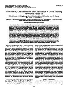
FIG. 7. Physical map of the 9.2-kb HindIII-HindIII region of S. gordonii DL1 chromosomal DNA showing the coding region for Hsa. Serine-rich region I (amino acid residues 138 to 219) and serine-rich region II (amino acid residues 450 to 2143) of Hsa are indicated by open bars. The cell wall anchoring domain (amino acid residues 2144 to 2178) is indicated by a closed bar. Nonrepetitive regions are indicated by hatched bars. The consensus sequence SASTSASVSASE is repeated 113 times between amino acid residues 488 and 1843.
SIALIC ACID-BINDING ADHESIN GENE OF S. GORDONII
VOL. 70, 2002
1217
FIG. 8. Comparison of Hsa of S. gordonii DL1 with Fap1 of S. parasanguis FW213, SrpA of S. crista CC5A, SP1772 of S. pneumoniae, TIGR4, and SA2447 of S. aureus Mu50. The consensus sequence and approximate total number of repeats are indicated above the large serine-rich region of each protein. Closed bars at the carboxyl termini (on the right side) of each protein denote the presence of cell wall anchoring domains.
secretion of Hsa into the growth medium by CM100(pAS8671), the construct that produces Hsa lacking the cell wall anchoring region (Fig. 5). The secretion of Hsa by such constructs would circumvent difficulties associated with extraction of this molecule from the streptococcal surface and thereby greatly facilitate further structural and functional characterization of the native adhesin. A molecular approach to test the hypothesis that receptor binding depends on the amino-terminal nonrepetitive region of Hsa is now also available through site-directed mutagenesis of the cloned gene. The further identification and molecular characterization of Hsa and its receptor binding site promises to provide new insights into the structural basis of oral microbial biofilm formation and also an improved understanding of the bacterial surface properties that contribute to the colonization of oral streptococci at nonoral sites such as damaged heart valves.
7. 8. 9.
10.
11. 12. 13. 14.
ACKNOWLEDGMENTS We thank T. Shiroza for providing plasmids for resident plasmid integration and J. P. Claverys for providing plasmid pR326. This work was supported by grants-in-aid for scientific research no. 08771590, 10771000, and 12771098 from the Ministry of Education, Culture, Sports, Science, and Technology, Tokyo, Japan.
15.
REFERENCES
16.
1. Andersen, R. N., N. Ganeshkumar, and P. E. Kolenbrander. 1993. Cloning of the Streptococcus gordonii PK488 gene, encoding an adhesin which mediates coaggregation with Actinomyces naeslundii PK606. Infect. Immun. 61: 981–987. 2. Baddour, L. M., G. D. Christensen, J. H. Lowrance, and W. A. Simpson. 1989. Pathogenesis of experimental endocarditis. Rev. Infect. Dis. 11:452– 463. 3. Baddour, L. M. 1994. Virulence factors among gram-positive bacteria in experimental endocarditis. Infect. Immun. 62:2143–2148. 4. Campbell, D. H., J. S. Garvey, N. E. Cremer, and D. H. Sussdorf. 1964. Methods in immunology. W. A. Benjamin, Inc., New York, N.Y. 5. Claverys, J. P., A. Dintilhac, E. V. Pestova, B. Martin, and D. A. Morrison. 1995. Construction and evaluation of new drug-resistance cassettes for gene disruption mutagenesis in Streptococcus pneumoniae, using an ami test platform. Gene 164:123–128. 6. Correia, F. F., R. Lamont, M. Bayer, B. Rosan, and J. M. DiRienzo. 1997. Cloning and sequencing of a mutated locus that affects fimbrial tuft organi-
17. 18. 19.
20. 21.
zation and corncob formation in Streptococcus crista CC5A. Int. J. Oral Biol. 22:241–248. Durack, D. T. 1995. Prevention of infective endocarditis. N. Engl. J. Med. 332:38–44. Foster, T. J., and M. Hook. 1998. Surface protein adhesins of Staphylococcus aureus. Trends Microbiol. 6:484–488. Gibbons, R. J., and J. van Houte. 1980. Bacterial adherence and the formation of dental plaques, p. 61–104. In E. H. Beachey (ed.), Bacterial adherence: receptors and recognition, series B, vol. 6. Chapman and Hall, London, United Kingdom. Gibbons, R. J., I. Etherden, and E. C. Moreno. 1983. Association of neuraminidase-sensitive receptors and putative hydrophobic interactions with high-affinity binding sites for Streptococcus sanguis C5 in salivary pellicles. Infect. Immun. 42:1006–1012. Gibbons, R. J. 1984. Adherent interactions which may affect microbial ecology in the mouth. J. Dent. Res. 63:378–385. Gibbons, R. J., I. Etherden, and E. C. Moreno. 1985. Contribution of stereochemical interactions in the adhesion of Streptococcus sanguis C5 to experimental pellicles. J. Dent. Res. 64:96–101. Hsu, S. D., J. O. Cisar, A. L. Sandberg, and M. Kilian. 1994. Adhesive properties of viridans streptococcal species. Microb. Ecol. Health Dis. 7:125– 137. Kuroda, M., T. Ohta, I. Uchiyama, T. Baba, H. Yuzawa, I. Kobayashi, L. Cui, A. Oguchi, K. Aoki, Y. Nagai, J. Lian, T. Ito, M. Kanamori, H. Matsumaru, A. Maruyama, H. Murakami, A. Hosoyama, Y. Mizutani-Ui, N. K. Takahashi, T. Sawano, R. Inoue, C. Kaito, K. Sekimizu, H. Hirakawa, S. Kuhara, S. Goto, J. Yabuzaki, M. Kanehisa, A. Yamashita, K. Oshima, K. Furuya, C. Yoshino, T. Shiba, M. Hattori, N. Ogasawara, H. Hayashi, and K. Hiramatsu. 2001. Whole genome sequencing of meticillin-resistant Staphylococcus aureus. Lancet 357:1225–1240. Laemmli, U. K. 1970. Cleavage of structural proteins during the assembly of the head of bacteriophage T4. Nature 227:680–685. Levine, M. J., M. C. Herzberg, M. S. Levine, S. A. Ellison, M. W. Stinson, H. C. Li, and T. van Dyke. 1978. Specificity of salivary-bacterial interactions: role of terminal sialic acid residues in the interaction of salivary glycoproteins with Streptococcus sanguis and Streptococcus mutans. Infect. Immun. 19:107–115. Maryanski, J. H., and C. L. Wittenberger. 1975. Mannitol transport in Streptococcus mutans. J. Bacteriol. 124:1475–1481. McBride, B. C., and M. T. Gisslow. 1977. Role of sialic acid in saliva-induced aggregation of Streptococcus sanguis. Infect. Immun. 18:35–40. Murray, P. A., M. J. Levine, L. A. Tabak, and M. S. Reddy. 1982. Specificity of salivary-bacterial interactions: II. Evidence for a lectin on Streptococcus sanguis with specificity for a NeuAc␣2,3Ga11,3Ga1NAc sequence. Biochem. Biophys. Res. Commun. 106:390–396. Navarre, W. W., and O. Schneewind. 1999. Surface proteins of gram-positive bacteria and mechanisms of their targeting to the cell wall envelope. Microbiol. Mol. Biol. Rev. 63:174–229. Ochman, H., A. S. Gerber, and D. L. Hartl. 1988. Genetic applications of an inverse polymerase chain reaction. Genetics 120:621–623.
1218
TAKAHASHI ET AL.
22. Perry, D., L. M. Wondrack, and H. K. Kuramitsu. 1983. Genetic transformation of putative cariogenic properties in Streptococcus mutans. Infect. Immun. 41:722–727. 23. Phillips, G. N., Jr., P. F. Flicker, C. Cohen, B. N. Manjula, and V. A. Fischetti. 1981. Streptococcal M protein: ␣-helical coiled-coil structure and arrangement on the cell surface. Proc. Natl. Acad. Sci. USA 78:4689–4693. 24. Ruhl, S., A. L. Sandberg, M. F. Cole, and J. O. Cisar. 1996. Recognition of immunoglobulin A1 by oral actinomyces and streptococcal lectins. Infect. Immun. 64:5421–5424. 25. Ruhl, S., J. O. Cisar, and A. L. Sandberg. 2000. Identification of polymorphonuclear leukocyte and HL-60 cell receptors for adhesins of Streptococcus gordonii and Actinomyces naeslundii. Infect. Immun. 68:6346–6354. 26. Sambrook, J., E. F. Fritsch, and T. Maniatis. 1989. Molecular cloning: a laboratory manual, 2nd ed. Cold Spring Harbor Laboratory Press, Cold Spring Harbor, N.Y. 27. Shiroza, T., and H. K. Kuramitsu. 1995. Development of a heterodimer plasmid system for the introduction of heterologous genes into streptococci. Plasmid 34:85–95. 28. Shiroza, T., N. Shinozaki, M. Hayakawa, T. Fujii, T. Oguma, M. Kobayashi, K. Fukushima, and Y. Abiko. 1998. Application of the resident plasmid integration technique to construct a strain of Streptococcus gordonii able to express the Bacillus circulans cycloisomaltooligosaccharide glucanotransferase gene, and secrete its active gene product. Gene 207:119–126. 29. Takahashi, Y., A. L. Sandberg, S. Ruhl, J. Muller, and J. O. Cisar. 1997. A specific cell surface antigen of Streptococcus gordonii is associated with bacterial hemagglutination and adhesion to ␣2,3-linked sialic acid-containing receptors. Infect. Immun. 65:5042–5051.
Editor: E. I. Tuomanen
INFECT. IMMUN. 30. Takamatsu, D., M. Osaki, and T. Sekizaki. 2000. Sequence analysis of a small cryptic plasmid isolated from Streptococcus suis serotype 2. Curr. Microbiol. 40:61–66. 31. Tettelin, H., K. E. Nelson, I. T. Paulsen, J. A. Eisen, T. D. Read, S. Peterson, J. Heidelberg, R. T. DeBoy, D. H. Haft, R. J. Dodson, A. S. Durkin, M. Gwinn, J. F. Kolonay, W. C. Nelson, J. D. Peterson, L. A. Umayam, O. White, S. L. Salzberg, M. R. Lewis, D. Radune, E. Holtzapple, H. Khouri, A. M. Wolf, T. R. Utterback, C. L. Hansen, L. A. McDonald, T. V. Feldblyum, S. Angiuoli, T. Dickinson, E. K. Hickey, I. E. Holt, B. J. Loftus, F. Yang, H. O. Smith, J. C. Venter, B. A. Dougherty, D. A. Morrison, S. K. Hollingshead, and C. M. Fraser. 2001. Complete genome sequence of a virulent isolate of Streptococcus pneumoniae. Science 293:498–506. 32. Towbin, H., T. Staehelin, and J. Gordon. 1979. Electrophoretic transfer of proteins from polyacrylamide gels to nitrocellulose sheets: procedure and some applications. Proc. Natl. Acad. Sci. USA 76:4350–4354. 33. Vieira, J., and J. Messing. 1987. Production of single-stranded plasmid DNA. Methods Enzymol. 153:3–11. 34. Wu, H., and P. M. Fives-Taylor. 1999. Identification of dipeptide repeats and a cell wall sorting signal in the fimbriae-associated adhesin, Fap1, of Streptococcus parasanguis. Mol. Microbiol. 34:1070–1081. 35. Yanisch-Perron, C., J. Vieira, and J. Messing. 1985. Improved M13 phage cloning vectors and host strains: nucleotide sequences of the M13mp18 and pUC19 vectors. Gene 33:103–119. 36. Yeung, M. K. 1999. Molecular and genetic analyses of Actinomyces spp. Crit. Rev. Oral Biol. Med. 10:120–138.