JOURNAL OF BACTERIOLOGY, Apr. 1995, p. 2074–2079 0021-9193/95/$04.0010 Copyright q 1995, American Society for Microbiology
Vol. 177, No. 8
Identification and Cloning of the Gene Encoding PenicillinBinding Protein 7 of Escherichia coli THOMAS A. HENDERSON,1 MARKUS TEMPLIN,2†
AND
KEVIN D. YOUNG1*
Department of Microbiology and Immunology, School of Medicine, University of North Dakota, ¨r Entwicklungsbiologie, Grand Forks, North Dakota 58202-9037,1 and Max-Planck-Institut fu Abteilung Biochemie, 72076 Tu ¨bingen, Germany2 Received 19 September 1994/Accepted 10 February 1995
Penicillin-binding protein (PBP) 7 of Escherichia coli is a poorly characterized member of the family of enzymes that synthesize and modify the bacterial cell wall. The approximate chromosomal position of the gene encoding this protein was determined by measuring the expression of PBPs during lytic infection of E. coli by each of the 476 miniset members of the Kohara l phage genomic library. Phages l363 and l364, encompassing the region from 47.7 to 48 min of the chromosome, overproduced PBP 7. One open reading frame, yohB, was present on both these phages and directed the expression of PBPs 7 and 8. The predicted amino acid sequence of PBP 7 contains the consensus motifs associated with other PBPs and has a potential site near the carboxyl terminus where proteolysis by the OmpT protein could occur, creating an appropriately sized PBP 8. The PBP 7 gene (renamed pbpG) was interrupted by insertion of a kanamycin resistance gene cassette and was moved to the chromosome of E. coli. No obvious growth defects were observed, suggesting that PBP 7 is not essential for growth under normal laboratory conditions. 25). We would like to know whether PBP 7 contributes to these functions in E. coli, and we report here the identification and cloning of the gene encoding PBP 7 and the construction of viable mutants unable to produce this protein.
Escherichia coli contains seven well-characterized penicillinbinding proteins (PBPs) designated PBPs 1a, 1b, 2, 3, 4, 5, and 6, in order of their decreasing molecular weights as determined with sodium dodecyl sulfate (SDS)-polyacrylamide gels. However, beginning with the earliest observations, other PBP bands have been observed. Two of these, PBPs 7 and 8, are smaller than the rest (molecular masses, approximately 31 and 29 kDa, respectively) and have a confusing history. One problem has been the lack of reproducibility with which they are observed in bacterial preparations (24, 25). Another source of confusion has been their nomenclature: PBP 7 was originally the name of the smaller of the two (25); they have been called PBPs 7a and 7b (5); and, in cases in which only one of the two was visible, their designation as PBP 7 or 8 has been arbitrary. While the seven larger PBPs are well characterized, PBPs 7 and 8 have been generally ignored. However, interest has been rekindled in these small PBPs. Tuomanen and Schwartz (27) found that PBP 7 was consistently bound by antibiotics that are able to lyse nongrowing E. coli cells, a property not normally associated with b-lactams. The nature of this sensitivity remains unclear. Also, an increase in the expression of PBP 8 has been correlated with increased resistance of E. coli to the antibiotics cephaloridine and ceftazidime (15). Recently, we clarified the relationship of the two proteins by showing that PBP 8 is a proteolytic product of PBP 7, an artifact of sample preparation due to the action of the OmpT protease (12). Also, Romeis and Ho ¨ltje have purified PBP 8 and shown that it is a DD-endopeptidase with a strict requirement for intact sacculi as a substrate (22). In addition, these authors showed that PBP 8 is not an integral membrane protein because it can be dissociated from membrane preparations by treatment with 1 M NaCl (22). The known PBPs play roles in cell wall synthesis and modification, and some have specific roles in cell division (10, 24,
MATERIALS AND METHODS Bacterial strains, plasmids, phage, media, and enzymes. Bacterial strains used as cloning hosts were as follows: E. coli CSQ (strain W1485, supE lacIq), E. coli SP1070 (his supF dacA::Km dacC; originally named JBS1002) (6) from J. Broome-Smith, E. coli XL1-Blue (Stratagene, La Jolla, Calif.), E. coli KS300 (F2 araD139 galE galK DlacX74 rpsL thi recA1 DphoA[PvuII]) (26) from J. Beckwith, and E. coli SF100 (KS300 recA1 DompT) (3) from G. Georgiou. Plasmid pHP45V-Km was received from J. Frey (9), and the pBCSK vectors were from Stratagene. The Kohara l phage miniset (13) was provided to us by Y. Kohara. Of the original 476 l clones in this set, 28 members did not correspond to the E. coli genomic map (7); the corrected versions of these clones were obtained from F. Blattner. Bacterial cells were grown in one of the following media (16): Luria-Bertani (LB) medium; LB-maltose medium (LB plus 0.2% maltose, 10 mM MgSO4, and 10 mM CaCl2); or minimal M9 medium plus 0.2% glucose and 40 mg of histidine per ml. Antibiotics were added as required at the following concentrations: tetracycline, 12.5 mg/ml; chloramphenicol, 50 mg/ml; and kanamycin, 50 mg/ml. Restriction enzymes and T4 DNA ligase were from New England Biolabs, Inc., Beverly, Mass. Protein assays were performed with the enhanced microBCA assay (Pierce Chemical Co., Rockford, Ill.). Expression of proteins encoded by the Kohara E. coli genomic library. Fresh, high-titer lysates of each phage in the Kohara miniset l library were prepared in the following manner. A 10-ml sample of stock lysate of each member of the l library was added to 75 ml of E. coli SP1070 (DdacA DdacC) that had been grown to late log phase in LB-maltose medium. This mixture was incubated without shaking for 20 min at 378C. Portions (3 ml) of LB-maltose were added to each tube, and the tubes were incubated overnight at 378C in a roller drum. The following morning, 30 ml of chloroform (CHCl3) was added, the tubes were incubated with rolling for 15 min at 378C, and whole cells and debris were removed from each culture by centrifugation at 4,000 3 g for 6 to 10 min. Samples (3 ml) of supernatant were moved to two 1.7-ml microcentrifuge tubes, each containing 8 ml of CHCl3, for storage at 48C. To express PBPs from genes cloned on the Kohara phages, E. coli SP1070 was grown overnight in LB-maltose and was inoculated into LB medium plus 10 mM MgSO4 to give an initial A600 of 0.02. When the A600 reached 0.200 to 0.250, 3 ml of the SP1070 culture was added to 500 ml of freshly prepared phage lysate (1010 to 1011 PFU/ml) and incubated with rotation for 45 min at 378C. Whole cells were collected from these samples by centrifugation at 16,000 3 g for 10 min. The supernatant was discarded, and the cell pellet was resuspended in 10 ml of 100 mM Tris-HCl (pH 8.0)–10 mM MgCl2–1% Triton X-100 prior to storage at 2208C. Each 10-ml sample was thawed and labeled with 125I-penicillin X as described by Henderson et al. (12). For rapid screening of PBP expression in
* Corresponding author. Phone: (701) 777-2624. Fax: (701) 7773894. Electronic mail address:
[email protected]. † Present address: Institute of Cell and Molecular Biology, University of Edinburgh, Edinburgh EH9 3JR, United Kingdom. 2074
VOL. 177, 1995 cells infected by the Kohara phages, 125I-labeled penicillin X samples were separated on precast SDS–12% polyacrylamide gel electrophoresis (PAGE) minigels (Bio-Rad, Hercules, Calif.) at 150 V for 70 min. For greater resolution, selected samples were separated on large-format SDS–12% PAGE gels (16.5 by 20 cm) (8) which were run at a 25-mA constant current per gel. Gels were dried and exposed to X-Omat AR film (Eastman Kodak, Rochester, N.Y.) for 1 to 10 days. PCR amplification and cloning of the PBP 7 gene. The gene for PBP 7 was amplified by the PCR procedure. Chromosomal DNA was prepared from E. coli MC1061 by the method of Wilson (29). To 50 ng of chromosomal DNA were added the primers 59-GCCCTCGAGGCCGCCAGCGCCACA-39 and 59-AGT GGATCCAAAATTACGGATGGCAGAGT-39, which were designed to introduce an XhoI and a BamHI site at the 59 and 39 ends of the amplified fragment, respectively. One unit of Taq polymerase (Boehringer Mannheim, Indianapolis, Ind.) was added to 50 ng of chromosomal DNA in 50 ml of buffer (10 mM Tris-HCl [pH 8.8], 50 mM KCl, 1.5 mM Mg2Cl, 100 mg of bovine serum albumin, 250 mM [each of the four] deoxynucleoside triphosphates, 600 ng of each primer). Amplification was performed by incubation of the reaction mixture for 5 min at 948C and cycling of the temperature 33 times (1 min at 948C, 1 min at 618C, and 2 min at 728C), followed by a final 3-min incubation at 728C. The resulting 2.1-kb fragment was purified, digested with XhoI and BamHI, and cloned into plasmid pBCSK1, yielding plasmid pMT34. Insertional mutagenesis of pbpG. A BamHI site was introduced by linker ligation into either the SmaI or the EcoRV site of the cloned pbpG gene, and a kanamycin resistance gene from pHP45V-Km (9) was inserted into the new BamHI site. These plasmid-borne pbpG mutants were transferred to the chromosome of E. coli CSQ by l-mediated transduction (14) using as an intermediate Kohara phage l363, which contains the wild-type pbpG gene. Sequence analysis. For the initial similarity search, DNA sequences were compared with other sequences in the GenBank and EMBL libraries, with the BLAST program (1), available as an electronic mail server at the National Center for Biotechnology Information (electronic mail address,
[email protected]. nih.gov). Additional analysis of sequence data was performed with the PC/Gene suite of programs (IntelliGenetics, Inc., Mountain View, Calif.) and with DNA Inspector IIe (Textco, West Lebanon, N.H.). Membrane association of PBPs 7 and 8. The membrane association of PBP 7 was determined by a modification of the method of Romeis and Ho ¨ltje (22). Alterations of the method included the substitution in buffer I of 10 mM TrisHCl (pH 8.0) for 10 mM Tris-maleate (pH 6.8), the use of DNase I of type II (Sigma, St. Louis, Mo.), and the processing of a sample of 2 ml. The locations of the PBPs were tested in two strains: E. coli SF100 (which produces only PBP 7) and E. coli KS300 (which produces PBPs 7 and 8). Purification of PBP 8. PBP 7 is cleaved by the OmpT protease to yield the proteolytic fragment PBP 8 (12), which was purified for peptide sequencing. E. coli SP1070 (DdacA DdacC) was grown in a 500-liter fermentor to mid-log phase in minimal M9 medium supplemented with 0.2% glucose and 40 mg of histidine per ml. Cells were grown, harvested, and frozen in liquid nitrogen by the staff of the University of Wisconsin Biochemistry Fermentation Facility (Madison, Wis.). A portion of the frozen cell paste (150 g) was thawed and suspended in 300 ml of 100 mM Tris-HCl (pH 8.0)–10 mM MgCl2 containing 40 mg of each of the following per ml: RNase A, DNase I (type I), and phenylmethylsulfonyl fluoride. The mixture was broken by two passes through a French pressure cell (American Instruments Co., Urbana, Ill.) at 16,000 lb/in2, and the lysate was incubated at 378C for 30 min. Cell debris was removed by centrifugation at 3,000 3 g for 10 min at 48C. Between 20 and 30% of the cellular PBP 8 was pelleted by this procedure, and this material was processed further. The resulting membrane pellet was suspended to a final volume of 100 ml in 100 mM Tris-HCl (pH 8.0)–10 mM MgCl2–100 mM NaCl–2% Triton X-100 (solubilization buffer) and was solubilized by being stirred overnight at 48C. Unsolubilized material was removed by centrifugation at 16,000 3 g for 20 min. Procion Blue H-EGN 125 (Pro Chemical and Dye, Somerset, Mass.) was coupled to Toyopearl HW65F resin (TosoHaas, Montgomeryville, Pa.) by the procedure of Mottl and Keck (17, 18). The solubilized 16,000 3 g supernatant (93 ml) was combined with an equal volume of dye-coupled resin that had been equilibrated with solubilization buffer. The sample and dye resin were mixed in a rotating water bath for 30 min at room temperature and poured into a sinteredglass (coarse grade) Bu ¨chner funnel. Proteins that remained unbound were removed from the resin by being washed with 1 liter of 100 mM Tris-HCl (pH 8.0)–10 mM MgCl2–100 mM NaCl–0.1% Triton X-100 (rinse buffer). Proteins bound to the dye resin were eluted with 300 ml of 1.0 M NaCl in rinse buffer. The eluted protein was concentrated at room temperature to 32 ml in an Amicon Model 202 stir cell (Amicon, Beverly, Mass.) that was fitted with a YM-10 membrane having a molecular mass cutoff of 10 kDa. Ampicillin affinity chromatography was performed by a modification of the procedure of Hara et al. (11). Ampicillin was bound to Affi-15 resin (Bio-Rad) and unreacted sites were blocked and equilibrated in 100 mM Tris-HCl (pH 8.0)–10 mM MgCl2–500 mM NaCl–0.1 mM dithiothreitol–0.1% Triton X-100 (affinity buffer). Dithiothreitol (final concentration, 0.1 mM) was added to the 32 ml of concentrated protein from the dye chromatography step. This solution was added to 6.2 ml of equilibrated Affi-15–ampicillin resin and was mixed gently on a rocking platform for 2 h at 378C. The mixture was poured into a 15-ml sintered-glass (coarse grade) Bu ¨chner funnel, and proteins that remained un-
GENE ENCODING E. COLI PBPs 7 AND 8
2075
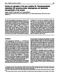
FIG. 1. Overexpression of E. coli PBP 7 from Kohara l phage. E. coli SP1070 (DdacA DdacC) (6) was infected with l Kohara phage. Infected cells were labeled with 125I-penicillin X, and the proteins were separated by SDS-PAGE. The positions of the PBPs are indicated on the left. Lane 1, uninfected E. coli SP1070; lane 2, E. coli infected with l363; lane 3, E. coli infected with l364.
bound were removed by being washed under a vacuum with 500 ml of affinity buffer at room temperature. The affinity resin was packed into a column (1-cm diameter), and bound proteins were eluted with hydroxylamine. Affinity buffer containing 1 M hydroxylamine was added to the resin and allowed to equilibrate for 30 min, and then a volume of 10 ml was eluted. This step was repeated twice more, when the resin had been in contact with the hydroxylamine for 60 and 120 min. Each 10-ml elution sample was dialyzed against six 1-liter changes of 100 mM Tris-HCl (pH 8.0)–10 mM MgCl2–500 mM NaCl. Each dialyzed sample was concentrated to 0.5 ml by centrifugation at 5,000 3 g through a Centricon-30 concentrator (Amicon) at 48C. N-terminal amino acid sequencing. PBP 8 (1.5 to 2 nmol) was separated by SDS–12% PAGE, the gel was stained with Coomassie blue G-250, and the proteins were electroblotted onto a ProBlott membrane (Applied Biosystems, Foster City, Calif.) by means of a Trans-Blot Cell (Bio-Rad). The N-terminal amino acid sequence was determined by Macromolecular Resources (Department of Biochemistry, Colorado State University, Fort Collins, Colo.) with an Applied Biosystems 473A protein sequencer with standard Edman chemistry.
RESULTS AND DISCUSSION Overexpression of PBPs by a l library of the E. coli genome. The Kohara miniset library is composed of 476 individual l phages that contain an ordered array of cloned DNA fragments representing approximately 99% of the chromosome of E. coli (13). Because genes cloned into l are often overexpressed during the lytic cycle of the phage (20), we infected E. coli with each l phage of the Kohara miniset and assayed for the overproduction of PBPs. The well-characterized E. coli PBPs were overexpressed from phage clones known to carry the appropriate genes (data not shown). The only exception was PBP 4, which was consistently expressed to a much lower degree. Infection with two Kohara phages, l363 and l364, directed overexpression of PBPs 7 and 8 (Fig. 1, lanes 2 and 3). Between PBPs 3 and 4 there is an unidentified protein band that is labeled with 125I-penicillin X (Fig. 1). This protein appears when cells are lysed by any of several means (data not shown), and we are attempting to determine whether it is a proteolytic product of a known PBP or whether it represents an additional, novel PBP. Identification and subcloning of the gene encoding PBPs 7 and 8. The chromosomal map position of the overlap between
2076
HENDERSON ET AL.
J. BACTERIOL.
FIG. 2. Map of the region from 47.6 to 48.2 min of the E. coli chromosome. The chromosomal area cloned in the l Kohara phages l363 and l364 is shown. The unidentified open reading frame yohB has been redesignated pbpG. Arrows indicate directions of transcription.
l363 and l364 corresponds to the region from 47.8 to 48 min on the E. coli chromosome (Fig. 2) (23). During the course of this work, the DNA sequence for the region from 47 to 48 min of the E. coli chromosome was reported (21). Richterich et al. (21) identified potential open reading frames in this area and one, yohB at 47.92 min (Fig. 2), yielded a predicted protein sequence similar to known carboxypeptidases and PBPs. Because yohB was contained on DNA cloned in l363 and l364, we used the sequence information to subclone this gene to determine whether it encoded PBPs 7 and 8. An 11.5-kb EcoRI DNA fragment was isolated from l364 and was cloned into the EcoRI site of pBCSK1. E. coli containing the resulting plasmid, pTAH100, overexpressed PBPs 7 and 8 (Fig. 3, lane 3). yohB was further subcloned by excising a 1.1-kb XbaI fragment from pTAH100 and ligating it into the XbaI site of pBCSK1, creating pTAH101, in which yohB was the only open reading frame present. E. coli containing pTAH101 overexpressed PBPs 7 and 8 (Fig. 3, lane 4), indicating that PBPs 7 and 8 were encoded by yohB. The yohB gene was also subcloned by PCR-mediated amplification directly from the chromosome. The resulting clone, pMT34, carried
FIG. 3. Overexpression of PBPs 7 and 8 from the cloned pbpG gene. E. coli XL1-Blue was transformed with various plasmids, whole cells were labeled with 125 I-penicillin X, and the proteins were separated by SDS-PAGE. Each lane shows the PBPs expressed by E. coli containing the following plasmids: lane 1, pBCSK1; lane 2, pBCSK2; lane 3, pTAH100; lane 4, pTAH101.
yohB and yohC on the amplified DNA fragment, and cells containing pMT34 also overexpressed PBPs 7 and 8 (data not shown). Therefore, yohB was redesignated pbpG (for penicillinbinding protein 7, with the number 7 represented by G, the seventh letter of the alphabet). Overexpression of PBP 7 did not affect bacterial growth (data not shown). Inactivation of pbpG. A kanamycin resistance gene cassette was inserted into the coding sequence of the cloned pbpG gene, and E. coli containing this plasmid no longer overproduced PBP 7 or 8 (data not shown). The insertion mutation was moved into the chromosome of E. coli by the l transduction method of Kulakauskas et al. (14). Kanamycin resistant transductants did not express PBP 7 or 8 (Fig. 4). Although the chromosomal pbpG gene had been disrupted, the bacteria grew normally, suggesting that PBP 7 is a nonessential protein when E. coli is grown in rich or minimal media under normal laboratory conditions. Characteristics of the pbpG gene product. The pbpG gene
FIG. 4. Deletion of PBPs 7 and 8 by interposon insertion mutagenesis of pbpG. A mutated pbpG gene was interrupted and transduced into the chromosome of E. coli CSQ. Whole cells of E. coli transductants were labeled with 125 I-penicillin X, and the proteins were separated by SDS-PAGE. The following E. coli strains were labeled (the restriction site into which the kanamycin resistance cassette was inserted is indicated in brackets): lane 1, CSQ; lane 2, TH105K (CSQ pbpG::VKm [EcoRV]); lane 3, TH103K (CSQ pbpG::VKm [SmaI]).
VOL. 177, 1995
GENE ENCODING E. COLI PBPs 7 AND 8
2077
FIG. 5. Nucleotide sequence and deduced amino acid sequence of the portion of the E. coli genome containing the pbpG gene. Nucleotide numbering is presented on the right, and amino acid numbering is presented at the left. The open reading frame (nt 170 to 1108) can potentially encode a polypeptide of 313 amino acids. The arrow between amino acids 21 and 11 indicates the predicted position at which signal peptidase can cleave the protein. Peptide sequencing confirmed that the underlined residues (amino acids 1 to 8) form the amino terminus of PBP 8. The probable site at which OmpT protease cleaves PBP 7 to form PBP 8 is also underlined (the two lysine residues at positions 268 and 269). The 235 and 210 regions of a potential promoter sequence are underlined (nt 72 to 77 and nt 96 to 101, respectively), as are sequences that can form the stem (nt 1119 to 1142) of a potential r-independent terminator, which includes a run of T residues just downstream of the stem (nt 1146 to 1150). Interposon antibiotic resistance cassettes were inserted into the EcoRV (nt 449) and SmaI (nt 768) restriction sites. These sites are shown in boldface type and overlined. The motifs SXXK, (S/Y)XN, and (K/H)(T/S)G, at amino acids 42 to 45, 99 to 101, and 206 to 208, respectively, are shown in boldface type and double underlined. The sequence is derived from that reported by Richterich et al. (21) and includes only the region surrounding the yohB open reading frame (here renamed pbpG).
begins at nucleotide (nt) 37649 and ends at nt 36708 on the complementary strand of the sequence reported by Richterich et al. (21). The sequence of the cloned XbaI-EcoRI DNA fragment is displayed in Fig. 5, with some downstream sequence appended. The first AUG codon of pbpG is located 170 nt downstream of the XbaI site (Fig. 5). If this is the actual start site for translation, then pbpG encodes a 313-amino acid, 34.2kDa protein. This is larger than the 31,000-molecular-weight PBP 7 observed by SDS-PAGE (12, 25). However, PBPs 7 and 8 are periplasmic enzymes (22), suggesting that they may possess a cleavable signal sequence. This would be consistent with the structure of other well-characterized low-molecular-weight PBPs, which are cleaved at their amino termini as the proteins mature, although these PBPs remain membrane associated (10). According to the algorithm of von Heijne (28), the sequence of the amino terminus of the predicted PbpG protein satisfies the criteria, including the 23 and 21 alanine rule, for the existence of a single peptidase cleavage site after the 28th amino acid. This predicts that PbpG should be cleaved between the alanine and lysine residues (Fig. 5).
To determine whether this cleavage occurs, PBP 8 was purified by dye affinity chromatography (17, 18) and ampicillin affinity chromatography. The amount of PBP 8 relative to that of total protein was determined at each step by separation of labeled proteins by SDS-PAGE and measurement of the intensity of the PBP 8 band on the autoradiogram. PBP 8 was enriched 81-fold after the dye affinity step and over 4,000-fold after the ampicillin affinity step (Table 1), giving a final yield that was ;2.5 times greater than that of a previous purification scheme (22). Approximately 1.5 to 2 nmol of PBP 8 was isolated by electroblotting the protein from a polyacrylamide gel onto a nylon membrane, and the N-terminal sequence of PBP 8 was determined. The sequence KTAAATTA (Fig. 5, underlined amino acids 1 to 8) confirmed that the lysine at the predicted site of cleavage was the true amino terminus of the PBP 8 protein. Thus, if PBPs 7 and 8 have a common amino terminus (see below), then the mature PBP 7 protein is predicted to contain 285 amino acids with a calculated molecular mass of 31,164 Da, consistent with the observed molecular weight for PBP 7. This result suggests that PBPs 7 and 8 are
2078
HENDERSON ET AL.
J. BACTERIOL. TABLE 1. Purification of PBP 8
Sample
Vol (ml)
Concn (mg/ml)
Total protein
Activity (107 U)a
Recovery (%)
Sp act (104 U)a
Purification (fold)
Whole cells 3,000 3 g pellet 16,000 3 g supernatant Procion Blue H-EGN Affi-15–ampicillin
460 100 93 32 1.5
35.0 6.6 3.6 0.39 0.04
16.1 g 0.66 g 0.34 g 12.5 mg 0.06 mg
20 5.4 2.5 1.6 0.39
100 20.7 9.7 6.2 1.5
1.2 8.1 7.5 138 6,500
0 5.1 4.7 81.2 4,060
a
Activity was measured by optical scanning of the radioactive protein bands separated by SDS-PAGE; the area under each protein peak reflects the amount of I-penicillin X bound. Activity is expressed in arbitrary units of area and equals the specific activity [area/(micrograms of protein per lane)] multiplied by the total amount (in micrograms) of protein in each sample. 125
probably processed in a manner similar to that of the classical low-molecular-weight PBPs. PBP 8 is an artifact derived from PBP 7 after cleavage by the OmpT protease (12). OmpT has been reported to cleave proteins between pairs of dibasic amino acids (Arg-Arg, Lys-Lys, Arg-Lys, and Lys-Arg) (4). There are two such sites in the 285-amino-acid sequence of PbpG: Arg-88–Lys-89 and Lys268–Lys-269. The latter site is located 17 amino acids from the carboxyl terminus of the protein. If the OmpT protease cleaves PBP 7 between these two lysines, then the resulting 268-aminoacid protein has a molecular mass of 29,452 Da, consistent with the 29-kDa size of PBP 8 as measured by SDS-PAGE (12, 25). Combined with the preceding results, this information suggests that PBPs 7 and 8 have a common amino terminus and that PBP 8 differs from PBP 7 by an OmpT-mediated truncation of the carboxyl end of PBP 7. Calculations for PBPs 7 and 8 gave a pI value of 10.4 for both proteins. This is significantly greater than 8.3, the value for PBP 8 as reported by Ayala et al. (2). We separated purified PBP 8 by two-dimensional nonequilibrium pH gel electrophoresis and compared the pI of PBP 8 with those of a set of standard proteins (Bio-Rad). PBP 8 had a pI greater than 8.6, the pI of rabbit muscle glyceraldehyde-3-phosphate dehydrogenase, by a factor that suggested a final pI value of approximately 10 (data not shown). Thus, the measured pI is consistent with the calculations from the predicted protein sequence. The difference between these values and those of Ayala et al. (2) may simply reflect the availability of accurate protein standards. PBP 7 is not an integral membrane protein. With the exception of PBP 4 (10), the PBPs of E. coli are membrane bound. Analysis of the amino acid sequence of PBP 7 revealed no defined transmembrane helix, amphiphilic helix, or lipid anchor. Romeis and Ho ¨ltje (22) found that PBP 8 could be removed from bacterial membranes by 1 M NaCl and that PBPs 7 and 8 could be released from cells by osmotic shock. They thus concluded that the proteins were located in the periplasm and were not integral membrane proteins. To confirm this possibility, membrane fractions were isolated from E. coli SF100 (ompT) and incubated in 1 M NaCl. PBP 7 was solubilized from membranes by exposure to 1 M NaCl (data not shown). Therefore, PBPs 7 and 8 appear to be soluble proteins that associate with the bacterial membrane in a saltdependent manner. PBP 7 contains the active-site motifs that characterize other PBPs. Four peptide motifs are shared among the PBPs and b-lactamases, which together make up a family of serine activesite penicillin-interacting proteins (10). Three-dimensional crystallographic analyses place the consensus-like sequences at the active sites of these enzymes, and the distance between the four motifs is somewhat conserved in the primary structure of the proteins (10). The active-site serine occurs in the first
motif, SXXK (where X is any amino acid), which is located 33 to 60 amino acids from the amino terminus of the smaller penicillin-interacting proteins. PBP 7 contains a potential active-site motif, SISK, within this range (Fig. 5, amino acids 42 to 45). The second motif, (S/Y)XN, is located approximately 48 to 93 amino acids from the active-site serine (10), except in the case of PBP 4, in which the two motifs are separated by 243 amino acids (19). The predicted amino acid sequence of PBP 7 also includes such a sequence, SEN (Fig. 5, amino acids 99 to 101), 57 amino acids from the active-site serine at position 42. The third motif is simply a peptide segment with one or two dicarboxylic acids (D and/or E), located approximately 30 to 60 amino acids after the (S/Y)XN sequence. In PBP 7, there are four potential dicarboxylic acid candidates between the SEN and KTG sequences: E-138, D-152, E-174, and D-175. However, the aspartic acid at position 152 is located in a run of 5 amino acids, S(T/S)ARD, that are completely conserved between PBPs 5, 6, and 7 (Fig. 6). The distance of this residue from the previous motif is 41 amino acids, consistent with the distance for other D/E motifs in penicillin-interacting proteins (10). Finally, the fourth PBP motif, (K/H)(T/S)G, occurs 40 to 90 amino acids from the D/E motif. In PBP 7, the KTG sequence is located 55 amino acids from D-152 (Fig. 5, amino acids 206 to 208). Thus, the predicted protein sequence of PBP 7 conforms in every respect to the consensus motifs and their distributions in previously characterized PBPs. Comparison of PBP 7 with other PBPs. Alignment of PBP 7 with known PBPs indicated that it was most similar to PBPs 5 and 6 of E. coli (Fig. 6 and data not shown). PBP 7 shares 67 identical amino acids with PBP 5 and 77 identical amino acids with PBP 6. Thus, over its 285-amino-acid length, PBP 7 is 24 and 27% identical to PBPs 5 and 6, respectively. Clusters of extended identity or similarity appear in the vicinity of the three major active-site motifs (Fig. 6, amino acids shown in boldface type at positions 42 to 45, 99 to 101, and 206 to 208). In addition, the three PBPs share 5 highly conserved amino acids (STARD) that appear in PBP 7 at positions 148 to 152, which may correspond to the fourth, dicarboxylic acid motif. If the sequence of PBP 4 is altered to conform to that of the class A b-lactamases, as in the analysis by Mottl et al. (19), the modified PBP 4 shares 43 (15%) identical amino acids with PBP 7. Two of the four motifs, SXXK and SXN, are perfectly aligned, and the KTG sequence is misaligned by only 1 amino acid (data not shown). Little or no similarity was observed when PBP 7 was compared to the E. coli MepA protein, another bacterial endopeptidase. Summary. We have identified and cloned the gene that encodes PBP 7, a poorly characterized protein in E. coli. PBP 7, though soluble, is at least partially associated with the bacterial membrane, and E. coli grows normally when the protein is overexpressed or when the pbpG gene is inactivated. Although the protein is not essential under usual laboratory con-
GENE ENCODING E. COLI PBPs 7 AND 8
VOL. 177, 1995
5.
6.
7. 8.
9.
10. 11.
12.
13.
14.
15.
16. 17. FIG. 6. Alignment of amino acid sequences of PBP 7 and E. coli PBPs 5 and 6. Asterisks indicate the positions of amino acids that are identical between PBP 7 and PBPs 5 or 6. Dots represent conservative amino acid replacements. Dashes are introduced to maximize alignment. Each PBP is numbered beginning from the first residue following the signal peptidase cleavage site.
18.
ditions, further investigation of PBP 7 should be valuable because of its correlation with the lytic action of the penem class of b-lactams (27). In addition, PBP 7 may play a specialized role in remodeling the cell wall since the enzyme exhibits unique substrate requirements (22).
20.
19.
21. 22. 23.
ACKNOWLEDGMENTS This work was supported by Public Health Service grant GM40947 from the National Institutes of Health and by SmithKline Beecham Pharmaceuticals. M.T. especially thanks J.-V. Ho ¨ltje, U. Schwarz, and T. Romeis for scientific direction and help.
24.
25. 26.
REFERENCES 1. Altschul, S. F., W. Gish, W. Miller, E. W. Myers, and D. J. Lipman. 1990. Basic local alignment search tool. J. Mol. Biol. 215:403–410. 2. Ayala, J. A., M. A. de Pedro, and D. Va ´zquez. 1984. Application of a charge/ size two-dimensional gel electrophoresis system to the analysis of the penicillin-binding proteins of Escherichia coli. FEBS Lett. 168:93–96. 3. Baneyx, F., and G. Georgiou. 1990. In vivo degradation of secreted fusion proteins by the Escherichia coli outer membrane protease OmpT. J. Bacteriol. 172:491–494. 4. Baneyx, F., and G. Georgiou. 1991. Construction and characterization of Escherichia coli strains deficient in multiple secreted proteases: protease III
27.
28. 29.
2079
degrades high-molecular-weight substrates in vivo. J. Bacteriol. 173:2696– 2703. Barbas, J. A., J. Dı´az, A. Rodrı´guez-Te´bar, and D. Va ´zquez. 1986. Specific location of penicillin-binding proteins within the cell envelope of Escherichia coli. J. Bacteriol. 165:269–275. Broome-Smith, J. K. 1985. Construction of a mutant of Escherichia coli that has deletions of both the penicillin-binding protein 5 and 6 genes. J. Gen. Microbiol. 131:2115–2118. Chuang, S.-E., D. L. Daniels, and F. R. Blattner. 1993. Global regulation of gene expression in Escherichia coli. J. Bacteriol. 175:2026–2036. Dreyfuss, G., S. A. Adam, and Y. D. Choi. 1984. Physical change in cytoplasmic messenger ribonucleoproteins in cells treated with inhibitors of mRNA transcription. Mol. Cell. Biol. 4:415–423. Fellay, R., J. Frey, and H. Krisch. 1987. Interposon mutagenesis of soil and water bacteria: a family of DNA fragments designed for in vitro insertional mutagenesis of gram-negative bacteria. Gene 52:147–154. Ghuysen, J.-M. 1991. Serine b-lactamases and penicillin-binding proteins. Annu. Rev. Microbiol. 45:37–67. Hara, H., Y. Nishimura, J.-I. Kato, H. Suzuki, H. Nagasawa, A. Suzuki, and Y. Hirota. 1989. Genetic analyses of processing involving C-terminal cleavage in penicillin-binding protein 3 of Escherichia coli. J. Bacteriol. 171:5882– 5889. Henderson, T. A., P. M. Dombrosky, and K. D. Young. 1994. Artifactual processing of penicillin-binding proteins 7 and 1b by the OmpT protease of Escherichia coli. J. Bacteriol. 176:256–259. Kohara, Y., K. Akiyama, and K. Isono. 1987. The physical map of the whole E. coli chromosome: application of a new strategy for rapid analysis and sorting of a large genomic library. Cell 50:495–508. Kulakauskas, S., P. M. Wikstro¨m, and D. E. Berg. 1991. Efficient introduction of cloned mutant alleles into the Escherichia coli chromosome. J. Bacteriol. 173:2633–2638. Malouin, F., S. Chamberland, N. Brochu, and J. Parr, Jr. 1991. Influence of growth media on Escherichia coli cell composition and ceftazidime susceptibility. Antimicrob. Agents Chemother. 35:477–483. Miller, J. H. 1972. Experiments in molecular genetics. Cold Spring Harbor Laboratory, Cold Spring Harbor, N.Y. Mottl, H., and W. Keck. 1991. Purification of penicillin-binding protein 4 of Escherichia coli as a soluble protein by dye-affinity chromatography. Eur. J. Biochem. 200:767–773. Mottl, H., and W. Keck. 1992. Rapid screening of a large number of immobilized textile dyes for the purification of proteins: use of penicillin-binding protein 4 of Escherichia coli as a model enzyme. Protein Expression Purif. 3:403–409. Mottl, H., P. Terpstra, and W. Keck. 1991. Penicillin-binding protein 4 of Escherichia coli shows a novel type of primary structure among penicillininteracting proteins. FEMS Microbiol. Lett. 78:213–220. Murray, N. E. 1991. Special uses of l phage for molecular cloning. Methods Enzymol. 204:280–301. Richterich, P., N. Lakey, G. Gryan, L. Jachn, L. Mintz, K. Robinson, and G. M. Church. GenBank accession number U00007. Romeis, T., and J.-V. Ho ¨ltje. 1994. Penicillin-binding protein 7/8 of Escherichia coli is a DD-endopeptidase. Eur. J. Biochem. 224:597–604. Rudd, K. E. 1992. Alignment of E. coli DNA sequences to a revised, integrated genomic restriction map, p. 2.3–2.43. In J. H. Miller (ed.), A short course in bacterial genetics: a laboratory manual and handbook for Escherichia coli and related bacteria. Cold Spring Harbor Laboratory Press, Cold Spring Harbor, N.Y. Spratt, B. G. 1975. Distinct penicillin binding proteins involved in the division, elongation, and shape of Escherichia coli K12. Proc. Nat. Acad. Sci. USA 72:2999–3003. Spratt, B. G. 1977. Properties of the penicillin-binding proteins of Escherichia coli K12. Eur. J. Biochem. 72:341–352. Strauch, K. L., and J. Beckwith. 1988. An Escherichia coli mutation preventing degradation of abnormal periplasmic proteins. Proc. Natl. Acad. Sci. USA 85:1576–1580. Tuomanen, E., and J. Schwartz. 1987. Penicillin-binding protein 7 and its relationship to lysis of nongrowing Escherichia coli. J. Bacteriol. 169:4912– 4915. von Heijne, G. 1986. A new method for predicting signal sequence cleavage sites. Nucleic Acids Res. 14:4683–4690. Wilson, K. 1990. Preparation of genomic DNA from bacteria, p. 2.4.1–2.4.5. In F. A. Ausubel, R. Brent, R. E. Kingston, D. D. Moore, J. G. Seidman, J. A. Smith, and K. Struhl (ed.), Current protocols in molecular biology. Greene Publishing, New York.