Mar. Drugs 2015, 13, 6759-6773; doi:10.3390/md13116759 OPEN ACCESS
marine drugs ISSN 1660-3397 www.mdpi.com/journal/marinedrugs Article
Identification of Antiviral Agents Targeting Hepatitis B Virus Promoter from Extracts of Indonesian Marine Organisms by a Novel Cell-Based Screening Assay Atsuya Yamashita 1, Yuusuke Fujimoto 1, Mayumi Tamaki 2, Andi Setiawan 3, Tomohisa Tanaka 1, Kaori Okuyama-Dobashi 1, Hirotake Kasai 1, Koichi Watashi 4, Takaji Wakita 4, Masaaki Toyama 5, Masanori Baba 5, Nicole J. de Voogd 6, Shinya Maekawa 7, Nobuyuki Enomoto 7, Junichi Tanaka 2,* and Kohji Moriishi 1,* 1
2
3
4
5
6
7
Department of Microbiology, Division of Medical Sciences, Graduate School of Interdisciplinary Research, University of Yamanashi, 1110 Shimokato, Chuo, Yamanashi 409-3898, Japan; E-Mails:
[email protected] (A.Y.);
[email protected] (Y.F.);
[email protected] (T.T.);
[email protected] (K.O.-D.);
[email protected] (H.K.) Department of Chemistry, Biology and Marine Science, University of the Ryukyus, 1 Senbaru, Nishihara, Okinawa 903-0213, Japan; E-Mail:
[email protected] Department of Chemistry, Faculty of Science, Lampung University, Jl. Sumantri Brodjonegoro No. 1, Bandar Lampung 35145, Indonesia; E-Mail:
[email protected] Department of Virology II, National Institute of Infectious Diseases, 1-23-1 Toyama, Shinjuku-ku, Tokyo 162-8640, Japan; E-Mails:
[email protected] (K.W.);
[email protected] (T.W.) Division of Antiviral Chemotherapy Center for Chronic Viral Disease, Graduate School of Medical and Dental Sciences, Kagoshima University, 8-35-1 Sakuragaoka, Kagoshima 890-8544, Japan; E-Mails:
[email protected] (M.T.);
[email protected] (M.B.) Naturalis, National Museum of Natural History, P.O. Box 9517, Leiden 2300 RA, The Netherlands; E-Mail:
[email protected] The First Department of Internal Medicine, Faculty of Medicine, University of Yamanashi, 1110 Shimokato, Chuo, Yamanashi 409-3898, Japan; E-Mails:
[email protected] (S.M.);
[email protected] (N.E.)
* Authors to whom correspondence should be addressed; E-Mails:
[email protected] (K.M.);
[email protected] (J.T.); Tel.: +81-55-273-9537 (K.M.); +81-98-895-8560 (J.T.); Fax: +81-55-273-6728 (K.M.); +81-98-895-8565 (J.T.).
Mar. Drugs 2015, 13
6760
Academic Editor: Peer B. Jacobson Received: 19 September 2015/ Accepted: 23 October 2015 / Published: 6 November 2015
Abstract: The current treatments of chronic hepatitis B (CHB) face a limited choice of vaccine, antibody and antiviral agents. The development of additional antiviral agents is still needed for improvement of CHB therapy. In this study, we established a screening system in order to identify compounds inhibiting the core promoter activity of hepatitis B virus (HBV). We prepared 80 extracts of marine organisms from the coral reefs of Indonesia and screened them by using this system. Eventually, two extracts showed high inhibitory activity (>95%) and low cytotoxicity (66% to 77%). Solvent fractionation, column chromatography and NMR analysis revealed that 3,5-dibromo-2-(2,4-dibromophenoxy)-phenol (compound 1) and 3,4,5-tribromo-2-(2,4-dibromophenoxy)-phenol (compound 2), which are classified as polybrominated diphenyl ethers (PBDEs), were identified as anti-HBV agents in the extracts. Compounds 1 and 2 inhibited HBV core promoter activity as well as HBV production from HepG2.2.15.7 cells in a dose-dependent manner. The EC50 values of compounds 1 and 2 were 0.23 and 0.80 µM, respectively, while selectivity indexes of compound 1 and 2 were 18.2 and 12.8, respectively. These results suggest that our cell-based HBV core promoter assay system is useful to determine anti-HBV compounds, and that two PBDE compounds are expected to be candidates of lead compounds for the development of anti-HBV drugs. Keywords: marine organism; hepatitis B virus; HBV; HBV core promoter; high-throughput screening; antiviral agent
1. Introduction Hepatitis B virus (HBV) infection is a serious public health problem worldwide, with more than 240 million people estimated to be chronically infected [1]. Chronic infection with HBV leads to liver cirrhosis and hepatocellular carcinoma, which are adverse outcomes seen in untreated patients [2,3]. HBV is an enveloped DNA virus that belongs to the genus Orthohepadnavirus of the Hepadnaviridae family [4]. The infectious virion of HBV contains incompletely double-stranded and relaxed circular DNA (rcDNA), surrounded with a lipid bilayer and viral surface proteins. Following virus entry into hepatocytes, rcDNA migrates into the nucleus and is then converted into a covalently closed circular DNA (cccDNA), which encodes overlapping open reading frames (ORFs). The viral genes are transcribed under the control of four promoters (core, preS1, preS2/S, and X promoters) and two enhancer regions (enhancer I and enhancer II also referred as the core upstream regulatory sequence: CURS), and translated into the core protein (Hepatitis B core antigen: HBcAg), precore protein (Hepatitis B e antigen: HBeAg), surface proteins (Large S, Middle S and Small S protein), polymerase (reverse transcriptase and DNA-dependent DNA polymerase) and X protein. These viral regulatory elements play a role in transcriptions of 3.5, 2.4, 2.1 and 0.7 kb mRNAs. The mRNA with a size of 3.5 kb, which is termed
Mar. Drugs 2015, 13
6761
pregenomic RNA (pgRNA), is packaged with the viral polymerase into a viral capsid consisting of core proteins. The pgRNA is enclosed with capsid proteins in cytoplasm and then reverse-transcribed into a negative-strand DNA in the cytoplasmic capsid. The transcription of pgRNA is regulated under the control of the core promoter, which consists of the basic core promoter and the upper regulatory region including negative regulatory region and CURS. Thus, the core promoter is responsible for HBV replication as well as the viral particle formation and is capable of being targeted for development of an effective HBV therapy [5–10]. The currently available antiviral agents for the treatment of chronic HBV infection are classified as follows: (1) immunomodulatory agents, such as conventional interferon-alpha and pegylated interferon-alpha; and (2) oral nucleoside/nucleotide analogues (NAs), such as three nucleoside (lamivudine, entecavir and telbivudine) and two nucleotide analogues (adefovir and tenofovir). Treatments with these agents are capable of preventing disease progression to liver cirrhosis and hepatocellular carcinoma, resulting in improvement of the survival rate of patients with chronic HBV infections [11–13]. However, interferon therapy is associated with major problems such as serious side effects, genotype-dependent treatment response and moderate antiviral activity, while long-term therapy using NAs promotes the emergence of drug-resistant viruses. In addition, the most serious problem is that currently available agents do not eradicate cccDNA, the template in transcription of HBV pgRNA and mRNA. Safer and more effective anti-HBV agents are still needed for efficient therapy [14,15]. Natural products including terrestrial plants and microbes have historically been sources for the development of various drugs targeting human diseases. Research on natural products has often included marine organisms because of the chemical and biological novelties of marine natural products. trabectedin (Yondelis®) and eribulin (Halaven®) are derived from chemical compounds isolated from marine organisms, and approved for anticancer therapy [16,17]. Ara-A (vidarabine) is a semisynthetic anti-herpes drug made from spongouridine isolated from the Caribbean sponge Tethya crypta [18,19]. In this study, we established a screening system to identify compounds inhibiting HBV core promoter activity and then screened 80 extracts of marine organisms collected from the coral reefs of Indonesia in order to identify anti-HBV agents. 2. Results and Discussion 2.1. Establishment of HBV Core Promoter Reporter Cell Line The core promoter consists of CURS and basal core promoter (BCP) (Figure 1A) and is responsible for transcription of 3.5 kb mRNA, pgRNA [4]. CURS negatively and positively regulates the promoter activity [4]. The region composed of both CURS and BCP or BCP only was cloned into pGL4.18 [luc2P/Neo] plasmid (Figure 1A). The resulting plasmids were designated as pGL4.18 CURS_BC_AeUS (CURS BCP) or pGL4.18 BC_AeUS (BCP) in this study (Figure 1A). The plasmid pGL4.18 CURS_BC_AeUS or pGL4.18 BC_AeUS was transfected with phRG-TK into human hepatoma cell line Huh7, human cervical cancer cell line HeLa, and human fibrosarcoma cell line HT-1080. The resulting cells were harvested 48 h post-transfection and suspended in lysis buffer in order to estimate luciferase activity. Previous findings suggested that HBV core promoter (CURS and BCP) is more active in hepatoma cell lines than other cell lines [6,20–23]. In this study, Huh7 cell line exhibited the highest
Mar. Drugs 2015, 13
6762
luciferase activity under the control of CURS BCP or BCP among tested cell lines (Figure 1B). Moreover, the Huh7 cells transfected with pGL4.18 CURS_BC_AeUS exhibited 5-time higher luciferase activity than the cells transfected with pGL4.18 BC_AeUS (Figure 1B). These results suggest its potential for establishment of a cell-based screening assay based on HBV promoter activity. The plasmid pGL4.18 CURS_BC_AeUS was introduced into Huh7 cells again for establishment of a stable cell line. The transfected cells were incubated in the presence of G418 until colony formation. The Huh7 cell line exhibiting the highest luciferase activity was selected by colony isolation, and designated as Huh7 GL4.18 CURS_BC_AeUS.
Figure 1. Development of Hepatitis B virus (HBV) core promoter reporter system. (A) Schematic representation of the firefly luciferase reporter plasmid pGL4.18 CURS_BC_AeUS and pGL4.18 BC_AeUS; (B) HBV core promoter activity in three cell lines. Each plasmid described above was transfected with phRG-TK into hepatic (Huh7) and non-hepatic (HeLa and HT-1080) cells. Luciferase activity was measured at 48 h post-transfection as described in the Experimental Section. Firefly luciferase activity was normalized with Renilla luciferase activity. Luciferase activity was expressed as a fold induction compared with the value of cells transfected with pGL4.18 [luc2P/Neo] empty control vector (control). The data shown in this panel are representative of three independent experiments. Error bars indicate standard deviation.
Mar. Drugs 2015, 13
6763
2.2. Validation of Cell-Based HBV Core Promoter Assay We calculated the Z′ factor in order to evaluate the Huh7 G4.18 CURS_BC_AeUS cell line for high-throughput screening. The Z′ factor is a useful tool for measurement of the quality or suitability of high throughput screening, and the value spanning from 0.5 to 1.0 exhibits an appropriate assay [24,25]. In this study, the value of Z′ factor was 0.79 (n = 48) using Huh7 GL4.18 CURS_BC_AeUS cells (Figure 2). The coefficient of variation (CV), which represents unevenness of the screening system, should be less than 10% for a correct assay [24]. The CV value of our system was 7.0% (Figure 2).
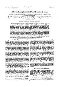
Figure 2. Validation of cell based HBV core promoter reporter assay. Huh7 GL4.18 CURS_BC_AeUS cells (positive control) and Huh7 GL4.18 cells (negative control) were harvested at 72 h. The luciferase activity was determined as described in the Experimental Section. The Z′ factor and coefficient of variation (CV) value was calculated as described in the Experimental Section. HepG2.2.15 cell line is generally used to screen for anti-HBV agents, although HepG2.2.15 cell-based drug screening assay requires at least 10 days for screening. However, cell-based HBV core promoter assay was completed for 3 days for screening. The cell-based HBV core promoter assay is more advantageous than the assay using HepG2.2.15 cell in high-throughput screening of anti HBV agents. Thus, the cell-based HBV core promoter assay was employed in this study for high-throughput screening of extracts prepared from marine organisms. 2.3. High-Throughput Screening for Extracts of Marine Organisms Inhibiting HBV Core Promoter Activity We collected marine organisms from coral reefs of Indonesia and prepared 80 extracts from them with methanol (MeOH). We then screened them in order to discover anti-HBV agents using our screening system. Each extract was added at a final concentration of 25 µg/mL to the culture supernatant of Huh7 GL4.18 CURS_BC_AeUS cells. Luciferase activity and cell viability were measured 48 h after treatment. Among them, extracts of samples code named 00A14 and 00X18 exhibited high inhibitory activity of more than 95% and low cytotoxicity of 66% to 77% (Table 1, Figure 3). The 00A14 extract was prepared from the marine sponge Dysidea granulosa collected from the coral reefs of Simua Island, while the 00X18 extract was prepared from the marine sponge Dysidea sp. collected from the coral reefs of Buton strait. Dysidea granulosa of 00A14 was similar to Dysidea sp. of 00X18 regarding morphological features.
Mar. Drugs 2015, 13
6764
The 00X18 extract, but not the 00A14 extract, was further analyzed in this study because of the much smaller amount of Dysidea granulosa than of Dysidea sp. Table 1. Effect of marine organism extracts on HBV core promoter activity and cell viability. Sample
Sample
No.
Code Name
1 2
Inhibitory
Cell Viability
Collection
Activity (%)
(% of Control)
Site
Porifera
0
101.5
Simua Island
Porifera
2.3
97.7
Simua Island
Ircinia ramosa
Porifera
0
177.4
Simua Island
00A08
Liosina sp.
Porifera
99.4
0
Simua Island
00A09
Clathria sp.
Porifera
98.6
0
Simua Island
Specimen
Phylum
00A01
Callyspongia sp.
00A05
Xestospongia sp.
3
00A07
4 5 6
00A10
Unidentified
Chordata
18.1
108.3
Simua Island
7
00A11
Hippospongia sp.
Porifera
40.3
97.7
Simua Island
8
00A12
Petrosia sp.
Porifera
6.9
92.8
Simua Island
9
00A13
Porifera
5.4
119.1
Simua Island
10
00A14
Porifera
96.5
77.3
Simua Island
11
00B15
Porifera
0
104.8
Kajuongia Island
Callyspongia cf. aerizusa Dysidea granulosa Spheciospongia vagabunda
12
00B16
Callyspongia sp.
Porifera
0
101.3
Kajuongia Island
13
00B17
Unidentified
Porifera
0
91.2
Kajuongia Island
14
00J85
Unidentified
Porifera
0
96.6
Buton Island
15
00J86
Unidentified
Porifera
0
97.6
Buton Island
16
00J87
Unidentified
Porifera
0
95.2
Buton Island
17
00J88
Phyllospongia sp.
Porifera
17.9
90.7
Buton Island
18
00J89
Unidentified
Porifera
0
101.3
Buton Island
19
00J90
Unidentified
Porifera
0
96.3
Buton Island
20
00J91
Parazoanthus sp.
Cnidaria
24.2
90.1
Buton Island
21
00K92
Unidentified
Porifera
14.8
95.9
Buton Island
22
00K94
Ianthella basta
Porifera
94.2
28.8
Tobea Island
23
00K95
Unidentified
Chordata
9.2
92.4
Tobea Island
24
00K97
Higginsia mixta
Porifera
16.1
84.5
Tobea Island
Porifera
0
85.7
Magintin Island
Chordata
3.1
76.6
Magintin Island
Thrinacophora
25
00L00
26
00L02
Unidentified
27
00M03
Gelliodes fibulata
Porifera
13.7
98.2
Masaloka Island
28
00M04
Clavularia viridis
Cnidaria
99.6
1.4
Masaloka Island
29
00M05
Coelocarteria sp.
Porifera
15.1
91.1
Masaloka Island
30
00M06
Mycale sp.
Porifera
37.6
87.6
Masaloka Island
28.2
94.2
Masaloka Island
cervicornis
31
00M07
Unidentified
Porifera
32
00M08
Unidentified
Porifera
96.3
24.3
Masaloka Island
33
00N09
Unidentified
Porifera
15.3
83.7
Buton strait
34
00N10
Porifera
0
92.9
Buton strait
35
00N11
Porifera
0
95.5
Buton strait
Myrmekioderma granulatum Callyspongia samarensis
Mar. Drugs 2015, 13
6765 Table 1. Cont.
36
00N12
Biemna sp.
Porifera
6.8
87.5
Buton strait
37
00N13
Biemna triraphis
Porifera
24.1
92.6
Buton strait
38
00N14
Xestospongia exigua
Porifera
99.4
1.6
Buton strait
39
00P16
Unidentified
Cnidaria
0
101.0
Muna Island
40
00Q17
Unidentified
Porifera
16.1
101.9
Buton strait
41
00Q18
Unidentified
Porifera
2.5
84.8
Buton strait
42
00Q19
Axinyssa sp.
Porifera
51.5
104.0
Buton strait
43
00Q20
Mycale sp.
Porifera
22.7
86.0
Buton strait
44
00R22
Clavularia inflate
Cnidaria
22.1
107.0
Buton Island
45
00R23
Paralemnalia sp.
Cnidaria
19.2
99.0
Buton Island
46
00R24
Junceella fragilis
Cnidaria
18.6
102.9
Buton Island
47
00R25
Nephthea sp.
Cnidaria
0
94.6
Buton Island
48
00S26
Svenzea sp.
Porifera
25.5
88.5
Tobea Island
49
00S27
Unidentified
Cnidaria
32.0
97.3
Tobea Island
50
00S28
Coelogorgia sp.
Cnidaria
0
97.7
Tobea Island
51
00T29
Theonella sp.
Porifera
35.3
85.4
Tobea Island
52
00T30
Unidentified
Porifera
47.6
93.6
Tobea Island
53
00T31
Higginsia cf. mixta
Porifera
14.7
142.0
Tobea Island
54
00T32
Paratelesto sp.
Cnidaria
0
94.7
Tobea Island
55
00U33
Pycnoclabella sp.
Chordata
25.8
91.9
Muna Island
56
00U34
Lissoclinum patella
Chordata
27.8
87.2
Muna Island
57
00X01
Polycarpa contecta
Chordata
25.9
93.6
Simua Island
58
00X02
Dysidea sp.
Porifera
56.8
73.4
Tobea Island
59
00X04
Nephthea sp.
Cnidaria
0
99.2
Beromasidi Island
60
00X05
Haliclona fascigera
Porifera
7.8
98.7
Torobulu
61
00X06
Axinyssa sp.
Porifera
98.9
4.9
Torobulu
62
00X07
Unidentified
Porifera
16.7
86.0
Torobulu
63
00X08
Unidentified
Cnidaria
12.9
87.0
Torobulu
64
00X10
Niphates olemda
Porifera
70.2
38.5
Buton Island
65
00X11
Unidentified
Porifera
99.7
1.1
Tobea Island
66
00X12
Unidentified
Porifera
70.6
63.9
Tobea Island
67
00X13
Unidentified
Porifera
13.9
108.7
Magintin Island
68
00X14
Xestospongia sp.
Porifera
21.1
105.6
Magintin Island
69
00X15
Dysidea/Euryspongia
Porifera
10.2
114.4
Magintin Island
70
00X16
Unidentified
Chordata
0
130.3
Buton strait
71
00X17
Dysidea cf. arenaria
Porifera
14.7
80.0
Buton strait
72
00X18
Dysidea sp.
Porifera
95.0
65.3
Buton strait
73
00X19
Unidentified
Porifera
23.0
92.5
Buton strait
74
00X21
Gelliodes/Niphates
Porifera
36.7
80.9
Buton strait
75
00X22
Amphimedon/Haliclona
Porifera
31.2
90.9
Buton strait
76
00X23
Dysidea cf. arenaria
Porifera
0
92.8
Buton strait
77
00X24
Unidentified
Porifera
0
99.2
Buton strait
78
00X26
Anthelia sp.
Cnidaria
61.5
107.4
Buton strait
79
00X27
Unidentified
Chordata
14.8
85.2
Tobea Island
80
00X28
Clathria sp.
Porifera
58.9
82.0
Tobea Island
Mar. Drugs 2015, 13
6766
Figure 3. Correlation between the inhibitory activity of each marine organism extract against HBV core promoter and the cell viability of each marine organism extract. Each closed circle represents one marine organism extract. The x-axis indicates inhibitory activity against HBV core promoter, while the y-axis indicates cell viability. 2.4. Identification of PBDEs as the Inhibitory Compounds of HBV Production via HBV Core Promoter Activity The extract of 00X18 was separated with several chromatographic steps to give two polybrominated diphenyl ethers (PBDEs), 3,5-dibromo-2-(2,4-dibromophenoxy)-phenol (compound 1) and 3,4,5-tribromo2-(2,4-dibromophenoxy)-phenol (compound 2) as major constituents (Figure 4A). The compounds were identified by comparing the NMR data with those published. Huh7 GL4.18 CURS_BC_AeUS cells were incubated with each of those compounds to evaluate their effects on the core promoter activity. Both compounds inhibited the core promoter activity in a dose-dependent manner (Figure 4B). IC50 values of compounds 1 and 2 are 2.3 µM and 4.9 µM, respectively, suggesting that compounds 1 and 2 included in the 00X18 extract inhibit HBV core promoter activity. We next addressed the effects of compounds 1 and 2 on HBV production and cell viability. HBV-producing cultured cells, HepG2.2.15.7, were incubated in culture medium containing various concentrations of compound 1 or 2. Entecavir was used as the positive control for anti-HBV activities of compound 1 and 2. The amount of supernatant HBV DNA and cell viability were measured by using real-time PCR and MTS assay, respectively. Treatment with compound 1 or 2 impaired production of HBV DNA in a dose-dependent manner (Figure 5). The IC50 and CC50 values of compound 1 were 0.23 µM and 4.19 µM, respectively, while the EC50 and CC50 values of compound 2 were 0.80 µM and 10.26 µM, respectively. Thus, the selectivity indexes of compounds 1 and 2 were 18.2 and 12.8, respectively (Table 2). These results suggest that compounds 1 and 2 possess anti-HBV activity. However, IC50 values of compound 1 and 2 were higher than that of entecavir, while compounds 1 and 2 were more toxic than entecavir (Table 2).
Mar. Drugs 2015, 13
Figure 4. Effect of polybrominated diphenyl ethers (PBDEs) on HBV core promoter activity. (A) Structure of 3,5-dibromo-2-(2,4-dibromo-phenoxy)-phenol (Compound 1) and 3,4,5-tribromo-2-(2,4-dibromo-phenoxy)-phenol (Compound 2); (B) Huh7 GL4.18 CURS_BC_AeUS cells were incubated for 48 h in the medium containing various concentrations of PBDEs. Luciferase and cytotoxicity assays were carried out by the method described in the Experimental section. Data are representative of three independent experiments. Error bars indicate standard deviation.
Figure 5. Effect of PBDEs on HBV production. HepG2.2.15.7 cells were incubated with various concentrations of compound 1 or 2. Supernatant HBV DNA and cytotoxicity were estimated by real-time qPCR and MTS assay, respectively, as described in the Experimental section. The data were representative of three independent experiments. Error bars indicate standard deviation.
6767
Mar. Drugs 2015, 13
6768
Table 2. Anti HBV activity and cytotoxicity of Compound 1, 2 and entecavir in HepG2.2.15.7 cells. Compound Compound 1 Compound 2 Entecavir
EC50 a (µM) 0.23 ± 0.07 0.80 ± 0.34 0.021 ± 0.003
CC50 b (µM) 4.19 ± 0.12 10.26 ± 3.69 >100
Selectivity c Index 18.2 12.8 >4761
a
Fifty percent effective concentration based on the inhibition of the HBV viral DNA release; b Fifty percent cytotoxicity concentration based on the reduction of cell viability; c Selectivity index (CC50/EC50).
HepG2.2.15 cells have generally been used to screen chemical compounds for anti-HBV agents, but the disadvantage of HepG2.2.15 cell-based drug screening assay requires at least 10 days for screening. However, cell-based HBV core promoter assay was completed for 2 days for screening. Thus, cell-based HBV core promoter assay offers an advantage in high-throughput screening of anti HBV agents. PBDEs were recently isolated from marine sponges and biologically synthesized by their associated microorganisms [26,27]. Several groups reported multifunctional properties containing antibacterial, antifungal, anti-microalgal and anti-inflammatory activities of PBDEs [28–30]. Treatment with PBDEs also inhibited the enzymatic activities of endogenous and viral proteins [31–33]. Compound 1 suppressed activity of Tie2 kinase, which is associated with angiogenesis essential for tumor growth and survival [34]. The data reported by Zhang et al., indicate that compound 1 induces G1 phase cell cycle arrest in MCF-7 cells (a breast cancer cell line) [35], although HBV could infect and replicate in non-dividing cells [36]. These reports indicate that endogenous factors are associated with the inhibitory effect of PBDEs on HBV propagation. Further studies will reveal the mechanism of PBDE-related suppression of HBV production, and will be required for the development of more effective and safe anti-HBV agents based on PBDEs. 3. Experimental Section 3.1. Cell Culture HepG2.2.15.7 cell line was subcloned from HepG2.2.15 cell line, which is stably transfected with the HBV genome (genotype D) [37,38]. HepG2.2.15.7 cell line produced HBV at a higher level than HepG2.2.15 cell line. This cell lines were maintained in Dulbecco’s Modified Eagle’s Medium/Ham’s Nutrient Mixture F12 medium supplemented with 10% fetal bovine serum, 100 U/mL Penicillin, 100 µg/mL Streptomycin, 2 mM L-Glutamine, 400 µg/mL G418, 50 µM hydrocortisone and 5 µg/mL Insulin. Huh7 cells, HeLa cells and HT-1080 cells were maintained in Dulbecco’s modified Eagle’s medium containing 10% fetal calf serum, 100 U/mL Penicillin and 100 µg/mL Streptomycin. 3.2. Plasmid Construction and Transient or Stable Expression The HBV CURS BCP and BCP fragment was amplified from pUC19 HBV AeUS plasmid [39] by PCR using the following primers: CURS BCP: 5′-GCTAGCGATCCTGCCCAAGGTCTTACATAA-3′ (the underlined region indicates Nhe I site) and 5′-AGATCTAAGAGATGATTAGGCAGAGGT-GAA-3′ (underlined region indicates Bgl II site); BCP: 5′-GCTAGCTGGGGGAGGAGATTAGGT-TAAAGG-3′ (the underlined region indicates Nhe I site) and 5′-AGATCTAAGAGATGATTAGGC-AGAGGTGAA-3′
Mar. Drugs 2015, 13
6769
(underlined region indicates Bgl II site). These PCR products were cloned into a TA cloning vector, pTA2 (TOYOBO, Osaka, Japan). After sequence confirmation, these PCR fragments were introduced between Nhe I and Bgl II sites of pGL4.18 [luc2P/Neo] (Promega, Madison, WI, USA). The resulting plasmid was designated as pGL4.18 CURS_BC_AeUS and pGL4.18 BC_AeUS in this study. Huh7, HeLa and HT-1080 cell lines were co-transfected with pGL4.18 CURS_BC_AeUS and phRG-TK using Lipofectamine LTX reagent (Thermo Fisher Scientific, Waltham, MA, USA). To standardize transfection efficiency and cell recovery, we used the phRG-TK plasmid (Promega, Madison, WI, USA) encoding Renilla luciferase under the control of the herpes simplex virus type 1 thymidine kinase promoter. The plasmid pGL4.18 [luc2P/Neo] was used as a negative control instead of pGL4.18 CURS_BC_AeUS. The transfected cells were harvested 48 h post-transfection and then were lysed in Passive lysis buffer (Promega, Madison, WI, USA). Luciferase activity was measured using a Dual-luciferase reporter assay system (Promega, Madison, WI, USA). The resulting luminescence was detected by a Luminescencer-JNR AB-2100 (ATTO, Tokyo, Japan). The pGL4.18 CURS_BC_AeUS or pGL4.18 [luc2P/Neo] plasmid was transfected into Huh7 cells using Lipofectamine LTX reagent (Thermo Fisher Scientific, Waltham, MA, USA). These transfected cells were seeded on the plate and then incubated until colonies formed. The stable cell lines were established by colony isolation. The clone exhibiting the highest activity among the isolated clone was designated as Huh7 GL4.18 CURS_BC_AeUS, and the negative control clone was designated as Huh7 GL4.18. 3.3. Validation of Screening Method Huh7 GL4.18 CURS_BC_AeUS and Huh7 cells were seeded at 2 × 10⁴ cells per well in a 48-well plate. Luciferase activity was measured after 72 h of incubation. The Z′ factor was calculated as follows:
.
.
_
_
. .
SD: Standard Deviation. The minimal acceptable value for a high-throughput screening assay is usually considered to be 0.5. The theoretical maximum is 1 [24,25]. The CV is calculated using the formula: %
.
_ .
_ _
_
100
SD: Standard Deviation. The acceptable value of CV for a high-throughput screening assay is less than 10% [24]. 3.4. Cell-Based HBV Promoter Assay Huh7 GL4.18 CURS_BC_AeUS cells were seeded at 2 × 10⁴ cells per well in a 48-well plate and then treated with 25 µg/mL each extracts 24 h after seeding cells. The treated cells were harvested 48 h post-treatment and then lysed with Cell culture lysis buffer (Promega, Madison, WI, USA).
Mar. Drugs 2015, 13
6770
Luciferase activity was measured by using Luciferase assay systems (Promega, Madison, WI, USA). The resulting luminescence was detected as described above. 3.5. Determination of Cytotoxicity Huh7 GL4.18 CURS_BC_AeUS cells were seeded at a density of 1 × 10⁴ cells per well in a 96-well plate and incubated at 37 °C for 24 h. Each extract was added at 25 µg/mL to the culture supernatant. The treated cells were harvested 48 h post-treatment. Cell viability was estimated by dimethylthiazol carboxymethoxy-phenylsulfophenyl tetrazolium (MTS) assay using a Celltiter 96 aqueous one-solution cell proliferation assay kit (Promega, Madison, WI, USA). 3.6. Preparation of Extracts from Marine Organisms The marine organisms were collected at coral reefs around Sulawesi, Muna, and Buton Islands, Indonesia, in August 2000. Marine sponge No. 10 was identified as Dysidea granulosa in this study and deposited at the Netherlands Centre for Biodiversity with code RMNH POR 10013. Each specimen was preserved with a small amount of ethanol until use. After decantation of ethanol solution, each specimen was extracted three times with MeOH. A crude extract was prepared by concentrating the combined solution under vacuum and then kept at −20 °C until use. A portion of each extract was solubilized in dimethyl sulfoxide (DMSO) after measuring its weight. 3.7. Separation of PBDEs A crude MeOH extract (1.24 g) of specimen No. 72 was partitioned between EtOAc and water. The lipophilic layer yielded 481 mg after concentration and it was applied to a silica gel column and separated into six fractions. A portion of the third fraction (185 mg) was subjected to silica HPLC (hexane-CH2Cl2 or hexane-EtOAc). Finally, two PBDEs, 3,5-dibromo-2-(2,4-dibromo-phenoxy)-phenol (Compound 1, 2.5 mg) and 3,4,5-tribromo-2-(2,4-dibromo-phenoxy)-phenol (Compound 2, 7.3 mg), were identified by checking NMR data as follows: Compound 1: 1H NMR (acetone-d6) δ 7.83 (1H, d, J = 2.4 Hz, H-3′), 7.43 (1H, dd, J = 8.8, 2.4 Hz, H-5′), 7.39 (1H, d, J = 2.2 Hz, H-4), 7.26 (1H, d, J = 2.2 Hz, H-6), 6.59 (1H, d, J = 8.8 Hz, H-6′). Compound 2: 1H NMR (acetone-d6) δ 7.83 (1H, d, J = 2.4 Hz, H-3′), 7.51 (1H, s, H-6), 7.42 (1H, dd, J = 8.8, 2.4 Hz, H-5′), 6.63 (1H, d, J = 8.8 Hz, H-6′). 3.8. Determination of Anti HBV Activity and Cytotoxicity in HepG2.2.15.7 Cells HepG2.2.15.7 cells were seeded at 1 × 10⁴ cells per well in a collagen coated 96-well plate (Corning, Corning, NY, USA) and incubated at 37 °C for 24 h before treatment. The tested compound was added to the culture medium at the indicated concentrations. The culture medium was exchanged every 3 days for fresh medium containing the compound. The treated cells and culture supernatants were harvested 9 days post-treatment and were subjected to MTS assay and estimation of HBV DNA, respectively. The culture supernatant was mixed with an equal volume of Sidestep lysis and stabilization buffer (Agilent Technologies, Santa Clara, CA, USA). The viral DNA included in the mixture was estimated by real-time quantitative PCR using the THUNDERBIRD Probe
Mar. Drugs 2015, 13
6771
qPCR Mix (TOYOBO, Osaka, Japan). The forward and reverse primers targeting HBV surface region are 5′-ACTCACCAACCTCCTGTCCT-3′ and 5′-GACAAACGGGCAACATACCT-3′, respectively. The fluorogenic probe was 5′-FAM-TATCGCTGGATGTGTCTGCGGCGT-TAMRA-3 [40]. 3.9. Reagents Entecavir and hydrocortisone were purchased from Sigma-Aldrich (St. Louis, MO, USA). G418 and insulin were purchased from Wako (Osaka, Japan). 4. Conclusions We developed a cell-based assay based on HBV core promoter activity, screened marine products using this assay system and finally identified two PBDEs as anti-HBV compounds. Acknowledgments We thank M. Furugori for her secretarial work and C. Endoh for technical assistance. We also thank M. Mizokami for kindly providing a plasmid. This work was supported by Grants-in-Aid from the Ministry of Health, Labor, and Welfare, Japan (H24-Bsou-kanen-012 and -005) and from the Ministry of Education, Sports, Culture, Science and Technology of Japan (26350973 and 15K08493). Author Contributions Atsuya Yamashita, Junichi Tanaka, Masaaki Toyama, Masanori Baba, and Kohji Moriishi designed all of the experiments. Mayumi Tamaki, Andi Setiawan, and Junichi Tanaka collected marine organisms, purified the materials and identified compounds. Nicole J. De Voogd identified marine sponges. Tomohisa Tanaka, Kaori Okuyama-Dobashi, Hirotake Kasai, Koichi Watashi, Takaji Wakita, Shinya Maekawa and Nobuyuki Enomoto analyzed the data. Atsuya Yamashita and Yuusuke Fujimoto conducted other experiments. Atsuya Yamashita, Junichi Tanaka and Kohji Moriishi wrote the manuscript. Conflicts of Interest The authors declare no conflicts of interest. References 1.
2. 3. 4. 5. 6.
Ott, J.J.; Stevens, G.A.; Groeger, J.; Wiersma, S.T. Global epidemiology of hepatitis B virus infection: New estimates of age-specific HBsAg seroprevalence and endemicity. Vaccine 2012, 30, 2212–2219. Liaw, Y.F.; Chu, C.M. Hepatitis B virus infection. Lancet 2009, 373, 582–592. Trepo, C.; Chan, H.L.; Lok, A. Hepatitis B virus infection. Lancet 2014, 384, 2053–2063. Locarnini, S.; Littlejohn, M.; Aziz, M.N.; Yuen, L. Possible origins and evolution of the hepatitis B virus (HBV). Semin. Cancer Biol. 2013, 23, 561–575. Beck, J.; Nassal, M. Hepatitis B virus replication. World J. Gastroenterol. 2007, 13, 48–64. Kramvis, A.; Kew, M.C. The core promoter of hepatitis B virus. J. Viral Hepat. 1999, 6, 415–427.
Mar. Drugs 2015, 13 7. 8. 9. 10. 11. 12. 13. 14. 15. 16. 17. 18. 19. 20. 21.
22. 23. 24.
25. 26.
6772
Moolla, N.; Kew, M.; Arbuthnot, P. Regulatory elements of hepatitis B virus transcription. J. Viral Hepat. 2002, 9, 323–331. Nassal, M. Hepatitis B virus replication: Novel roles for virus-host interactions. Intervirology 1999, 42, 100–116. Seeger, C.; Mason, W.S. Hepatitis B virus biology. Microbiol. Mol. Biol. Rev. 2000, 64, 51–68. Tang, H.; Banks, K.E.; Anderson, A.L.; McLachlan, A. Hepatitis B virus transcription and replication. Drug News Perspect. 2001, 14, 325–334. Lok, A.S. Personalized treatment of hepatitis B. Clin. Mol. Hepatol. 2015, 21, 1–6. You, C.R.; Lee, S.W.; Jang, J.W.; Yoon, S.K. Update on hepatitis B virus infection. World J. Gastroenterol. 2014, 20, 13293–13305. Zoulim, F.; Durantel, D. Antiviral therapies and prospects for a cure of chronic hepatitis B. Cold Spring Harb. Perspect. Med. 2015, 5, doi:10.1101/cshperspect.a021501. Gish, R.; Jia, J.D.; Locarnini, S.; Zoulim, F. Selection of chronic hepatitis B therapy with high barrier to resistance. Lancet Infect. Dis. 2012, 12, 341–353. Grimm, D.; Thimme, R.; Blum, H.E. HBV life cycle and novel drug targets. Hepatol. Int. 2011, 5, 644–653. Cragg, G.M.; Newman, D.J. Natural products: A continuing source of novel drug leads. Biochim. Biophys. Acta 2013, 1830, 3670–3695. Molinski, T.F.; Dalisay, D.S.; Lievens, S.L.; Saludes, J.P. Drug development from marine natural products. Nat. Rev. Drug Discov. 2009, 8, 69–85. Donia, M.; Hamann, M.T. Marine natural products and their potential applications as anti-infective agents. Lancet Infect. Dis. 2003, 3, 338–348. Sagar, S.; Kaur, M.; Minneman, K.P. Antiviral lead compounds from marine sponges. Mar. Drugs 2010, 8, 2619–2638. Guo, W.; Chen, M.; Yen, T.S.; Ou, J.H. Hepatocyte-specific expression of the hepatitis B virus core promoter depends on both positive and negative regulation. Mol. Cell. Biol. 1993, 13, 443–448. Honigwachs, J.; Faktor, O.; Dikstein, R.; Shaul, Y.; Laub, O. Liver-specific expression of hepatitis B virus is determined by the combined action of the core gene promoter and the enhancer. J. Virol. 1989, 63, 919–924. Yee, J.K. A liver-specific enhancer in the core promoter region of human hepatitis B virus. Science 1989, 246, 658–661. Yuh, C.H.; Ting, L.P. The genome of hepatitis B virus contains a second enhancer: Cooperation of two elements within this enhancer is required for its function. J. Virol. 1990, 64, 4281–4287. Fatokun, A.A.; Liu, J.O.; Dawson, V.L.; Dawson, T.M. Identification through high-throughput screening of 4′-methoxyflavone and 3′,4′-dimethoxyflavone as novel neuroprotective inhibitors of parthanatos. Br. J. Pharmacol. 2013, 169, 1263–1278. Zhang, J.H.; Chung, T.D.; Oldenburg, K.R. A simple statistical parameter for use in evaluation and validation of high throughput screening assays. J. Biomol. Screen. 1999, 4, 67–73. Agarwal, V.; El Gamal, A.A.; Yamanaka, K.; Poth, D.; Kersten, R.D.; Schorn, M.; Allen, E.E.; Moore, B.S. Biosynthesis of polybrominated aromatic organic compounds by marine bacteria. Nat. Chem. Biol. 2014, 10, 640–647.
Mar. Drugs 2015, 13
6773
27. Teuten, E.L.; Xu, L.; Reddy, C.M. Two abundant bioaccumulated halogenated compounds are natural products. Science 2005, 307, 917–920. 28. Handayani, D.; Edrada, R.A.; Proksch, P.; Wray, V.; Witte, L.; van Soest, R.W.; Kunzmann, A.; Soedarsono. Four new bioactive polybrominated diphenyl ethers of the sponge Dysidea herbacea from west sumatra, indonesia. J. Nat. Prod. 1997, 60, 1313–1316. 29. Hanif, N.; Tanaka, J.; Setiawan, A.; Trianto, A.; de Voogd, N.J.; Murni, A.; Tanaka, C.; Higa, T. Polybrominated diphenyl ethers from the indonesian sponge Lamellodysidea herbacea. J. Nat. Prod. 2007, 70, 432–435. 30. Sionov, E.; Roth, D.; Sandovsky-Losica, H.; Kashman, Y.; Rudi, A.; Chill, L.; Berdicevsky, I.; Segal, E. Antifungal effect and possible mode of activity of a compound from the marine sponge Dysidea herbacea. J. Infect. 2005, 50, 453–460. 31. Fu, X.; Schmitz, F.J.; Govindan, M.; Abbas, S.A.; Hanson, K.M.; Horton, P.A.; Crews, P.; Laney, M.; Schatzman, R.C. Enzyme inhibitors: New and known polybrominated phenols and diphenyl ethers from four indo-pacific Dysidea sponges. J. Nat. Prod. 1995, 58, 1384–1391. 32. Salam, K.A.; Furuta, A.; Noda, N.; Tsuneda, S.; Sekiguchi, Y.; Yamashita, A.; Moriishi, K.; Nakakoshi, M.; Tani, H.; Roy, S.R.; et al. Pbde: Structure-activity studies for the inhibition of hepatitis C virus NS3 helicase. Molecules 2014, 19, 4006–4020. 33. Yamazaki, H.; Sumilat, D.A.; Kanno, S.; Ukai, K.; Rotinsulu, H.; Wewengkang, D.S.; Ishikawa, M.; Mangindaan, R.E.; Namikoshi, M. A polybromodiphenyl ether from an indonesian marine sponge Lamellodysidea herbacea and its chemical derivatives inhibit protein tyrosine phosphatase 1B, an important target for diabetes treatment. J. Nat. Med. 2013, 67, 730–735. 34. Xu, Y.M.; Johnson, R.K.; Hecht, S.M. Polybrominated diphenyl ethers from a sponge of the Dysidea genus that inhibit Tie2 kinase. Bioorg. Med. Chem. 2005, 13, 657–659. 35. Zhang, H.; Skildum, A.; Stromquist, E.; Rose-Hellekant, T.; Chang, L.C. Bioactive polybrominated diphenyl ethers from the marine sponge Dysidea sp. J. Nat. Prod. 2008, 71, 262–264. 36. Cohen, D.; Adamovich, Y.; Reuven, N.; Shaul, Y. Hepatitis B virus activates deoxynucleotide synthesis in nondividing hepatocytes by targeting the R2 gene. Hepatology 2010, 51, 1538–1546. 37. Ogura, N.; Watashi, K.; Noguchi, T.; Wakita, T. Formation of covalently closed circular DNA in Hep38.7-Tet cells, a tetracycline inducible hepatitis B virus expression cell line. Biochem. Biophys. Res. Commun. 2014, 452, 315–321. 38. Sells, M.A.; Chen, M.L.; Acs, G. Production of hepatitis B virus particles in Hep G2 cells transfected with cloned hepatitis B virus DNA. Proc. Natl. Acad. Sci. USA 1987, 84, 1005–1009. 39. Sugiyama, M.; Tanaka, Y.; Kato, T.; Orito, E.; Ito, K.; Acharya, S.K.; Gish, R.G.; Kramvis, A.; Shimada, T.; Izumi, N.; et al. Influence of hepatitis B virus genotypes on the intra- and extracellular expression of viral DNA and antigens. Hepatology 2006, 44, 915–924. 40. Liu, Y.; Hussain, M.; Wong, S.; Fung, S.K.; Yim, H.J.; Lok, A.S. A genotype-independent real-time PCR assay for quantification of hepatitis B virus DNA. J. Clin. Microbiol. 2007, 45, 553–558. © 2015 by the authors; licensee MDPI, Basel, Switzerland. This article is an open access article distributed under the terms and conditions of the Creative Commons Attribution license (http://creativecommons.org/licenses/by/4.0/).