Plant Physiology, May 1999, Vol. 120, pp. 205–215, www.plantphysiol.org © 1999 American Society of Plant Physiologists
Identification of the Soluble Starch Synthase Activities of Maize Endosperm1 Heping Cao, Jennifer Imparl-Radosevich, Hanping Guan, Peter L. Keeling, Martha G. James, and Alan M. Myers* Department of Biochemistry, Biophysics, and Molecular Biology, Iowa State University, Ames, Iowa 50011 (H.C., M.G.J., A.M.M.); and ExSeed Genetics, L.L.C., and Department of Agronomy, Food Sciences Building, Iowa State University, Ames, Iowa 50011 (J.I.-P., H.G., P.L.K.) nah et al., 1993; Nelson and Pan, 1995; Ball et al., 1996; Nakamura, 1996; Preiss and Sivak, 1996; Smith et al., 1997). SS enzymes add glucosyl units at the nonreducing end of linear chains through new a(134) linkages. BEs cleave interior a(134) bonds and form a new glycoside bond between the C1 reducing end of the released glucan and a C6 elsewhere within a linear chain; thus, BEs introduce branch linkages. Debranching enzymes hydrolyze branch linkages and are needed for determination of the normal branching pattern in amylopectin. Multiple isoforms exist for each enzyme, although their specific roles in starch biosynthesis generally are not known. In maize (Zea mays), at least five different genes code for predicted SSs. The wx locus codes for the major granulebound SS, GBSSI (Shure et al., 1983; Klo¨sgen et al., 1986), and the du1 gene codes for a protein similar in sequence to known SSs (Gao et al., 1998). In addition to these two genetically defined loci, three different cDNAs are known that code for the SSs designated zSSI, zSSIIa, and zSSIIb (Harn et al., 1998; Imparl-Radosevich et al., 1998; Knight et al., 1998). Genetic and biochemical analyses reveal that individual SS isozymes have specific effects on starch structure, influencing both chain length and branchlinkage formation. GBSSI is known to synthesize the amylose component of starch (Martin and Smith, 1995; Denyer et al., 1996; van de Wal et al., 1998). Alteration of DU1 causes changes in amylopectin structure, including increased branching frequency (Shannon and Garwood, 1984; Wang et al., 1993a, 1993b), and mutations affecting a Chlamydomonas reinhardtii SS have similar effects (Fontaine et al., 1993). Mutations of the pea rug5 locus, which affects SSII, alter starch granule structure and chain-length distribution (Craig et al., 1998). Maize du1- mutations and pea rug5 mutations also condition pleiotropic effects on other starch biosynthetic enzymes (Boyer and Preiss, 1981; Craig et al., 1998). Thus, various SSs and BEs may act in a concerted manner to determine starch structure. An important step in understanding the specific functions of each SS isozyme in maize starch biosynthesis would be to determine which of the known cDNAs code for the major soluble enzyme activities present during
This study identified the complement of soluble starch synthases (SSs) present in developing maize (Zea mays) endosperm. The product of the du1 gene, DU1, was shown to be one of the two major soluble SSs. The C-terminal 450 residues of DU1 comprise eight sequence blocks conserved in 28 known or predicted glucan synthases. This region of DU1 was expressed in Escherichia coli and shown to possess SS activity. DU1-specific antisera detected a soluble endosperm protein of more than 200 kD that was lacking in du1- mutants. These antisera eliminated 20% to 30% of the soluble SS activity from kernel extracts. Antiserum against the isozyme zSSI eliminated approximately 60% of the total soluble SS, and immunodepletion of du1- mutant extracts with this antiserum nearly eliminated SS activity. Two soluble SS activities were identified by electrophoretic fractionation, each of which correlated specifically with zSSI or DU1. Thus, DU1 and zSSI accounted for the great majority of soluble SS activity present in developing endosperm. The relative activity of the two isozymes did not change significantly during the starch biosynthetic period. DU1 and zSSI may be interdependent, because mutant extracts lacking DU1 exhibited a significant stimulation of the remaining SS activity.
Plant starches are composed almost entirely of glucosyl units joined by either a(134) or a(136) glycoside bonds. In the amylopectin component of starch, these two types of linkage are located with a specific spatial distribution, and this defined branching pattern in turn determines higherorder structures (Gallant et al., 1997). The number of glucosyl units in each a(134)-linked chain and the locations of a(136) branch linkages that join the linear chains are expected to be determined by inherent properties of the starch biosynthetic enzymes. The specific mechanisms by which these enzymes determine architectural specificity, however, are not well understood. Three enzymatic activities are involved in starch synthesis from the glucosyl donor ADPGlc (for reviews, see Han1 This work was supported by the U.S. Department of Agriculture (grant no. 96-35300-3779 to A.M.M. and M.G.J.) and the National Science Foundation (grant no. DIR-9113593 to the Iowa State University Signal Transduction Training Group). This is journal paper no. J-18330 of project no. 3197 of the Iowa Agriculture and Home Economics Experiment Station (Ames). This paper is dedicated to the memory of Dr. Bruce Wasserman. * Corresponding author; e-mail
[email protected]; fax 1– 515–294 – 0453.
Abbreviations: BE, branching enzyme; DAP, days after pollination; GBSS, granule-bound starch synthase; GS, glycogen synthase; IPTG, isopropyl-1-thio-b-d-galactopyranoside; SS, starch synthase. 205
206
Cao et al.
endosperm development. Two SS-activity peaks, designated SSI and SSII, have been fractionated biochemically from soluble extracts of maize endosperm (Boyer and Preiss, 1981; Preiss and Sivak, 1996). The enzyme responsible for the SSI-activity peak was purified and identified as the product of the Ss1 cDNA; this enzyme is designated zSSI (Mu et al., 1994; Knight et al., 1998). The protein responsible for the SSII-activity peak has not yet been identified, however, characterization of the Du1 cDNA provided indirect evidence that DU1 is this enzyme (Gao et al., 1998). Specifically, the SSII-activity peak is significantly reduced in du1- mutants (Boyer and Preiss, 1981), and the predicted size of DU1 and the tissue-specific expression pattern of the Du1 cDNA match the characteristics of this enzyme (Dang and Boyer, 1988; Mu et al., 1994; Gao et al., 1998). Whether DU1 accounts for a significant portion of soluble SS activity is not clear, however, because du1mutations do not cause decreased total soluble SS activity in endosperm extracts (Singletary et al., 1997). This study used recombinant proteins and antibodies generated through application of the cloned Du1 and Ss1 cDNAs to investigate in greater detail the correlation between known cDNAs and the specific soluble SSs present in maize endosperm during starch biosynthesis. DU1 was shown directly to possess SS activity and to account for a significant proportion of the total activity in soluble kernel extracts. Essentially all of the soluble SS activity was accounted for by the combination of DU1 and zSSI. The fact that du1- mutations do not affect total SS activity was explained by the observation that zSSI is more active in mutant endosperm extracts than in nonmutant extracts. MATERIALS AND METHODS Biological Materials, Nomenclature, and Media Maize (Zea mays) nomenclature follows the standard of Beavis et al. (1995); in addition, cDNA names are not italicized to distinguish them from gene names. Maize inbreds used were W64A and Oh43. Plants homozygous for du1-Ref (Mangelsdorf, 1947) or du1-R4059 (Gao et al., 1998) were the progeny of at least four successive backcrosses to W64A. Escherichia coli strains used for cloning and recombinant protein expression were XL-1-Blue, DH5a, and BL21(DE3) (Ausubel et al., 1989). Standard Luria-Bertani medium (Ausubel et al., 1989) was supplemented with 50 mg/mL kanamycin or 100 mg/mL ampicillin to make Luria-Bertani K or Luria-Bertani A medium, respectively. Computational Methods Multiple sequence alignment used the PILEUP program of the GCG Sequence Analysis Software Package (Genetics Computer Group, Madison, WI). Conserved sequence motifs were assigned based on the presence of at least one invariant residue in the 28 polypeptides analyzed and conservative substitutions of several nearby residues in the same positions in the alignment among all of the sequences. Consensus sequences were determined by votes
Plant Physiol. Vol. 120, 1999
according to the method of Posfai et al. (1989). Abbreviations as used in Table I and accession numbers are as follows: Aae (Aquifex aeolicus) GS, AE000704; At (Arabidopsis thaliana) SS, AL021713; Atu (Agrobacterium tumefaciens) GS, L24117; Bst (Bacillus stearothermophilus) GS, D87026; Bsu (Bacillus subtilis) GS, Z25795; Eco (E. coli) GS, J02616; Hin (Hemophilus influenzae) GS, P45179; Hv (Hordeum vulgare; barley) GBSSI, X07932; Ib (Ipomoea batatas; sweet potato) GBSSI, U44126; Me (Manihot esculenta; cassava) GBSSI, X74160; Os (Oryza sativa; rice) GBSSI, X53694; SSS, D16202; Ps (Pisum sativum; pea) GBSSI, X88789; SSII, X88790; Sb (Sorghum bicolor; sorghum) GBSSI, U23945; St (Solanum tuberosum; potato) GBSSI, X58453; GBSSII, X87988; SSSI, Y10416; SSSIII, X94400 and X95759; Syn (Synechocystis sp. PCC6803) GS, D90899; Ta (Triticum aestivum; wheat) GBSSI, X57233; SS, U66377; SSS, U48227; and Zm (Z. mays; maize) DU1, AF023159; GBSSI, M24258; SSI, AF036891; SSIIa, AF019296; SSIIb, AF019297. Construction of Expression Plasmids Plasmid pHC1 was constructed as an intermediate in the generation of the antigens used to raise the antisera antiDU1N and anti-DU1F. This plasmid contains the entire Du1 cDNA coding region delineated by two EcoRI sites, one located immediately upstream of the presumed initiation codon, and the second 225 bp downstream of the termination codon. A 1.5-kb fragment was PCR amplified from the partial Du1 cDNA clone pMgf10 (Gao et al., 1998) using primers HCp1 and M13F. HCp1 (59-AAACCCGGGAATTCGATGGAGATGGTCCTACG-39) contains SmaI and EcoRI sites located upstream of the presumed initiation codon (restriction sites and the initiation codon are underlined), and M13F is located downstream of the cDNA insert on the noncoding strand. The amplified fragment was cleaved at the SmaI site of the primer and the unique AgeI site within the cDNA sequence. This fragment was cloned in the SmaI and AgeI sites of pMg10-6, which contains the Du1 cDNA extending from 125 bp upstream of the presumed initiation codon to the downstream EcoRI site (M. Gao, unpublished results). The resulting plasmid is pHC1. DNA sequence analysis indicated that no mutations arose in the Du1 cDNA during construction of this plasmid. Plasmid pHC2 expresses a fusion protein containing the Schistosoma japonicum glutathione-S-transferase protein at its N terminus and DU1 residues 1 to 648 at its C terminus; this polypeptide was used as the DU1N antigen. pHC2 was constructed by cloning the EcoRI-SalI fragment from pHC1 into pGEX4T-3 (Pharmacia) digested with the same enzymes. Plasmid pHC4 expresses a fusion protein containing thioredoxin at its N terminus and full-length DU1 at its C terminus; this protein was used as the DU1F antigen. pHC4 was constructed by cloning the EcoRI fragment from pHC1 into pET-32b(1) (Novagen, Madison, WI). Plasmids pHC5 and pHC6 express the C-terminal region of DU1 (DU1C) in E. coli. The Du1 cDNA from codon 1226 to termination codon 1675 was PCR amplified using pHC1 as the template. The upstream primer was HCp2 (59GCAGAATTCGATGCACATTGTCCAC-39), which places an EcoRI site adjacent to codon 1226 (the EcoRI site and
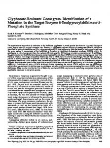
Table I. Conserved sequence motifs of the ADPGlc-dependent a-1,4-glucosyltransferases from plants and prokaryotes The plant enzymes are divided arbitrarily into three classes based on their known subcellular locations or, in instances of uncharacterized proteins, their high degree of sequence similarity to such characterized enzymes. Soluble enzymes are those such as DU1 that are exclusively or nearly exclusively located in the soluble phase. Granule-bound enzymes are those such as GBSSI, i.e. the product of a wx gene, that are exclusively found in the granule fraction. Dual-location enzymes are those such as zSSI that are present in significant amounts in both the granule-associated and soluble fractions. Conservative substitutions are noted when they fall into the functional groups defined by Dayhoff and Orcutt (1979), which are AGPST, ILMV, HKR, DENQ, FWY, and C. Invariant residues are denoted by asterisks under the plant SS consensus sequence, and rare exceptions to the designated consensus are underlined. Numerals refer to amino acid positions beginning at the first ATG codon of the open reading frame; exceptions are Ta SSS and Ta SS, for which the complete cDNA sequences are not available. The number of nonconserved amino acids adjacent to each conserved motif is indicated. Full references for each sequence are listed in “Materials and Methods.”
208
Cao et al.
codon 1226 are underlined). The downstream primer was M13F. The amplified fragment digested with EcoRI was cloned into pET-29b(1) and pET-32b(1) (Novagen) to form pHC5 and pHC6, respectively. The sequence of the entire DU1C insert and the junction with the T7 promoter was determined in clones with correct restriction maps. These data confirmed that no mutations arose in the Du1 cDNA sequence during construction of the plasmids.
Plant Physiol. Vol. 120, 1999
10% glycerol, 10 mm EDTA, 5 mm DTT, and 3% 103 proteinase inhibitor cocktail [Sigma no. P8465]), and broken by sonication. Lysates were cleared by centrifugation in a microfuge, and the supernatants were used for subsequent analyses. The S-tag Rapid Assay kit (Novagen) was used for detection of S-tag sequences by measurement of reconstituted RNase A activity. Zymogram Analysis
Antigen and Antibody Production To produce the DU1N antigen, 1-L exponential-phase cultures of E. coli cells containing pHC2 were grown for 2 h at 37°C in the presence of 0.1 mm IPTG. Cells were collected by centrifugation, and the pellet (7 g wet weight, from 2 L of culture) was suspended in 100 mL of 140 mm NaCl, 2.7 mm KCl, 10 mm Na2HPO4, 1.8 mm KH2PO4, 1 mm PMSF, 0.01 mm trans-epoxysuccinyl-l-leucylamido-(4guanidino)butane, 10 mm EDTA, 5 mm DTT, and 1 mg/mL lysozyme; all subsequent treatments were at 0°C. Cells were lysed by sonication. The DU1N fusion protein was affinity purified using glutathione-agarose beads, essentially as described previously (Rahman et al., 1998). The fusion protein was eluted in 100 mm Tris-HCl, pH 8.0, 120 mm NaCl, and 20 mm glutathione. To produce the DU1F antigen, 0.5-L exponential-phase cultures of E. coli cells containing pHC4 were grown for 1.5 h at 37°C in the presence of 0.5 mm IPTG. Cells were collected by centrifugation, suspended in 25 mL of 50 mm Tris-HCl, pH 7.0, 1 mm PMSF, 10 mm EDTA, 5 mm DTT, 10% glycerol, and 3% 103 proteinase inhibitor cocktail (Sigma no. P8465), and broken by sonication. Lysates were centrifuged at 10,000g for 10 min, and the pellets were dissolved by boiling for 10 min in 13 SDS-PAGE sample buffer. A band of more than 200 kD was observed during SDS-PAGE that was specific to cells containing pHC4 and reacted with anti-DU1N in immunoblot analysis (data not shown). This protein, therefore, was identified as the DU1F antigen. The DU1F antigen band was cut out of large-scale 6% polyacrylamide gels, crushed to a powder, and used for immunization. Antisera were raised in rabbits by standard procedures (Harlow and Lane, 1988). For the initial immunization with the DU1N antigen, 300 mg of protein was injected in complete Freund’s adjuvant. Booster immunizations of 200 mg of fusion protein were supplied three times at 3-week intervals. Immunization with DU1F followed a similar protocol, except that approximately 50 mg of antigen was supplied in all four injections. Expression of DU1C in E. coli E. coli strain BL21(DE3) containing pHC5 or pHC6 was grown in Luria-Bertani K or Luria-Bertani A medium, respectively. Overnight cultures were inoculated into fresh medium at a 1:10 dilution and grown at 37°C until the density was 0.8 A600/mL. IPTG was added to 0.5 mm and the cultures were grown for 5 h at 25°C. Cells were collected by centrifugation, suspended in one-twentieth culture volume of sonication buffer (50 mm Tris-HCl, pH 7.0,
Zymogram analysis was performed essentially as described by Bule´on et al. (1997) with a few modifications. Endosperm from three to four kernels was frozen in liquid N2, crushed to a fine powder, and suspended by vortexing in 50 mm Tris acetate, pH 8.0, 10 mm EDTA, and 5 mm DTT (1 mL/g kernel fresh weight). The crude homogenate was cleared by centrifugation at 10,000g for 10 min at 4°C, and protein concentration in the supernatant was determined. Protein samples (225 mg) were boiled in SDS-PAGE buffer (65 mm Tris-HCl, pH 6.8, 2% SDS, 10% glycerol, and 5% 2-mercaptoethanol) and loaded onto an 8% acrylamide gel containing 0.1% glycogen. Electrophoresis was performed under denaturing conditions (25 mm Tris-HCl, pH 8.3, 192 mm Gly, 0.1% SDS, and 5 mm DTT) for 3 h at 4°C at 80 V in a Bio-Rad Mini-Protean II cell. The gel was washed four times for 30 min each at room temperature in 40 mm Tris-HCl, pH 7.0, and 5 mm DTT to remove SDS and allow proteins to renature. The gel was then incubated in reaction buffer (100 mm Bicine, pH 8.0, 0.5 m citrate, 25 mm potassium acetate, 0.5 mg/mL BSA, 5 mm ADPGlc, 5 mm 2-mercaptoethanol, and 20 mg/mL glycogen) for 36 h at room temperature. Enzyme activities were detected by the addition of iodine stain (0.2% iodine and 2% potassium iodide in 10 mm HCl), and the zymograms were photographed immediately. Fractionation of Maize Kernel Extracts and Glucan Synthase Assays Kernels were collected from developing ears, immediately frozen in liquid N2, and stored at 280°C. Frozen kernels were ground on ice with a mortar and pestle in homogenization buffer (50 mm Tris-HCl, pH 7.0, 10% glycerol, 10 mm EDTA, 5 mm DTT, 1 mm PMSF, and 50 mL/g tissue 103 proteinase inhibitor cocktail [Sigma no. P2714]; total, 2.5 mL/g tissue). The homogenate was centrifuged at 10,000g for 10 min, and the supernatant was used for SS assays and determination of protein concentration. To obtain starch granules, the 10,000g pellet was vortexed vigorously in homogenization buffer and centrifuged again. The pellet from the third wash was suspended in homogenization buffer and used as the starch granule fraction. Glucan synthase assays were performed in microfuge tubes in a total volume of 0.1 mL. The standard reactions contained 100 mm Bicine-NaOH, pH 8.0, 5 mm EDTA, 0.5 m sodium citrate, 0.5 mg/mL BSA, 10 mg/mL glycogen, 1 mm ADP-[14C]Glc (150 cpm/nmol; catalog no. CFB144, Amersham), and various amounts of total soluble extract. Reactions were initiated by the addition of the labeled ADPGlc, incubated for 30 min at 30°C, and terminated by
Identification of Maize Soluble Starch Synthase Activities the addition of 1 mL of 75% methanol/1% KCl. Incorporation of radioactive label into methanol-insoluble glucan was determined according to the method of Cao and Preiss (1996). All assays were performed in duplicate or triplicate, and the maximal observed variation was approximately 10%. Preliminary experiments demonstrated that the amount of 14C incorporated into methanol-precipitable glucan is linear with the amount of protein in the assay. Furthermore, approximately 10% of the 14C in the assay was recovered in insoluble glucan. Thus, the assays were performed in conditions of substrate excess. Some assays varied from the standard procedure by the omission of glycogen and/or sodium citrate. When glycogen was omitted from the assay, it was added to the standard concentration after the reaction was stopped by the addition of methanol. Immunoblot and Immunodepletion Methods Protein concentrations were determined according to the method of Bradford (1976). SDS-PAGE and transfer of protein from the gels to nitrocellulose filters followed standard methods (Sambrook et al., 1989). The primary antisera were anti-SSI (Mu et al., 1994) diluted 1:1,000 or 1:3,000, antiDU1N diluted 1:10,000 or 1:75,000, and anti-DU1F diluted 1:2,000. The secondary antibody was goat anti-rabbit IgGalkaline phosphatase conjugate (Bio-Rad) diluted 1:3,000, which was detected using the 5-bromo-4-chloro-3-indolyl phosphate/nitroblue tetrazolium reagent system (BioRad). Fusion proteins containing the S-tag amino acid sequence were detected by the same procedure, except that S-protein-alkaline phosphatase conjugate (Novagen) diluted 1:5,000 was used instead of a primary antibody. Immunodepletion experiments were performed as follows. Total soluble kernel extracts (50 mL) were mixed with an equal volume of serum. The solutions were incubated on ice for 90 min with gentle mixing every 10 to 15 min, after which 10% (w/v) protein A-Sepharose CL-4B (Sigma) was added. The mixtures were then gently shaken continuously for 30 min and centrifuged for 10 min at 10,000g, and the supernatants were assayed for SS activity. The pellets were washed with buffer three times before immunoblot analysis of the precipitated proteins. Preliminary experiments in which the volume of immune serum added to the assays was varied were used to ensure that these conditions were saturating for the amount of antibody present (data not shown). SS-activity values obtained after treatment with preimmune serum were taken as 100%.
209
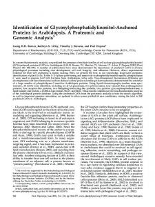
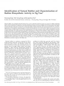
quence motifs were identified in addition to the three noted previously. The eight conserved sequence blocks are designated motifs I to VIII, in order from the N to the C termini; according to this notation, motifs I, VII, and VIII correspond to regions I, II, and III, respectively, as designated previously (Preiss and Sivak, 1996). The conserved sequences are distributed in the 359 residues of DU1 between positions 1237 and 1595. Recombinant DU1 Exhibits SS Activity The 449 C-terminal residues of DU1 (positions 1226– 1674; designated DU1C) were expressed in E. coli from plasmids pHC5 or pHC6. These plasmids are based in the expression vector pET-29b(1) or pET-32b(1), respectively, and thus produce DU1C fusion proteins containing either 35 or 167 plasmid-derived residues at their N termini. Expression of the fusion proteins was monitored by enzymatic and immunoblotting analyses that detected the S-tag sequence present in these N-terminal extensions. Proteins of the expected sizes were expressed specifically when the DU1C coding region was present (Fig. 1). Increased glucan synthase activity was observed in total soluble extracts of E. coli cells expressing DU1C. Cells containing pHC5 or pHC6 were exposed to IPTG to induce expression of the DU1C proteins, and total soluble extracts were tested for glucan synthase activity. DU1C expression resulted in approximately 5-fold increased SS activity compared with control cells lacking the maize coding region (Table II). A similar increase also occurred in the reconstituted RNase A activity conferred by the S-tag sequence of the N-terminal extension (data not shown). Nearly identical results were obtained when DU1C was expressed in pET-29b(1) or pET-32b(1). The activity increase relative to the endogenous level was relatively modest, although similar levels were detected also for zSSI expressed in E. coli
RESULTS Sequence Motifs Conserved in DU1 and SSs Three conserved sequence blocks identified previously in comparisons of various GBSSI proteins and E. coli GS (van der Leij et al., 1991; Preiss and Sivak, 1996) were present in the DU1 C-terminal region (Gao et al., 1998). This comparative analysis was extended to include 28 SS or GS sequences from 17 species (Table I). Thirty-three residues are conserved in all 28 enzymes. Five conserved se-
Figure 1. Expression of DU1C in E. coli. Gene expression from the T7 promoter of the indicated plasmid was induced in exponentialphase E. coli cells. Total soluble lysates were fractionated by SDSPAGE, and specific proteins containing the S-tag sequence (specified by the pET plasmid) were detected by S-protein-alkaline phosphatase conjugate. Lane 1, pET-32b; lane 2, pHC6 (DU1C in pET-32b); lane 3, pET-29b; and lane 4, pHC5 (DU1C in pET-29b). Asterisks indicate polypeptides of approximately the size predicted from the plasmid and Du1 cDNA sequences, which are present only when the DU1C coding region is contained within the plasmid.
210
Cao et al.
Table II. Glucan synthase activity in E. coli soluble extracts Gene expression was induced for 5 h in the exponential phase of E. coli cells transformed with the indicated plasmid. Total soluble extracts were assayed for SS activity in the presence of citrate and glycogen primer. Values indicate means 6 SE (n 5 4). Plasmida
Insert
Glucan Synthase Activity Total units
Relative unitsb
nmol Glc incorporated min21 mg21
pET-29b(1) pHC5 pET-32b(1) pHC6
None DU1Cc None DU1C
2.10 6 0.29 10.41 6 1.93 2.88 6 0.18 14.19 6 1.60
1.0 5.0 1.0 4.9
Plant Physiol. Vol. 120, 1999
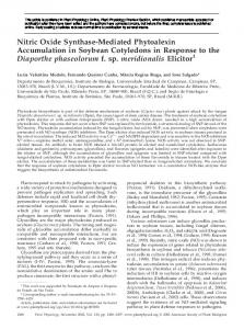
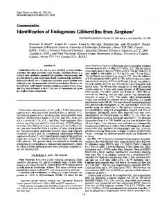
(Fig. 2b), which is known to be both granule associated and soluble (Mu et al., 1994). The amount of DU1 present in the granule and soluble fractions was determined by immunoblot analysis of protein samples standardized based on kernel fresh weight. In contrast to zSSI, the anti-DU1N signal was found almost exclusively in the soluble fraction (Fig. 2b), indicating that DU1 is not stably associated with starch granules in 20-DAP endosperm. The temporal expression pattern of DU1 and SSI in kernels at various times after pollination was monitored. DU1
a pET-29b(1) and pET-32b(1) are from Novagen. pHC5 and pHC6 b are based in these two vectors, respectively. Total activity units obtained for the appropriate plasmid vector with no insert are asc signed a value of 1. DU1C, DU1 residues 1226 to 1674.
(Knight et al., 1998). In addition, the level of recombinant enzyme activity observed for DU1C was comparable to that of potato GBSSII expressed in a similar system (Edwards et al., 1995). These data provide direct evidence that DU1 is a SS and that its C-terminal 449 residues are sufficient to provide this enzymatic activity. Immunological Detection of DU1 in Kernel Extracts Detection of DU1 in kernel extracts revealed the apparent size of this SS, its temporal expression pattern, and its lack of association with starch granules. The polyclonal antiserum anti-DU1N was raised in rabbits against the N-terminal 648 residues of DU1. This region of DU1 is unique among known protein sequences (Gao et al., 1998), so anti-DU1N is expected to react specifically with DU1 and not with other SSs. Figure 2a shows that in immunoblot analysis of total soluble kernel extracts (i.e. the 10,000g supernatant) from nonmutant kernels, anti-DU1N detected a protein that migrated at an apparent molecular mass of more than 200 kD. This protein was missing in two different du1- mutants. In kernels homozygous for the reference mutation du1-Ref, a smaller immunoreactive protein was detected, whereas in kernels homozygous for the presumed transposon-induced allele du1-R4059 (Gao et al., 1998), the protein was completely eliminated (Fig. 2a). Identical results were obtained using a different antiserum, anti-DU1F, which was raised against full-length DU1 (data not shown). Thus, both anti-DU1N and anti-DU1F recognized DU1, the product of the du1 gene. The zSSI protein of apparently 76 kD also was identified in immunoblot analysis of these same kernel extracts, using anti-SSI antiserum (Fig. 2a). Anti-DU1N did not recognize SSI, and anti-SSI did not recognize DU1. Therefore, in this assay, both antisera reacted specifically with a distinct isozyme. DU1 was found to be located primarily in the soluble fraction of kernel extracts, as opposed to being associated with starch granules. Kernels harvested 20 DAP were fractionated into soluble and granule fractions. The identity of the granule fraction was verified by enrichment for zSSI
Figure 2. Immunological detection of DU1 and SSI in kernel extracts. a, Total soluble extracts from 20-DAP kernels of the W64A genetic background homozygous for the indicated allele were fractionated by SDS-PAGE and probed with anti-DU1N or anti-SSI. An equal amount of protein was loaded in each lane. du1-M5 indicates the allele du1-R4059. The asterisk indicates full-length DU1. b, Extracts of nonmutant W64A kernels and congenic du1-Ref mutant kernels collected 20 DAP were separated into granule (i.e. 10,000g pellet) and total soluble fractions (i.e. 10,000g supernatant). An equal volume of each fraction was separated by SDS-PAGE; therefore, each pair of lanes is standardized to kernel fresh weight. The samples were probed with anti-DU1N or anti-SSI, as indicated. c, Total soluble extracts of W64A kernels harvested at various times after pollination (as indicated) were analyzed by SDS-PAGE and immunoblot analysis using anti-DU1N or anti-SSI.
Identification of Maize Soluble Starch Synthase Activities
211
was detected first at 12 DAP and was maintained at a nearly constant level throughout the period of starch biosynthesis for up to at least 32 DAP (Fig. 2c). Anti-SSI produced a signal in the 8-DAP kernel extract (Fig. 2c), indicating that in these tissue samples zSSI was expressed earlier than DU1. Immunodepletion of SS Activity in Kernel Extracts Immunodepletion experiments investigated the amount of SS activity in endosperm provided by DU1 and zSSI. Total soluble extracts of kernels harvested 20 DAP were treated with anti-DU1N, anti-DU1F, anti-SSI, or preimmune serum. Immune complexes were then removed from solution after binding to protein A-Sepharose beads. Residual SS activity remaining in the supernatant was determined in the presence of citrate and exogenous primer, conditions known to yield maximal activity of these enzymes (Preiss and Sivak, 1996). Preliminary experiments titrated the amount of serum; the following data were obtained in conditions of antibody excess. Nonmutant extracts of either the W64A or Oh43 background were depleted of approximately 28% of their total SS activity by either of the two anti-DU1 sera (Fig. 3). Anti-SSI depleted 66% and 61% of the total SS activity in the two genotypes, respectively. Treatment of a du1- mutant extract with either of the two anti-DU1 sera had virtually no effect on the total SS activity, suggesting that the particular enzyme affected by these antibodies is specifically that coded for by Du1. In contrast, anti-SSI treatment depleted virtually all of the SS activity present in the du1- mutant extract, which suggests that the great majority of SS activity in the soluble fraction
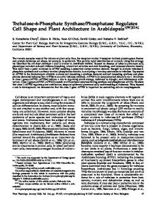
Figure 4. Specific identification of SS isozymes. a, SS activity zymogram. Proteins in total soluble endosperm extracts were separated based on molecular mass by SDS-PAGE and then allowed to renature in the gel. SS substrates were provided to the entire gel, and positions of glucan synthesis were detected by staining with iodine. Two congenic strains in the W64A genetic background were analyzed, one bearing the nonmutant allele Du1 and the other containing du1-Ref (indicated as du1-). Two SS activities are evident in the nonmutant endosperm, one of which is missing from the du1-Ref extract. b, Immunoblot analysis. Proteins in duplicates of the gel shown in a were transferred to nitrocellulose paper and probed with the indicated antiserum. A polypeptide of the same mobility and genetic specificity as the larger SS activity is recognized by antiDU1N, whereas a protein of the same mobility as the smaller SS activity is recognized by anti-SSI.
of 20-DAP endosperm is provided by a combination of zSSI and DU1. This analysis was applied to kernel extracts from nonmutant W64A plants collected 12, 18, or 32 DAP and also to an independently prepared 20-DAP extract. In these experiments, the range of total SS activity neutralized by antiDU1N ranged from 17% to 24%, and anti-SSI treatment depleted 47% to 66% of the activity (data not shown). The relative abundance of the DU1 and zSSI activities, therefore, did not change significantly during the period of starch biosynthesis. Figure 3. Immunodepletion of SS activity. Total soluble extracts from kernels of the indicated genotype collected 20 DAP were treated with preimmune serum or saturating amounts of the indicated antiserum, and residual SS activity was assayed after removal of the immune complexes. The du1-Ref mutant was in the W64A genetic background. SS activity remaining after treatment with preimmune serum was defined as 100%. These values were 7.0 nmol min21 mg21 for W64A, 12.9 nmol min21 mg21 for the du1-Ref mutant, and 16.4 nmol min21 mg21 for Oh43.
Fractionation of SS Activities in Total Endosperm Extracts The SS activities present in 20-DAP endosperm also were correlated with particular cloned cDNAs by a combination of zymogram, immunoblot, and mutational analyses. These SSs were fractionated by SDS-PAGE and detected by their activity in gels after protein renaturation. Two activity bands were observed, one of more than 200 kD and the other of approximately 76 kD (Fig. 4a). The sizes of these
212
Cao et al.
isozymes correlate roughly with those predicted by the Du1 cDNA and the Ss1 cDNA, respectively (Gao et al., 1998; Knight et al., 1998). Immunoblot analysis of the same protein samples revealed that the .200-kD isozyme reacted with anti-DU1N, whereas the 76-kD isozyme reacted with anti-zSSI (Fig. 4b). Extracts from du1- mutant endosperm entirely lacked activity of the .200-kD isozyme. These results support the conclusions of the previous section: (a) that there are two major soluble SSs present in developing endosperm cells, and (b) that one of these is DU1, the product of the du1 gene, and the other is zSSI, the product of the Ss1 cDNA.
Plant Physiol. Vol. 120, 1999
not shown). The immunodepletion data described above indicated that the only SS remaining in du1- mutants was zSSI (Fig. 3). Thus, it appears that the activity of zSSI was increased in du1- mutants. The level of zSSI protein detected by immunoblot analysis appeared to be slightly increased in du1- mutant kernels relative to nonmutants, either when the extracts were compared on a total protein basis (Fig. 2a) or when they were standardized by the fresh weight of the tissue used to prepare the extracts (Fig. 2b). This apparent increase, however, is significantly less than the 8-fold increase in enzyme activity. Thus, the elevated zSSI activity in du1- mutant extracts most likely results in part from the increased specific activity of the enzyme.
Increased Total SS Activity in du1- Mutant Extracts The conclusion that du1 specifies a SS appears to be inconsistent with a previous report indicating that soluble SS activity is not decreased in a du1- mutant. To the contrary, the total soluble SS activity was found to be increased approximately 2-fold in du1-Ref mutant extracts (Singletary et al., 1997); this observation was confirmed independently in the current study (Fig. 5). Congenic strains were analyzed, ruling out genetic background differences as the explanation for the different total SS levels. A possible explanation for this phenomenon is that a SS other than DU1 is hyperactive in du1- mutants. To test this possibility, SS activity in total soluble kernel extracts was assayed in the presence or absence of citrate and/or exogenous glucan primer. These experiments were intended to differentiate between zSSI, which is known to be stimulated significantly by citrate and to be independent of exogenous primer, and the enzyme accounting for the SSII-activity peak, which is primer dependent and largely citrate independent (Boyer and Preiss, 1981). Citrate-stimulated, primer-independent SS activity was increased approximately 8-fold in du1-Ref mutant extracts compared with nonmutant extracts (Fig. 5). Similar results were obtained for six other independent du1- alleles (data
Figure 5. SS activity in total soluble kernel extracts. Total soluble extracts from kernels of the indicated genotype collected 20 DAP were assayed for SS activity in the presence or absence of exogenous primer (10 mg/mL glycogen) and 0.5 M citrate, as indicated. The du1-Ref mutant was in the W64A genetic background.
DISCUSSION Important goals in the understanding of the mechanisms of starch biosynthesis include identification of the particular SS isozymes active during endosperm development and determination of the specific functions of each enzyme. Multiple soluble SSs are present in endosperm, as was shown initially by biochemical fractionation. Two activity peaks were observed, designated SSI, which does not require exogenous glucan primer and is stimulated by citrate, and SSII, which is dependent on exogenous primer and is largely insensitive to citrate (enzyme designations according to Boyer and Preiss, 1981). Five different cDNA clones are known that code for SSs, however, so it is necessary to correlate each enzymatic activity with a particular genetic element. The cDNA and protein that account for the SSI-activity peak were identified recently; however, the protein(s) responsible for the second SS had not been clearly assigned before this study. An apparent 76-kD protein copurified with the SSI activity (Mu et al., 1994). Sequence information indicated that the Ss1 cDNA codes for this polypeptide, and this cDNA directed expression of an active SS that is immunologically cross-reactive with the purified enzyme (Imparl-Radosevich et al., 1998; Knight et al., 1998). Thus, the genetic element responsible for synthesis of zSSI has now been identified. Presumably, at least one other protein provides additional SS activity in the soluble fraction, because of the distinct enzymatic characteristics and apparent Mr of the enzyme responsible for the SSII-activity peak. Detailed characterization of this second enzyme is lacking because it has proven difficult to purify. The gene du1 was proposed to code for a soluble SS activity based in part on the facts that du1- mutants lack the SSII activity (Boyer and Preiss, 1981) and that Du1 codes for a protein similar in sequence to known SSs (Table I). This study confirms the identification of DU1 as an SS active in developing endosperm. Expression of the DU1 C terminus correlated with induction of SS activity, and DU1-specific antibodies immunodepleted a significant portion of the enzyme present in kernel extracts. Furthermore, a specific SS enzyme activity identified by zymogram analysis migrated in SDS-PAGE at the same rate as DU1 and was missing in a du1- mutant. The SS activity of DU1 resides within the C-terminal 450 residues; the function(s) of the remaining 1224 residues remains to be determined.
Identification of Maize Soluble Starch Synthase Activities Inferences drawn from the immunodepletion data presume that anti-DU1N is specific for DU1. Immunological specificity was indicated by three observations. First, in immunoblot analysis, anti-DU1N failed to detect zSSI (and anti-SSI failed to detect DU1). Second, when du1- mutant extracts were treated with anti-DU1N, there was no decrease in residual SS activity, even though anti-SSI treatment of the same extracts reduced the activity almost completely. Thus, anti-DU1N did not neutralize zSSI. Third, the anti-DU1 and anti-SSI immunoprecipitates were analyzed by immunoblotting using both antisera; the anti-DU1N complexes did not contain zSSI, and visa versa (H. Cao, unpublished results). DU1 and zSSI most likely account for all of the soluble SS activity in developing kernels. Only two enzymes were observed in zymograms, and each of these could be correlated with either DU1 or zSSI. The fact that the combined effects of a du1- mutation and anti-zSSI immunodepletion caused nearly complete loss of SS activity suggests that these are the only two isozymes present in the soluble fraction. Consistent with this conclusion is the fact that antibodies reactive with either of the remaining known SSs, zSSIIa or zSSIIb (Harn et al., 1998), failed to detect polypeptides in soluble extracts of 20-DAP kernels (J. ImparlRadosavich and H. Cao, unpublished results). There is one inconsistency with the idea that DU1 and zSSI account for all soluble SS activity, which is the fact that the amount of enzyme neutralized by anti-DU1 and anti-SSI sera together is about 90% of the total (Fig. 3). A possible explanation for this observation is that the anti-DU1 sera are not capable of binding all of the DU1 present, perhaps as a result of partial degradation of the protein. Alternatively, one or more additional soluble SSs might exist that are not detected in the zymograms, either because they fail to recover activity after denaturation or they comigrate with zSSI. We consider the latter possibility unlikely, because if an additional enzyme exists, it should be evident after treatment of the du1- mutant extract with anti-SSI. Despite these arguments, the data in this study certainly do not exclude the possibility that SSs in addition to DU1 and zSSI exist as minor activities in the soluble fraction of maize endosperm. This study supports the hypothesis that DU1 accounts for the SSII-activity peak (Boyer and Preiss, 1981). The fact that there are two SS peaks in anion-exchange chromatography and two enzymes in the zymograms is most simply explained by a direct correspondence. Such a correspondence is further indicated by molecular mass comparisons: immunoblots indicated that DU1 has a mass of more than 200 kD, and the native size of the protein responsible for the SSII-activity peak was estimated to be 180 kD (Mu et al., 1994). The .200-kD protein detected by anti-DU1N was not present in du1- mutants. Most tellingly, zymogram analysis revealed the existence of a .200-kD SS that is missing in du1- mutants, as is the case also for the SSIIactivity peak (Boyer and Preiss, 1981). All of these diverse observations would be explained if DU1 were the active SS enzyme present in the SSII-activity peak. The correspondence between the molecular mass determined for the native enzyme in the SSII-activity peak, the size of the SS identified as DU1 separated in denaturing zymograms, and
213
the size of DU1 predicted by cDNA cloning indicates that this enzyme functions as a monomer. The proteolytically labile nature of DU1 may explain the facts that purification of the native SS present in the SSII-activity peak has been problematic and that different molecular masses (180 and 95 kD) have been reported (Mu et al., 1994; Preiss and Sivak, 1996). Assignment of DU1 as a soluble protein of more than 200 kD was supported by an independent study (Yu et al., 1998). A protein of this size was present in the purified amyloplast stromal fraction from nonmutant plants but was lacking in a du1- mutant. This protein most likely is the same as the .200-kD SS and the .200-kD anti-DU1N reactive protein shown here to be absent in du1- kernels. Taken together, these data indicate that DU1, as expected, is located within plastids. DU1 and zSSI share the property that their mobility in SDS-PAGE is slower than predicted from their cDNA sequences. The Ss1 cDNA predicts a 64-kD protein, whereas zSSI runs in gels at 76 kD (Knight et al., 1998). The Du1 cDNA predicts a 188-kD protein; however, DU1 in kernel extracts runs significantly slower than the 200-kD marker. Anomalous migration in SDS-PAGE is thought to be an intrinsic property of zSSI (Knight et al., 1998) and other SSs (Edwards et al., 1995, 1996). The same phenomenon may apply to DU1, or it could be posttranslationally modified. Removal of DU1 from the soluble endosperm fraction apparently causes some change that results in increased activity of zSSI. A possible explanation is that DU1 deficiency causes accumulation of a glucan not present normally, and this provides an efficient primer for zSSI. This observation explains the fact that total SS activity is not reduced in du1- mutant extracts (Singletary et al., 1997), even though a specific SS isozyme is lacking. The emerging characterization of the complement of maize SSs warrants some discussion here of their nomenclature in relation to that of the starch biosynthesis enzymes of other higher plants. The situation is straightforward with regard to GBSSI, which is the product of the wx locus and is highly conserved in all plant tissues that produce storage starch (for review, see Ball et al., 1998). The original nomenclature for the maize soluble SSs was based on DEAE chromatography fractions (Boyer and Preiss, 1981; Preiss and Sivak, 1996), and in this paper we refer accordingly to the SSI- and SSII-activity peaks. The enzyme responsible for the SSI-activity peak, termed zSSI, has been characterized by both biochemical purification and cDNA cloning (Mu et al., 1994; Harn et al., 1998; Imparl-Radosevich et al., 1998; Knight et al., 1998). zSSI is very similar in primary sequence to a soluble SS of rice (Baba et al., 1993). It exists both bound to starch granules and in a soluble form in the amyloplast stroma, and in this regard it is similar to the enzymes of pea embryos and potato tubers designated as SSII. However, in terms of amino acid sequence, zSSI is not the counterpart of pea SSII or potato SSII. The sequences of the maize enzymes coded for by the zSSIIa and zSSIIb cDNAs (Harn et al., 1998; Knight et al., 1998) are significantly closer to those of pea or potato SSII (Dry et al., 1992; Edwards et al., 1995) than is the zSSI sequence. Thus, zSSI-type enzymes have been
214
Cao et al.
reported so far only in monocots, whereas zSSIIa and zSSIIb are members of a more generally conserved class, along with pea SSII and potato SSII. It is not known whether this sequence conservation extends to functional conservation. zSSIIa and zSSIIb are expressed in developing endosperm not at all or only at very low levels, whereas the SSII enzymes of pea and potato clearly are present at significant levels during storage-starch biosynthesis. We argue above that the SS designated here by virtue of its genetic identification as DU1 is the enzyme responsible for the SSII-activity peak. This enzyme is the evolutionary counterpart of potato SSIII (Abel et al., 1996; Marshall et al., 1996) based on both the very high sequence identity between these two enzymes and the fact that both enzymes are exclusively soluble. We propose that the most logical nomenclature for these enzymes is one based on evolutionary sequence conservation and species; therefore, we suggest that DU1 should also be known as zSSIII. In summary, the enzyme coded for by the du1 locus, zSSIII/DU1, is most likely responsible for the maize SSII-activity peak and is the apparent evolutionary counterpart of potato SSIII. Based on sequence identity, zSSIIa and zSSIIb appear to be the evolutionary counterparts of pea SSII and potato SSII, although they are not highly expressed in developing maize endosperm. zSSI accounts for the SSI-activity peak, and as yet a distinct evolutionary counterpart from pea or potato has not been reported. zSSI is distinct from GBSSI, the product of the wx locus. Finally, the lowercase “z” in the names designated here indicates that the species of origin of each enzyme is maize. The reason that multiple soluble SSs are used in storagestarch biosynthesis is not known at present. DU1 clearly is distinct from zSSI in that it is located almost entirely within the soluble phase of endosperm cells, whereas zSSI is abundant in both the granule and the soluble fractions (Fig. 2b). The fact that du1- mutations alter starch structure indicates that DU1 provides a specific function(s) that cannot be compensated for by zSSI. Similarly, severe reduction of potato SSIII by antisense RNA expression causes significant changes in granule structure that cannot be compensated for by the remaining soluble SS activity (Abel et al., 1996; Marshall et al., 1996). Although the specific functions of each soluble SS remain to be determined, identification of the genetic sources of the two major isoforms in maize will provide significant new tools for such investigations. ACKNOWLEDGMENTS We thank Afroza Rahman and Tracie Bierwagen for technical advice and assistance. Received October 6, 1998; accepted January 23, 1999. LITERATURE CITED Abel GJW, Springer F, Willmitzer L, Kossmann J (1996) Cloning and functional analysis of a cDNA encoding a novel 139 kDa starch synthase from potato (Solanum tuberosum L.). Plant J 10: 981–991 Ausubel FM, Brent R, Kingston RE, Moore DD, Smith JA, Seidman JG, Struhl K (1989) Current Protocols in Molecular Biology. John Wiley & Sons, New York
Plant Physiol. Vol. 120, 1999
Baba T, Nishihara M, Mizuno K, Kawasaki T, Shimada H, Kobayashi E, Ohnishi S, Tanaka K, Arai Y (1993) Identification, cDNA cloning, and gene expression of soluble starch synthase in rice (Oryza sativa L.) immature seeds. Plant Physiol 103: 565–573 Ball S, Guan H-P, James M, Myers A, Keeling P, Mouille G, Bule´on A, Colonna P, Preiss J (1996) From glycogen to amylopectin: a model for the biogenesis of the plant starch granule. Cell 86: 349–352 Ball SG, van de Wal MHBJ, Visser RGF (1998) Recent progress in understanding the biosynthesis of amylose. Trends Plant Sci 3: 462–467 Beavis W, Berlyn M, Burr B, Chandler V, Coe E, Fauron C, Nelson O, Polacco M, Rodermel S, Sachs M, and others (1995) A standard for maize genetics nomenclature. Maize Genet Coop Newslett 69: 182–184 Boyer CD, Preiss J (1981) Evidence for independent genetic control of the multiple forms of maize endosperm branching enzymes and starch synthases. Plant Physiol 67: 1141–1145 Bradford MM (1976) A rapid and sensitive method for the quantitation of microgram quantities of protein utilizing the principle of protein-dye binding. Anal Biochem 72: 248–254 Bule´on A, Gallant DJ, Bouchet B, Mouille G, D’Hulst C, Kossman J, Ball S (1997) Starches from A to C. Chlamydomonas reinhardtii as a model microbial system to investigate the biosynthesis of the plant amylopectin crystal. Plant Physiol 115: 949–957 Cao H, Preiss J (1996) Evidence for essential arginine residues at the active sites of maize branching enzymes. J Protein Chem 15: 291–304 Craig J, Lloyd JR, Tomlinson K, Barber L, Edwards A, Wang TL, Martin C, Hedley CL, Smith AM (1998) Mutations in the gene encoding starch synthase II profoundly alter amylopectin structure in pea embryos. Plant Cell 10: 413–426 Dang PL, Boyer CD (1988) Maize leaf and kernel starch synthases and starch branching enzymes. Phytochemistry 27: 1255–1259 Dayhoff MO, Orcutt BC (1979) Methods for identifying proteins by using partial sequences. Proc Natl Acad Sci USA 76: 2170–2174 Denyer K, Clarke B, Hylton C, Tatge H, Smith AM (1996) The elongation of amylose and amylopectin chains in isolated starch granules. Plant J 10: 1135–1143 Dry I, Smith AM, Edwards EA, Bhattacharyya M, Dunn P, Martin C (1992) Characterization of cDNAs encoding two isoforms of granule-bound starch synthase which show differential expression in developing storage organs. Plant J 2: 193–202 Edwards A, Marshall J, Denyer K, Sidebottom D, Visser RGF, Martin C, Smith AM (1996) Evidence that a 77-kilodalton protein from the starch of pea embryos is an isoform of starch synthase that is both soluble and granule bound. Plant Physiol 112: 89–97 Edwards A, Marshall J, Sidebottom C, Visser RGF, Smith AM, Martin C (1995) Biochemical and molecular characterization of a novel starch synthase from potato tubers. Plant J 8: 283–294 Fontaine T, Hulst CD, Maddelein M-L, Routier F, Pepin TM, Decq A, Wieruszeski J-M, Delrue B, Van den Koornhuyse N, Bossu J-P, and others (1993) Toward an understanding of the biogenesis of the starch granule: evidence that Chlamydomonas soluble starch synthase II controls the synthesis of intermediate size glucans of amylopectin. J Biol Chem 268: 16223–16230 Gallant DJ, Bouchet B, Baldwin PM (1997) Microscopy of starch: evidence of a new level of granule organization. Carbohydr Polym 32: 177–191 Gao M, Wanat J, Stinard PS, James MG, Myers AM (1998) Characterization of dull1, a maize gene coding for a novel starch synthase. Plant Cell 10: 339–412 Hannah LC, Giroux M, Boyer CD (1993) Biotechnological modification for sweet corn and maize improvement. Sci Hortic 55: 177–197 Harlow E, Lane D (1988) Antibodies: A Laboratory Manual. Cold Spring Harbor Laboratory Press, Cold Spring Harbor, NY
Identification of Maize Soluble Starch Synthase Activities Harn C, Knight M, Ramakrishnan A, Guan H, Keeling PL, Wasserman BP (1998) Isolation and characterization of the zSSIIa and zSSIIb starch synthase cDNA clones from maize endosperm. Plant Mol Biol 37: 639–649 Imparl-Radosevich JM, Li P, Zhang L, McKean AL, Keeling PL, Guan H (1998) Purification and characterization of maize starch synthase I and its truncated forms. Arch Biochem Biophys 353: 64–72 Klo¨sgen RB, Gierl A, Schwarz-Sommer Z, Saedler H (1986) Molecular analysis of the waxy locus of Zea mays. Mol Gen Genet 203: 237–244 Knight ME, Harn C, Lilley CER, Guan HP, Singletary G, MuForster C, Wasserman BP, Keeling PL (1998) Molecular cloning of starch synthase I from maize (W64A) endosperm and expression in E. coli. Plant J 14: 613–622 Mangelsdorf PC (1947) The inheritance of amylaceous sugary endosperm and its derivatives in maize. Genetics 32: 448–458 Marshall J, Sidebottom C, Debet M, Martin C, Smith AM, Edwards A (1996) Identification of the major starch synthase in the soluble fraction of potato tubers. Plant Cell 8: 1121–1135 Martin C, Smith AM (1995) Starch biosynthesis. Plant Cell 7: 971–985 Mu C, Harn C, Ko Y-T, Singletary GW, Keeling PL, Wasserman BP (1994) Association of a 76 kDa polypeptide with soluble starch synthase I activity in maize (cv B73) endosperm. Plant J 6: 151–159 Nakamura Y (1996) Some properties of starch debranching enzymes and their possible role in amylopectin biosynthesis. Plant Sci 121: 1–18 Nelson OE, Pan D (1995) Starch synthesis in maize endosperms. Annu Rev Plant Physiol Plant Mol Biol 46: 475–496 Posfai J, Bhagwat AS, Posfai G, Roberts RJ (1989) Predictive motifs derived from cytosine methyltransferases. Nucleic Acids Res 17: 2421–2435 Preiss J, Sivak M (1996) Starch synthesis in sinks and sources. In E Zamski, AA Schaffer, eds, Photoassimilate Distribution in Plants and Crops. Marcel Dekker, New York, pp 63–96
215
Rahman A, Wong K-S, Jane J-L, Myers AM, James MG (1998) Characterization of SU1 isoamylase, a determinant of storage starch structure in maize. Plant Physiol 117: 425–435 Sambrook J, Fritsch EF, Maniatis T (1989) Molecular Cloning: A Laboratory Manual, Ed 2. Cold Spring Harbor Laboratory Press, Cold Spring Harbor, NY Shannon JC, Garwood DL (1984) Genetics and physiology of starch development. In RL Whistler, JN BeMiller, EF Paschall, eds, Starch: Chemistry and Technology. Academic Press, San Diego, CA, pp 25–86 Shure M, Wessler S, Federoff N (1983) Molecular identification and isolation of the waxy locus in maize. Cell 35: 225–233 Singletary GW, Banisadr R, Keeling PL (1997) Influence of gene dosage on carbohydrate synthesis and enzymatic activities in endosperm of starch-deficient mutants of maize. Plant Physiol 113: 293–304 Smith AM, Denyer K, Martin C (1997) The synthesis of the starch granule. Annu Rev Plant Physiol Plant Mol Biol 48: 67–87 van der Leij FR, Visser RG, Ponstein AS, Jacobsen E, Feenstra WJ (1991) Sequence of the structural gene for granule-bound starch synthase of potato (Solanum tuberosum L.) and evidence for a single point deletion in the amf allele. Mol Gen Genet 228: 240–248 van de Wal M, D’Hulst C, Vincken J-P, Bule´on A, Visser R, Ball S (1998) Amylose is synthesized in vitro by extension of and cleavage from amylopectin. J Biol Chem 273: 22232–22240 Wang Y-J, White P, Pollak L, Jane J-L (1993a) Characterization of starch structures of 17 maize endosperm mutant genotypes with Oh43 inbred line background. Cereal Chem 70: 171–179 Wang Y-J, White P, Pollak L, Jane J-L (1993b) Amylopectin and intermediate materials in starches from mutant genotypes of the Oh43 inbred line. Cereal Chem 70: 521–525 Yu Y, He Mu H, Mu-Forster C, Wasserman BP (1998) Polypeptides of the maize amyloplast stroma. Stromal localization of starch-biosynthetic enzymes and identification of an 81kilodalton amyloplast stromal heat-shock cognate. Plant Physiol 116: 1451–1460