Identification of Three Urease Accessory Proteins That Are Required for Urease Activation in Arabidopsis1 Claus-Peter Witte*, Mario G. Rosso, and Tina Romeis Freie Universita¨t Berlin, Institut fu¨r Biologie, Abteilung Biochemie der Pflanzen, 14195 Berlin, Germany (C.-P.W., T.R.); and GABI-Kat at Max-Planck-Institute for Plant Breeding Research, 50829 Cologne, Germany (M.G.R.)
Urease is a nickel-containing urea hydrolase involved in nitrogen recycling from ureide, purine, and arginine catabolism in plants. The process of urease activation by incorporation of nickel into the active site is a prime example of chaperonemediated metal transfer to an enzyme. Four urease accessory proteins are required for activation in Klebsiella aerogenes. In plants urease accessory proteins have so far been only partially defined. Using reverse genetic tools we identified four genes that are necessary for urease activity in Arabidopsis (Arabidopsis thaliana; ecotypes Columbia and No¨ssen). Plants bearing T-DNA or Ds element insertions in either the structural gene for urease or in any of the three putative urease accessory genes AtureD, AtureF, and AtureG lacked the corresponding mRNAs and were defective in urease activity. In contrast to wild-type plants, the mutant lines were not able to support growth with urea as the sole nitrogen source. To investigate whether the identified accessory proteins would be sufficient to support eukaryotic urease activation, the corresponding cDNAs were introduced into urease-negative Escherichia coli. In these bacteria, urease activity was observed only when all three plant accessory genes were coexpressed together with the plant urease gene. Remarkably, plant urease activation occurred as well in cell-free E. coli extracts, but only in extracts from cells that had expressed all three accessory proteins. The future molecular dissection of the plant urease activation process may therefore be performed in vitro, providing a powerful tool to further our understanding of the biochemistry of chaperone-mediated metal transfer processes in plants.
The first step of the hydrolysis of urea to ammonium and carbonic acid is catalyzed by the nickel metalloenzyme urease, which is found in many microbes (Mulrooney and Hausinger, 2003) and plants (Frankenberger and Tabatabai, 1982; Witte and Medina-Escobar, 2001). In plants, urea originates from the breakdown of Arg, especially important during germination (Zonia et al., 1995; Goldraij and Polacco, 1999), and from purines or ureides. Because ureides serve as nitrogen-transport compounds in nitrogenfixing legumes their degradation has received special attention (Stebbins and Polacco, 1995; Todd and Polacco, 2004). Externally applied urea as fertilizer is also made accessible to plants by urease (Witte et al., 2002a). Since urea is one of the most used nitrogen fertilizers worldwide (http://faostat.fao.org), its enzymatic hydrolysis is a process of great agricultural importance. Activation of urease is best studied in Klebsiella aerogenes. It requires the carbamoylation of a Lys 1 This work was supported by the Program for Investment in the Future from the German Ministry of Education and Science and the Alexander von Humboldt Foundation. * Corresponding author; e-mail
[email protected]; fax 49–30–83853372. The author responsible for distribution of materials integral to the findings presented in this article in accordance with the policy described in the Instructions for Authors (www.plantphysiol.org) is: Claus-Peter Witte (
[email protected]). Article, publication date, and citation information can be found at www.plantphysiol.org/cgi/doi/10.1104/pp.105.070292.
residue and the incorporation of two nickel ions per active site (Kuchar and Hausinger, 2004). For in vivo urease activation, three urease accessory proteins, UreD, UreF, and UreG, are absolutely necessary and a fourth accessory protein, UreE, facilitates this process. The precise role of the accessory proteins is not yet clearly understood. Apo-urease and UreD, UreF, and UreG form a complex that is competent to incorporate CO2 and nickel upon GTP hydrolysis carried out by UreG (Soriano and Hausinger, 1999). Nickel is delivered by UreE, a nickel-binding metallochaperone (Soriano et al., 2000). After activation the complex dissociates. Prokaryotic ureases are mostly encoded by three genes that are collinearly fused to one gene in eukaryotes (Mulrooney and Hausinger, 2003). Plant urease genes have first been characterized from soybean (Glycine max). The soybean genome contains an embryo-specific urease encoded by the gene Eu1 (Meyer-Bothling and Polacco, 1987) and a ubiquitous urease encoded by Eu4 (Torisky et al., 1994). In contrast, potato (Solanum tuberosum), tomato (Lycopersicon esculentum), several other solanaceous species, and also Arabidopsis (Arabidopsis thaliana; ecotype Columbia [Col]) possess only a single urease gene (Witte et al., 2005). Using microbial accessory protein sequences, putative plant orthologs for UreD, UreF, and UreG were identified in the databases and the corresponding cDNAs cloned (Freyermuth et al., 2000; Witte et al., 2001; Bacanamwo et al., 2002). Sequence identities determined from pair-wise alignments of K. aerogenes
Plant Physiology, November 2005, Vol. 139, pp. 1155–1162, www.plantphysiol.org Ó 2005 American Society of Plant Biologists
1155
Witte et al.
UreD and UreF with Arabidopsis UreD and UreF candidates are about 20% in both cases, providing only a weak indication that these proteins may be orthologous. A plant gene with similarity to bacterial ureE was not found, at least in the genome of Arabidopsis. It was hypothesized that the function of the bacterial metallochaperone UreE may have been taken over in plants by the UreG protein. In comparison to bacterial UreG proteins, plant UreGs contain an extended N terminus rich in His and Asp residues and were shown to bind nickel (Freyermuth et al., 2000; Witte et al., 2001). A function for the putative plant urease accessory proteins in plant urease activation has so far been demonstrated only for an UreG ortholog from soybean. A mutation in the corresponding gene Eu3 eliminated both urease activities of this plant (Freyermuth et al., 2000). Functional complementation of defective microbial urease activation complexes indicated a possible role of other putative plant accessory protein orthologs in urease activation. A K. aerogenes urease operon carrying a mutation in ureG could be partially complemented by a potato ureG ortholog (Witte et al., 2001) and a Schizosaccharomyces pombe ureF mutant was partially complemented by a soybean protein with similarity to microbial UreF (Bacanamwo et al., 2002). However, a role in plant urease activation still needs to be established for possible plant UreF orthologs. Attempts to complement ureD mutants of K. aerogenes or S. pombe with putative plant UreD orthologs have failed so far (Witte, 2001; Bacanamwo et al., 2002). A further mutant lacking all urease activities in soybean is encoded by the Eu2 gene (Meyer-Bothling et al., 1987). However, the Eu2 mutant could not be assigned to any of soybean accessory protein candidate genes so far (Bacanamwo et al., 2002). Here we report that Arabidopsis null mutants of the genes encoding At2g35035, At1g21840, and At2g34470 lack urease activity in vivo, identifying these proteins as genuine urease accessory proteins AtUreD, AtUreF, and AtUreG. Simultaneous coexpression of Aturease, AtUreD, AtUreF, and AtUreG in Escherichia coli led to activation of plant urease in bacterial cells and in cellfree extracts, showing that these proteins are the core components for plant urease activation. RESULTS Isolation of Mutants
Using the insertion mutant collections of the Salk Institute Genomic Analysis Laboratory (Alonso et al., 2003), the German Plant Genomics Research Program (GABI-Kat lines; Rosso et al., 2003), and the Plant Functional Genomics Research Group at the RIKEN Genomic Science Centre (Kuromori et al., 2004), we isolated a homozygous mutant in the urease gene, two homozygous mutants in the putative ureF and ureD genes, respectively, and three homozygous mutants in the ureG gene of Arabidopsis (Fig. 1; Table I). Urease, 1156
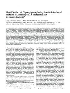
Figure 1. Schematic overview of Arabidopsis urease (A), ureD (B), ureF (C), and ureG (D) unspliced coding sequences. Exons are shown in gray, introns are shown in white. Primer positions are indicated by arrows. The positions of insertions in the mutant alleles are marked by the triangles. The arrow on the triangle is drawn near the left border for T-DNAs and on the G-side for Ds transposons indicating the orientation of the insertions. PCR product sizes for primer pairs used in RT-PCR experiments are given above the respective drawings. Product sizes without introns are given in parenthesis. The black triangle in B indicates the position of the stop codon in alternatively spliced ureD*.
ureG1 and 2, and both ureD mutants were obtained from T-DNA insertion collections of the Salk Institute and GABI-Kat in ecotype Col. Both ureF mutants and mutant ureG-3 were obtained from the Ds transposon insertion collection of RIKEN in ecotype No¨ssen (Table I). As recommended by RIKEN, the Ds donor line DS5392-12 (Kuromori et al., 2004) instead of No¨ssen wild type was used as control in all following experiments. All four genes are single copy in Col; the copy numbers in No¨ssen are unknown. The positions of the insertions were confirmed by sequencing the PCR products amplified with a primer binding to the left border of the T-DNA or the G-side of the Ds element in combination with a gene-specific primer, respectively (see ‘‘Materials and Methods’’). Except for mutant lines ureD-2 and ureG-2, the insertions had occurred in exons, likely preventing the generation of an intact mRNA. The insertion in ureD-2 was found in intron 1 8 bp downstream of the intron-exon boundary, possibly compromising correct splicing. In ureG-2 the T-DNA inserted exactly at the boundary of exon 3 to intron 4, eliminating the 5# splice site of intron 4. The ureF gene encodes a protein of 240 amino acids and does not contain introns. In both ureF mutants the reading frame is truncated and altered at the 3# end. UreF-1 encodes a protein of 217 amino acids, the Plant Physiol. Vol. 139, 2005
Identification of Plant Urease Accessory Proteins
Table I. Mutant lines
a
Gene
Gene Code
Insertion Type
Mutant Collection
Ecotype
Line ID
Mutant Name
urease ureD ureD ureF ureF ureG ureG ureG
At1g67550 At2g35035
T-DNA T-DNA T-DNA Ds Ds T-DNA T-DNA Ds
Salk Salk GABI-Kat RIKEN RIKEN GABI-Kat GABI-Kat RIKEN
Col Col Col No¨ssen No¨ssen Col Col No¨ssen
N538002 N605718 399D03 15-1020-1 15-4563-1 294B06 386A08 16-2845-1
ure-1 ureD-1 ureD-2 ureF-1 ureF-2 ureG-1 ureG-2 ureG-3
At1g21840 At2g34470
Insertion position in the unspliced coding sequence of the respective gene, 5# or 3# of the last unaltered base.
Insertion Positiona
5# 3# 5# 3# 3# 5# 5# 5#
of of of of of of of of
base base base base base base base base
3,909 (exon 18) 407 (exon 3) 108 (intron 1) 600b 564b 821 (exon 4) 615 (intron 3) 572 (exon 3)
b
ureF does not contain introns.
C-terminal 17 amino acids being altered, and ureF-2 encodes a 190-amino acid protein with two changed residues at the C terminus. None of the mutants displayed a visible phenotype under standard growth conditions used for Arabidopsis (see ‘‘Materials and Methods’’).
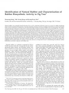
consistently observed (Fig. 2B). These products were ureD specific because they were absent in reactions performed with RNA from the ureD insertion mutants (lanes 4 and 6). Control RT-PCR reactions without RT were negative (lanes 7–12), demonstrating that the products amplified from an RNA template.
Reverse Transcription-PCR Analysis of Mutants
Urease Activity Assays
Insertion mutants were characterized by reverse transcription (RT)-PCR experiments testing for the presence and integrity of mRNA transcribed from the genes affected by the insertions. For each mutant two primer pairs were used, one spanning the insertion and one in an unaltered region of the gene but always spanning an intron where possible (Fig. 1). To test the RT reactions, control PCRs with primers for actin 2 (At3g18780) were carried out (Fig. 2D). Control reactions without RT to test for the presence of genomic DNAwere negative in all cases (Fig. 2B, lanes 7–12; data not shown). Reactions were repeated at least twice. These experiments demonstrated that all mutants lacked an intact wild-type mRNA for the respective mutant gene. In detail, for none of the mutants except ureG-1 an RT-PCR product was obtained with primers spanning the insertion (lanes marked ‘‘s’’ in Fig. 2), while the corresponding wild-type controls were positive yielding products of the expected size (for expected product sizes see Fig. 1). With RNA from ureG-1 plants, a product of reduced length was obtained, possibly because the corresponding template in this mutant is subject to aberrant splicing due to the insertion (Fig. 2A, lane 8). With the primer pairs binding in unaltered regions of the respective genes, RT-PCR products were obtained in all cases albeit in strongly reduced amounts for ureF-1, ureF-2, and ureG-3 (Fig. 2C, lanes 3, 5, and 9). For ureG-1 the obtained product was of increased size (Fig. 2A, lane 7) again arguing for altered splicing of the template in this mutant. Also, for ureG-2 splicing may be partially affected (lane 9). UreD transcript was previously described to be alternatively spliced in part leading to the incorporation of a stop codon exactly 3# of exon 4 (Fig. 1B, black triangle; accession no. AJ312203; Witte, 2001). RT-PCR experiments presented here indicated that ureD transcript is subject to further alternative or inefficient splicing because several higher Mr products were
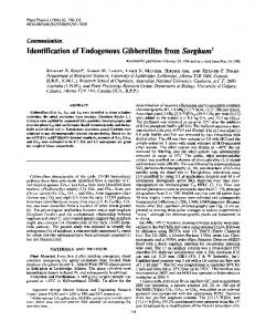
All mutant plants were unable to sustain growth on urea as sole nitrogen source (Fig. 3). In contrast, both wild-type accessions grew well either with nitrate or with urea. This experiment was repeated three times with identical results. For urease activity quantification, protein extracts were prepared from pooled leaves of four to five plants for each line, respectively. The plants were grown on soil supplemented with NiCl2 to allow efficient urease activation. For the wild-type plants, leaf urease activities of 13.5 6 0.7 mU mg21 (No¨ssen Ds donor line DS5-392-12) and 17.9 6 1.6 mU mg21 (Col 0) were determined (errors are confidence intervals for P 5 95%). Comparable activities were reported from leaves of other plants (Witte and Medina-Escobar, 2001). For none of the mutants could urease activity be detected. Thus, urease activity in all mutants was absent or at least strongly reduced below detection limit of the assay. These findings demonstrate that the Arabidopsis urease accessory proteins AtUreD, AtUreF, and AtUreG are required in vivo to generate a functional urease enzyme.
Plant Physiol. Vol. 139, 2005
Reconstitution of Plant Urease Activation in E. coli
To investigate whether AtUreD, AtUreF, and AtUreG are not only necessary but also sufficient for plant urease activation at least in a heterologous system, we coexpressed these accessory proteins together with Aturease in E. coli and measured urease activity in bacterial extracts. While some Enterobacteria like K. aerogenes contain a urease operon and are urease positive, E. coli generally does not possess ureolytic activity (Mobley et al., 1995). When Aturease and all three Arabidopsis accessory proteins were simultaneously expressed, ureolytic activity was observed in E. coli extracts. Lack of AtureD, AtureF, or AtureG eliminated urease activation 1157
Witte et al.
the assay (Fig. 4A). Alternatively, if the activation occurred in the cells but continued after extraction in vitro, the activity would be expected to increase during the enzyme assay. This was observed in the presence of the full-length ureD gene. To confirm that the nonlinear increase of ammonium production was in fact due to in vitro urease activation, an experiment
Figure 2. Characterization of mutants by RT-PCR. Lanes labeled with ‘‘S’’ show results obtained with primers spanning the T-DNA or Ds transposon insertion region. Unmarked lanes show results with primers that generate products either upstream or downstream of the insertion region (for primer positions and expected product sizes see Figure 1). A, Results for Col 0 wild type, the urease mutant, and mutants ureG-1 and ureG-2. B, Results for Col 0 and the mutants ureD-1 and ureD-2. Lanes 1 to 6, with RT; lanes 7 to 12, without RT. C, Results for the Ds element donor line DS5-392-12 (No¨) and ureF-1, ureF-2, and ureG-3. D, Control reactions with actin 2 primers.
in this system (Fig. 4A). Presence of the corresponding mRNAs was shown by RT-PCR (Fig. 4B). Thus, the three accessory proteins are not only necessary but also comprise the core components required for the activation of plant urease. Replacing ureD with the alternative splice variant ureD* (stop codon directly 3# of exon 4; Fig. 1) also led to urease activation but again only in the presence of the other accessory proteins. If the activation occurred only inside the E. coli cells but not after extraction, a constant activity would be measured in the extract. This appeared to be the case in the presence of truncated ureD* where a linear increase of ammonium concentration was observed in 1158
Figure 3. Growth test on agar plates with nitrate or urea as nitrogen source. Left column, plates supplemented with nitrate; right column, plates supplemented with urea. Photographs were taken 4 weeks after germination. Plant Physiol. Vol. 139, 2005
Identification of Plant Urease Accessory Proteins
Figure 4. Activation of Aturease by Arabidopsis urease accessory proteins coexpressed in E. coli. A, Ammonium generation from urea per milligram total protein in extracts of E. coli cultures that had expressed urease and different sets of urease accessory proteins: UreD (D); UreF (F); UreG (G), and UreD* (D*: truncated UreD from alternatively spliced mRNA). B, RT-PCR results from RNA extracted 2 h after gene induction with IPTG. The accessory proteins expressed in the respective cultures are indicated on the left. The mRNAs amplified are indicated above: Urease (U), ureD (D), ureF (F), and ureG (G).
combining different E. coli extracts was performed. Extracts of cells that had expressed urease in combination with UreF and UreG (1), or UreD and UreG (2), or UreD and UreF (3) did not display activity, whereas the combined extracts of (1) and (3) or (2) and (3) allowed urease activation in vitro (Fig. 5). These experiments also provided a control showing that yellow fluorescent protein (YFP), which was always present when one of the accessory proteins was missing (see ‘‘Materials and Methods’’), did not interfere with urease activation. Interestingly, combining extracts (1) and (2) did not lead to in vitro urease activation (Fig. 5). If either UreF or UreD had been lacking in the cell, extracts were not competent for in vitro urease activation even if extracts from cells that had expressed the missing protein were added. DISCUSSION
Urease allows plants to recycle urea-nitrogen originating from Arg breakdown and purine or ureide Plant Physiol. Vol. 139, 2005
catabolism. Since nitrogen availability is generally growth limiting for plants (Bray, 1983), efficient recycling is likely to give plants an ecological advantage. We demonstrate in this study that Arabidopsis requires three accessory proteins to generate urease activity. Soybean plants with defects in urease activity were previously shown to contain mutations in the urease and ureG genes (Meyer-Bothling and Polacco, 1987; Torisky et al., 1994; Freyermuth et al., 2000). Transgenic ureG-antisense potato plants also displayed reduced urease activity (C.P. Witte, M.A. Taylor, and H.V. Davies, unpublished data). These data could be corroborated by our results for urease and ureG mutants in Arabidopsis. Comparing accessory protein sequences from Arabidopsis and K. aerogenes shows that UreG is conserved best (42.8% identity) while UreD and UreF are only 21.8% and 19.4% identical, respectively. However, structural conservation of urease accessory proteins UreF from soybean and UreG from potato is sufficient to allow partial complementation of defective S. pombe and K. aerogenes urease activation complexes, respectively (Witte, 2001; Bacanamwo et al., 2002). In contrast, ureD mutants of S. pombe or K. aerogenes were not able to activate urease in the presence of UreD from soybean or Arabidopsis. Also, Arabidopsis UreF did not complement K. aerogenes urease activation in the absence of bacterial UreF (Witte, 2001; Bacanamwo et al., 2002). Our results show that the proteins used in these complementation studies, in particular plant UreF and UreD, are genuine urease accessory proteins. Thus, the structural conservation of the urease activation complex across kingdoms is only partial. Especially UreD but also UreF are not compatible with any type of urease although the primary sequence and the metallocentre of urease enzymes were strongly conserved during evolution (Mulrooney and Hausinger, 2003). In K. aerogenes UreD and UreF form a stable complex with apo-urease, to which UreG associates more
Figure 5. Arabidopsis urease activation in vitro. Ammonium generation from urea per unit total protein in single (white symbols) or combined (black symbols) extracts from E. coli cultures that had expressed urease and different combinations of urease accessory proteins. Legend as in Figure 4. 1159
Witte et al.
weakly and UreE binds only transiently (Soriano and Hausinger, 1999). Binding of UreD is necessary for association of UreF to the complex (Park and Hausinger, 1995). UreD may be very specific for its respective urease because it forms a tight complex with urease that can be chemically cross-linked at several residues (Chang et al., 2004). UreF in K. aerogenes was proposed to prevent nickel binding before carbamoylation (Moncrief and Hausinger, 1996) and to induce conformational change of the complex (Park and Hausinger, 1995; Chang et al., 2004). UreF masks UreD epitopes (Moncrief and Hausinger, 1996) and prevents chemical modification of UreD (Chang et al., 2004) upon binding to the complex, indicating that both proteins are closely associated and may act in a collaborative fashion. Interestingly, combining two E. coli extracts from cells that had expressed Aturease and an incomplete but complementary set of Arabidopsis accessory proteins led to in vitro plant urease activation. However, in vitro activation was observed only if one of the extracts was obtained from cells that had expressed AtUreD and AtUreF together (Fig. 5). A possible interpretation is that AtUreD and AtUreF form a complex with apo-urease in the E. coli cells but not in the cell-free extracts, while AtUreG can join the complex in vivo as well as in vitro. Similar to activation complex formation in K. aerogenes there may be an order of plant accessory protein association with urease, or plant UreD and UreF binding may only be possible during apo-urease protein synthesis in vivo. While improved tools, like, for example, antibodies for the individual accessory proteins, are needed to experimentally test these interpretations, the findings highlight the future potential of analyzing the plant urease activation process in vitro. An alternatively spliced variant of AtureD mRNA incorporating a premature stop codon directly 3# of exon 4 has been described (accession no. AJ312203; Witte, 2001). RT-PCR results in this study indicate that AtureD transcript is subject to further differential splicing (Fig. 2B). Differential or ineffective splicing may be a mechanism for reducing the intact mRNA level of plant ureD. In K. aerogenes, ureD possesses an ineffective GTG start codon, and UreD overexpression hinders bacterial urease activation in vivo (Park et al., 1994). However, AtUreD* generated from differentially spliced ureD* mRNA and comprising only the first four exons is still sufficient to promote plant urease activation in E. coli (Fig. 4). It needs to be tested whether AtUreD* is also active in planta. Arabidopsis UreD, UreF, and UreG are necessary and sufficient to activate Aturease in E. coli. In planta further proteins may be required. The soybean mutant Eu2 eliminates both urease activities of this plant (Meyer-Bothling et al., 1987) but could not be assigned to any of the accessory protein candidates and was not affected in nickel uptake and movement (Holland and Polacco, 1992). Eu2 may represent a UreE ortholog or a urease activation component specific to eukaryotes. 1160
The possibility to assemble a functional plant urease in a heterologous system and in vitro will allow a detailed biochemical analysis of the plant (eukaryotic) urease activation process. Great advances in understanding this process using the urease operon of K. aerogenes expressed in E. coli have been made, but the precise function of the urease accessory proteins remains in the dark. The comparison of the prokaryotic to the eukaryotic urease activation system may further our understanding of protein-mediated metal transfer processes.
MATERIALS AND METHODS Plant Material and Growth Conditions Mutants of Arabidopsis (Arabidopsis thaliana) from the Salk Institute collection (Alonso et al., 2003) and the Ds donor line DS5-392-12 (Kuromori et al., 2004) were ordered from the European Arabidopsis Stock Centre. GABI-Kat T-DNA mutants (Rosso et al., 2003) were received from the GABI-Kat mutant collection at the Max-Planck-Institute for Plant Breeding Research. Ds transposon mutants in ecotype No¨ssen were obtained from RIKEN (Kuromori et al., 2004). See Table I for details of the mutants. For genetic characterization and to isolate homozygous mutants, plants were grown on turf-based compost (Mini Tray; Balster Einheitserdewerk) in a controlled growth chamber (16 h light of 150 mmol m22 s21, 22°C day, 18°C night, 60% relative humidity). Agar plates were prepared with half-strength Murashige and Skoog nutrients without vitamins, sugars, or amino acids. Ammonium nitrate was omitted and cobalt chloride was replaced by 0.5 mM nickel chloride (Witte et al., 2002b). Plates contained either 9.35 mM KNO3 or 4.68 mM urea as sole nitrogen source. To urea plates 9.35 mM KCl was added to maintain the same molarity of potassium ions as on the nitrate plates. Plates were incubated for 4 weeks in a controlled growth room in short-day conditions (see below) inside a transparent plastic bag to avoid water loss.
Mutant Characterization Mutants from the Salk Institute and the GABI-Kat collections were screened with a primer binding at the left border of the T-DNA insert (TGGACCGCTTGCTGCAAC for the Salk Institute collection and ATATTGACCATCATACTCATTGC for GABI-Kat). For transposon mutants from RIKEN a primer binding at the G-side of the Ds element was used: TACCTCGGGTTCGAAATC. The following gene-specific primers were used, respectively: for urease, primer u5: ATCCTCTAGTCTAACAACATTG; for ureD-1, primer d1: ATGGCGACAGGGAAAG; for ureD-2, primer d2: TTGATCCTATTGCCTTGTACAC; for both ureF mutants, primer f1: ATGGAAGAAGACGAAAG; and for all ureG mutants, primer g5: CTAGTTCTCTACTGAAATTAGCAG (Fig. 1). The PCR products from the mutants were cloned and sequenced to confirm the position of the insertions (Table I).
RT-PCR RNA from plants was prepared using TRI reagent (Sigma) and treated with DNaseI (Sigma) following the manufacturer’s instructions. RT using 1.0 mg total RNA was performed with Moloney murine leukemia virus reverse transcriptase (Invitrogen) and a poly-T primer. PCR reactions employed the following primers (see Fig. 1): for urease: u1 1 u2 (GACAGCTGACAAGATGAAG; CTCCTTTGATTATCATTTCTG) and u3 1 u4 (GTGATATCAAGACCTATGTTTG; AAAGAGGAAATAGTTCCG); for ureD: d1 1 d2 (see above) and d3 1 d4 (CTCTTTTGGTTGTGATACCAG; TGATAGTCTTGCATCCGTTC); for ureF: f1 1 f2 (above; TGTCTCTATATCAGGAGATTTG) and f3 1 f4 (CTTGTATGTGGTTTACTCG; AGAGCAAAACAGTCTAGAAAACAAG); for ureG: g1 1 g2 (ATGGCATCACACGACCAC; GCCAATACCGACGGTAAAAG) and g3 1 g4 (CTTTTACCGTCGGTATTGGC; CACCGGATTCACAAAGAAGC); and for actin 2 (At3g18780): a1 1 a2 (GTGAACGATTCCTGGACCTGCCTC; GAGAGGTTACATGTTCACCACAAC), spanning an intron. For urease, ureF, and ureG, a PCR of 35 cycles (50°C annealing temperature) was performed, while for actin 2 only 30 cycles were used. For ureD a reaction with 40 cycles (55°C annealing temperature) was carried out.
Plant Physiol. Vol. 139, 2005
Identification of Plant Urease Accessory Proteins
RNA from Escherichia coli was extracted with TRI reagent from a cell pellet of 1 mL culture (optical density 5 0.8) two hours after iso-propyl-thiogalactoside (IPTG) induction. RNA was treated with DNaseI following the manufacturer’s instructions. Four-tenths of a microgram of RNA was used in the RT reaction with Moloney murine leukemia virus reverse transcriptase employing the gene-specific primers u2, d2, f2, and g2, respectively. The RT reaction was diluted 20 times in the PCR reactions that contained the primer pairs u1 1 u2, d1 1 d2, f1 1 f2, and g1 1 g2 (24 cycles; 50°C annealing temperature). Control RT-PCR reactions were carried out without adding reverse transcriptase.
Urease Assay Homozygous mutant plants and controls were grown on turf-based compost (Mini Tray; Balster Einheitserdewerk) in a controlled-climate chamber (8 h light of 150 mmol m22 s21, 22°C day, 18°C night, 60% relative humidity). The compost was supplemented twice during growth with 5 mL 1 mM NiCl2 per plant. The second supplement was given 1 week before the experiment to ensure optimal activation of urease. Four-tenths of a gram of leaf material from 6-week-old plants was pooled from leaves of four to five plants of each line, and proteins were extracted with 2.5 mL sodium phosphate buffer (50 mM, pH 7.5) containing 50 mM NaCl, 1 mM EDTA, 0.1 mM 4-(2-aminoethyl)benzenesulfonyl fluoride, and 5 mM dithiothreitol. Extracts were centrifuged (20,000 g; 15 min; 4°C), and 1.5 mL of the supernatants were passed through a 5-mL HiTrap G25 desalting gel filtration column (GE Biosciences) equilibrated with half-strength extraction buffer without 4-(2-aminoethyl)benzenesulfonyl fluoride and dithiothreitol. Desalted extracts were subjected to urease assay as described previously (Witte and Medina-Escobar, 2001). Ammonium accumulation from urea hydrolysis was determined with the phenol hypochloride reaction in 10-min intervals during a total of 50 min (generating six data points per assay). Linear regression analysis and a statistical test to analyze whether the slope of the regression line was significantly divergent from zero were performed with the GraphPad Prism software package (www.graphpad.com). One unit was defined as the production of 1 mmol ammonium per minute. Protein concentrations were determined with a commercial Bradford assay (Bio-Rad).
Cloning in Bacterial Expression Vectors and Expression in E. coli Urease from Arabidopsis was amplified by RT-PCR from leaf RNA introducing an NdeI site at the start codon and a SmaI site directly 3# of the stop codon. The urease was cloned into a derivate of the pET30 expression vector (Novagen, Merck) denominated pET30-CTH (Glinski et al., 2003) via the NdeI and EcoRV restriction sites of the vector. UreD and the alternative splice variant of ureD (accession no. AJ312203) were amplified from clones obtained previously (Witte, 2001; Bacanamwo et al., 2002) introducing an NcoI site at the start codon and a HindIII site directly 3# of the stop codon (the second stop codon in case of the alternative splice variant). YFP was amplified from pMON999-YFP (Shah et al., 2001), introducing an NcoI site at the start codon and a BglII site directly 3# of the stop codon. UreD and the alternative splice variant of ureD were cloned into the NcoI and HindIII sites of the vector pAlterEx2 (Promega). YFP was cloned into the NcoI and BamHI sites of this vector. These cloning steps generated the expression vectors pAlter-ureD (1), pAlterureD* (2), and pAlter-YFP (3). UreG was amplified from pUni51 (clone 61276; Yamada et al., 2003) introducing an NcoI site at the start codon and a BglII site directly 3# of the stop codon. UreF was excised with NdeI from a clone obtained previously (Witte, 2001) containing a NdeI site at the start codon and a second NdeI site 3# of the stop codon. YFP was amplified again from pMON999-YFP, introducing an NdeI site at the start codon and a BglII site directly 3# of the stop codon. These genes were cloned into the expression vector pCDF-Duet-1 (Novagen, Merck) containing two multiple cloning sites (mcs) generating the constructs pCDF-ureG-ureF (4), pCDF-YFP-ureF (5), and pCDF-ureG-YFP (6). UreG was cloned into the NcoI and BamHI sites of mcs1 and ureF was cloned into the NdeI site of mcs2. YFP was cloned into mcs1 via NcoI and BamHI for construct pCDF-YFP-ureF or into mcs2 via NdeI and BglII for construct pCDF-ureG-YFP. All constructs were confirmed by sequencing when PCR steps were involved. The vectors pET30-CTH, pAlter-Ex2, and pCDF-Duet1 possess the compatible origins of replication ColE1, P15A, and CDF, respectively, allowing the simultaneous presence of all vectors in the same cell. The E. coli expression strain BL21(DE3) was transformed with the pET30-CTH-urease construct. The resulting cells were transformed sequentially with constructs (1) 1 (4), (1) 1 (5), (1) 1 (6), (2) 1 (4), (2) 1 (5), (2) 1 (6),
Plant Physiol. Vol. 139, 2005
and (3) 1 (4), allowing the coexpression of urease with all three accessory proteins or coexpressing urease with any two of the accessory proteins and YFP. All transformants were resistant to kanamycin (50 mg mL21), streptomycin (50 mg mL21), and tetracycline (5 mg mL21) on plate. In liquid culture antibiotic concentrations were reduced by 40% each. For expression, 20 mL E. coli cultures (Luria-Bertani medium) supplemented with 100 mM NiCl2 were grown at 30°C to an optical density of 0.6 and induced with 1 mM IPTG for two hours. Cells were washed twice with 20 mL 25 mM phosphate buffer (pH 7.5) containing 25 mM NaCl and 0.5 mM EDTA and finally resuspended in 1.5 mL of this buffer. Cells were ruptured by sonification and urease assays were performed with the clarified extracts at 50°C in the presence of 50 mM urea (Witte and Medina-Escobar, 2001). For extract combination experiments equal volumes of extracts were mixed and subjected to the urease assay. Ammonium production was referred to total protein content of the extract determined by a commercial Bradford assay (Bio-Rad). Sequence data from this article can be found in the GenBank/EMBL data libraries under accession numbers NM_105422 for the urease gene, NM_179908 for the ureD gene, NM_102032 for the ureF gene, and NM_128999 for the ureG gene.
ACKNOWLEDGMENTS We would like to thank the Salk Institute, GABI-Kat, and RIKEN mutant collections for providing insertion mutants of Arabidopsis, and Renate Gru¨bnau and Gabriele Erzigkeit for technical assistance. Received August 24, 2005; revised September 15, 2005; accepted September 19, 2005; published October 21, 2005.
LITERATURE CITED Alonso JM, Stepanova AN, Leisse TJ, Kim CJ, Chen H, Shinn P, Stevenson DK, Zimmerman J, Barajas P, Cheuk R, et al (2003) Genome-wide insertional mutagenesis of Arabidopsis thaliana. Science 301: 653–657 Bacanamwo M, Witte CP, Lubbers MW, Polacco JC (2002) Activation of the urease of Schizosaccharomyces pombe by the UreF accessory protein from soybean. Mol Genet Genomics 268: 525–534 Bray CM (1983) Nitrogen Metabolism in Plants. Longman, New York Chang ZZ, Kuchar J, Hausinger RP (2004) Chemical cross-linking and mass spectrometric identification of sites of interaction for UreD, UreF, and urease. J Biol Chem 279: 15305–15313 Frankenberger W, Tabatabai M (1982) Amidase and urease activities in plants. Plant Soil 64: 153–166 Freyermuth SK, Bacanamwo M, Polacco JC (2000) The soybean Eu3 gene encodes an Ni-binding protein necessary for urease activity. Plant J 21: 53–60 Glinski M, Romeis T, Witte CP, Wienkoop S, Weckwerth W (2003) Stable isotope labeling of phosphopeptides for multiparallel kinase target analysis and identification of phosphorylation sites. Rapid Commun Mass Spectrom 17: 1579–1584 Goldraij A, Polacco JC (1999) Arginase is inoperative in developing soybean embryos. Plant Physiol 119: 297–303 Holland MA, Polacco JC (1992) Urease-null and hydrogenase-null phenotypes of a phylloplane bacterium reveal altered nickel metabolism in two soybean mutants. Plant Physiol 98: 942–948 Kuchar J, Hausinger RP (2004) Biosynthesis of metal sites. Chem Rev 104: 509–525 Kuromori T, Hirayama T, Kiyosue Y, Takabe H, Mizukado S, Sakurai T, Akiyama K, Kamiya A, Ito T, Shinozaki K (2004) A collection of 11,800 single-copy Ds transposon insertion lines in Arabidopsis. Plant J 37: 897–905 Meyer-Bothling LE, Polacco JC (1987) Mutational analysis of the embryospecific urease locus of soybean. Mol Gen Genet 209: 439–444 Meyer-Bothling LE, Polacco JC, Cianzio SR (1987) Pleiotropic soybean mutants defective in both urease isozymes. Mol Gen Genet 209: 432–438 Mobley HLT, Island MD, Hausinger RP (1995) Molecular-biology of microbial ureases. Microbiol Rev 59: 451–480
1161
Witte et al.
Moncrief MBC, Hausinger RP (1996) Purification and activation properties of UreD-UreF-urease apoprotein complexes. J Bacteriol 178: 5417–5421 Mulrooney SB, Hausinger RP (2003) Nickel uptake and utilization by microorganisms. FEMS Microbiol Rev 27: 239–261 Park IS, Carr MB, Hausinger RP (1994) In vitro activation of urease apoprotein and role of urea as a chaperone required for nickel metallocenter assembly. Proc Natl Acad Sci USA 91: 3233–3237 Park IS, Hausinger RP (1995) Evidence for the presence of urease apoprotein complexes containing UreD, UreF, and UreG in cells that are competent for in vivo enzyme activation. J Bacteriol 177: 1947–1951 Rosso MG, Li Y, Strizhov N, Reiss B, Dekker K, Weisshaar B (2003) An Arabidopsis thaliana T-DNA mutagenized population (GABI-Kat) for flanking sequence tag-based reverse genetics. Plant Mol Biol 53: 247–259 Shah K, Gadella TW, van Erp H, Hecht V, de Vries SC (2001) Subcellular localization and oligomerization of the Arabidopsis thaliana somatic embryogenesis receptor kinase 1 protein. J Mol Biol 309: 641–655 Soriano A, Colpas GJ, Hausinger RP (2000) UreE stimulation of GTPdependent urease activation in the UreD-UreF-UreG-urease apoprotein complex. Biochemistry 39: 12435–12440 Soriano A, Hausinger RP (1999) GTP-dependent activation of urease apoprotein in complex with the UreD, UreF, and UreG accessory proteins. Proc Natl Acad Sci USA 96: 11140–11144 Stebbins NE, Polacco JC (1995) Urease is not essential for ureide degradation in soybean. Plant Physiol 109: 169–175 Todd CD, Polacco JC (2004) Soybean cultivars ‘‘Williams 82’’ and ‘‘Maple Arrow’’ produce both urea and ammonia during ureide degradation. J Exp Bot 55: 867–877
1162
Torisky RS, Griffin JD, Yenofsky RL, Polacco JC (1994) A single-gene (Eu4) encodes the tissue-ubiquitous urease of soybean. Mol Gen Genet 242: 404–414 Witte CP (2001) Modifying nitrogen use efficiency: molecular manipulation of urea metabolism in leaves of Solanum tuberosum. PhD thesis. Scottish Crop Research Institute, University of Dundee, Dundee, UK Witte CP, Isidore E, Tiller SA, Davies HV, Taylor MA (2001) Functional characterisation of urease accessory protein G (ureG) from potato. Plant Mol Biol 45: 169–179 Witte CP, Medina-Escobar N (2001) In-gel detection of urease with nitroblue tetrazolium and quantification of the enzyme from different crop plants using the indophenol reaction. Anal Biochem 290: 102–107 Witte CP, Tiller S, Isidore E, Davies HV, Taylor MA (2005) Analysis of two alleles of the urease gene from potato: polymorphisms, expression, and extensive alternative splicing of the corresponding mRNA. J Exp Bot 56: 91–99 Witte CP, Tiller SA, Taylor MA, Davies HV (2002a) Leaf urea metabolism in potato: urease activity profile and patterns of recovery and distribution of N-15 after foliar urea application in wild-type and ureaseantisense transgenics. Plant Physiol 128: 1129–1136 Witte CP, Tiller SA, Taylor MA, Davies HV (2002b) Addition of nickel to Murashige and Skoog medium in plant tissue culture activates urease and may reduce metabolic stress. Plant Cell Tiss Organ Cult 68: 103–104 Yamada K, Lim J, Dale JM, Chen H, Shinn P, Palm CJ, Southwick AM, Wu HC, Kim C, Nguyen M, et al (2003) Empirical analysis of transcriptional activity in the Arabidopsis genome. Science 302: 842–846 Zonia LE, Stebbins NE, Polacco JC (1995) Essential role of urease in germination of nitrogen-limited Arabidopsis thaliana seeds. Plant Physiol 107: 1097–1103
Plant Physiol. Vol. 139, 2005