Identification of Variants of the High Plains virus Infecting Wheat in Kansas Dallas L. Seifers, Kansas State University, Agricultural Research Center–Hays, Hays, KS 67601-9228; T. J. Martin, Kansas State University, Agricultural Research Center–Hays, Hays, KS 67601-9228; Tom L. Harvey, Department of Entomology, Kansas State University, Manhattan, KS 66506; S. Haber, Cereal Research Centre, Agriculture & Agri-Food Canada, Winnipeg, MB, Canada; O. Krokhin, V. Spicer, S. Ying, and K. G. Standing, Department of Physics and Astronomy, University of Manitoba, Winnipeg, MB R3T2N2, Canada
ABSTRACT Seifers, D. L., Martin, T. J., Harvey, T. L., Haber, S., Krokhin, O., Spicer, V., Ying, S., and Standing, K. G. 2009. Identification of variants of the High Plains virus infecting wheat in Kansas. Plant Dis. 93:1265-1274. The properties of two virus isolates (U04-82 and U04-83) obtained from two wheat (Triticum aestivum) plants expressing mosaic symptoms were investigated using enzyme-linked immunosorbent assay (ELISA), sodium dodecyl sulfate polyacrylamide gel electrophoresis (SDSPAGE), time-of-flight mass spectrometry (TOFMS), and infection of wheat with resistance to Wheat streak mosaic virus (WSMV). The coat protein mass was estimated by SDS-PAGE as approximately 32 kDa for U04-82 and 30 kDa for U04-83. The amino acid sequence of the coat protein of U04-82 was 99.6 and 85.5% identical to two isolates, ABC58222 and TX96, respectively, of High Plains virus (HPV) described from Texas. U04-82 was transmitted by wheat curl mites and caused significant yield reductions in wheat resistant to WSMV. U04-83 was actually two distinct virus isolates whose capsid protein amino acid sequences were only 57 and 50% similar to that of TX96. Antiserum prepared to a synthetic peptide from the sequence of the U04-83 isolate recognized the two U04-83 isolates, but not the U04-82 isolate.
The High Plains disease of wheat (Triticum aestivum L.) and maize (Zea mays L.) was first reported in the United States in 1993–1994 from Texas and Kansas and later from Colorado, Nebraska, Idaho, and Utah (6). The disease appeared during late spring and summer; infected wheat developed mosaic symptoms and maize plants developed severe mosaic symptoms and necrosis (6). Symptomatic maize and wheat tissue was consistently demonstrated by sodium dodecyl sulfate polyacrylamide gel electrophoresis (SDS-PAGE) to contain a 32-kDa protein (6,21,23,31). Antiserum made to this “32 kDa” protein extracted from symptomatic plants from Texas reacted positively in enzyme-linked immunosorbent assay (ELISA) with samples obtained from symptomatic plants growing
Corresponding author: D. L. Seifers E-mail:
[email protected] Contribution No. 09-124-J from the Kansas Agricultural Experiment Station. Research in Kansas was supported in part by a grant from Kansas Wheat Commission. Research in Manitoba was supported by grants from Genome Canada and from NSERC (Canada). The High Plains virus nucleoprotein sequence of UO4-82 isolate of HPV will appear in the UniProt Knowledgebase under Accession No. P85309. Accepted for publication 20 July 2009.
doi:10.1094 / PDIS-93-12-1265 © 2009 The American Phytopathological Society
in Colorado, Kansas, Texas, Idaho, Utah, Montana, and South Dakota, and found to contain a protein of similar size (6,21, 23,31). This antiserum also preferentially labeled virus-like particles with a threadlike morphology observed in symptomatic maize leaves, providing evidence for the viral nature of the pathogen (1). The name High Plains virus (HPV) was initially proposed (6), although the name Wheat mosaic virus was subsequently suggested (37). The wheat curl mite (WCM) (Aceria tosichella Keifer) is the vector of HPV (21,23,38), Wheat streak mosaic virus (WSMV) (39), and Triticum mosaic virus (TriMV) (25). HPV and WSMV are often transmitted simultaneously, resulting in doubly infected plants (6,23). However, unlike WSMV, HPV is not mechanically transmissible in the traditional manner (6,23), although maize can be mechanically infected by vascular puncture inoculation of seed (12,13,31). Differential transmission of HPV isolates can occur depending upon the WCM source and the source of the HPV isolate (21). Seed transmission of the HPV has been demonstrated in maize (3,10). A limited host range has been identified for HPV that includes: barley (Hordeum vulgare L.), cheat (Bromus secalinus L.), rye (Secale cereale L.), green foxtail (Setaria viridis (L.) P. Beauv.), and yellow foxtail (Setaria glauca (L.) P. Beauv.) (24). Time-of-flight mass spectrometry (TOFMS) analyses of the “32 kDa” protein of HPV isolates obtained from in-
fected plants from Colorado, Kansas, Idaho, Texas, and Utah determined that the amino acid sequence varied by less than 10% (31,32), and they were similar to the deduced amino acid sequence of an HPV isolate (GenBank accession U60141) reported by Jensen and Hall (5). However, TOFMS data predicted an additional 18 amino acids at the N-terminus of each of the isolates that were not included in the U60141 sequence (31,32). The actual mass of the “32 kDa” protein was calculated to be 33,285 Da. Wheat plants growing at the Kansas State University Agricultural Research Center–Hays (KSU-ARCH) at Hays, KS, and observed with mosaic symptoms during March and April 2004, were found to contain a 32-kDa protein, but they reacted only weakly with an antiserum to HPV. Taken together, these observations caused us to hypothesize that these isolates might be a serotype variant of HPV, or possibly a new virus. MATERIALS AND METHODS Sources of antiserum. Antiserum to Agropyron mosaic virus (AgMV) (16), Johnsongrass mosaic virus (JGMV) (17), Maize dwarf mosaic virus (MDMV) (17), WSMV (20), Sugarcane mosaic virus strain MDB (SCMV-MDB) (17), Wheat American striate mosaic virus (WASMV) (18), Wheat yellow head virus (WYHV) (22), and Zea mosaic virus (ZeMV) (30) were described previously. Antiserum for Sorghum mosaic virus (SrMV) was obtained from K. Scholtoff (Texas A&M University, College Station, TX). Antiserum sources made to the HPV were: a Texas isolate HPV-SJ prepared in 1993 as described previously (6); HPV-BT, from Jacob Price (Texas A&M, Bushland, TX); and HPV-WO (13), made in 1998 from an HPV isolate infecting corn in Idaho in 1995 (obtained from John Abt, Ohio State University, Wooster, OH). Infectivity assays. ‘Tomahawk’ wheat was mechanically inoculated at the singleleaf stage with an extract prepared from leaves of symptomatic wheat plants by finger-rub inoculation as described previously (29). Inoculated plants were held in a greenhouse with natural lighting and rated for numbers of symptomatic plants at 2 weeks postinoculation. Indirect ELISA. The procedure used for the indirect ELISA has been described Plant Disease / December 2009
1265
previously (29). The antiserum to HPV-SJ was used at a 1:50,000 (vol/vol) dilution prepared from a 1:100 (vol/vol) dilution cross-absorbed stock in ELISA blocking buffer (26), the WASMV antiserum at a 1:600 (vol/vol) dilution, AgMV, HPV-WO, MDMV, JGMV, SCMV-MDB, SrMV, WSMV, and ZeMV each at a 1:1,000 (vol/vol) dilution, and WYHV at 1:800 (vol/vol) dilution, and the anti-rabbit antibody alkaline phosphatase conjugate at 1:3,000 vol/vol (Sigma Chemical Co., St. Louis, MO). Absorbance (405nm) was measured after 30 min (Titertek Multiskan Microelisa Plate Reader, Flow Laboratories, Inc., McLean, VA). The minimum positive absorbance threshold for all antisera was arbitrarily set at twice the healthy control. Minipurification and SDS-PAGE of proteins. The proteins from infected plants and mock-inoculated controls were extracted and minipurified using the protocol of Lane (9), as described previously (20). The method of Laemmli (8) was used for SDS-PAGE. Proteins separated in the 10% gels and were stained with Coomassie blue R-250 (19). Molecular weight standards (Sigma-Aldrich, Milwaukee, WI) were: myosin, rabbit muscle (205 kDa); βgalactosidase (116 kDa); phosphorylase b, rabbit muscle (97.5 kDa); albumin bovine (66 kDa); albumin, egg (45 kDa); and carbonic anhydrase, bovine erythrocyte (29 kDa). SDS-PAGE analysis was conducted twice on U04-82 and U04-83 infected tissue. Analysis of other field collected samples was conducted one time. Source of WCM. A colony of Ellis county, KS WCM was used that transmitted HPV, WSMV, and TriMV (21,25). This aviruliferous source of WCM was determined to be free of WSMV and HPV by testing with indirect ELISA (29).
TOFMS. The “32 kDa” protein bands (U04-82 sample) were excised from an SDS-PAGE separation of extracts of partially purified virus from the original wheat tissue and sent to the University of Manitoba time-of-flight mass spectrometry laboratory, where they were digested with endoproteinases as described previously for trypsin (33), using sequencing grade trypsin, Lys-C, Glu-C, and Asp-N (Roche Diagnostic Corp., Indianapolis, IN), either 10 ng trypsin, 50 ng Lys-C or Glu-C, in 25 mM ammonium bicarbonate, or else 50 ng Asp-N in 10 mM Tris-HCl (pH 7.6) solution. The “30 kDa” band slices (U04-83 sample) were subjected to digests using trypsin and Asp-N, both in “normal” mode (as above), and with a 50-50 mixture of normal and O-18 labeled water to distinguish peptide ions that contain the C-terminus (34). The Lys-C and Glu-C digests on the U04-82 sample had been unproductive, so they were not repeated on U04-83. In preliminary examinations, each of the digests was measured directly, without prior reversed phase high pressure liquid chromatography (RP-HPLC) fractionation, in the Manitoba MALDI-QqTOF instrument, using 2,5-dihydroxybenzoic acid as the matrix (11). After this preliminary measurement, the proteolyzed fragments were separated into 40 1-min fractions by RP-HPLC and deposited onto a MALDI target by a robot previously constructed in the Manitoba laboratory (7). The mass-tocharge (m/z) spectrum for each spot was then acquired in the same way on the TOFMS instrument. The most intense peaks from each TOFMS experiment set were then subjected to tandem MS (MS/MS) measurements after collisioninduced dissociation in the QqTOF collision cell.
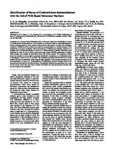
Fig. 1. Sequencing the peptide LAMSAGFSK (amino acids 154 to 162 in Fig. 3) by mass spectrometry. This shows a simple example: a case where a complete set of y ions is observed. Amino acids are defined by differences in masses of observed ions. 1266
Plant Disease / Vol. 93 No. 12
A total of 108 peaks were selected from three “32 kDa” band separations, and a total of 202 peaks from four “30 kDa” band separations. Peptide and protein sequences were then deduced by manual interpretation of the MS and MS/MS measurements; see a simple example of this procedure in Figure 1. Proteins were then identified by database searching with MS-TAG, BLAST, or FASTA (35). In addition to sequencing the U04-82 isolate from original infected wheat tissue, the coat protein of the isolate (U04-82-1) was sequenced again after being maintained on wheat for 18 months by WCM transfer (as described above). Preparation of a synthetic peptide and antiserum for the U04-83 isolate. A 20 amino acid peptide (SNFKYAKLELKGLNYGQLLE) was synthesized by Affinity BioReagents (Golden, CO) that is identical to amino acids 41 to 50 and 251 to 260 (Fig. 1). This peptide was coupled to Keyhole limpet hemocyanin (KLH) as a carrier molecule using glutaraldehyde according to procedures used by Affinity BioReagents to enhance the immune response, and was then used as the antigen to prepare antiserum. On days 1, 22, 36, and 50, 500 µg of antigen was administered to a New Zealand white rabbit. Blood was collected on days 46, 60, and 64, and raw serum was tested in indirect ELISA against original remaining frozen wheat tissue (collected in April 2004) infected with the U04-82 and U04-83 isolates. Field sampling of symptomatic wheat and corn for presence of samples with differential reaction to different antisera. On 24 April 2004, leaf tissue from 10 wheat plants (U04-77, U04-78, U04-80, U04-81, U04-82, U04-83, U04-84, U0485, U04-86, and U04-87) with mosaic symptoms was sampled, placed in labeled self-sealing plastic bags, and analyzed by ELISA against AgMV, WSMV, HPV, WYHV, and WASMV antisera. Extracts from these plants were also minipurified, and the proteins were separated by SDSPAGE (as described above). Leaf tissue other than that used for the initial ELISA and SDS-PAGE analyses was frozen at –80°C. Plants U04-82 and U04-83 were dug up on 26 April 2004, transplanted into clay pots, and held at 15°C with 8 h of fluorescent light (250 µE·s1·m-2) for 2 weeks until WCM transfers were made (see below), and then the remaining tissue was frozen at –80°C. Symptomatic maize (open pollinated sweet maize) plants were sampled from the field at KSU-ARCH on June 2004 (U04143) and June 2006 (U06-Maize A). The samples were analyzed by ELISA for infection with WSMV, HPV, MDMV, SCMV-MDB, JGMV, SrMV, and ZeMV. Leaf tissue from samples were frozen at –80°C for use as the HPV-positive controls.
WCM acquisition and maintenance of the U04-82 and U04-83 HPV isolates. To determine if WCM could transmit the U04-82 and U04-83 isolates, a symptomatic leaf from a plant infected with each isolate was checked for the presence of WCM under a microscope (×20) prior to infesting it with aviruliferous WCM (AvWCM). The ends of the excised leaves (approximately 12 × 150 mm) were placed in water. The first and second leaves from 10 Tomahawk plants infested with AvWCM were attached via spring-loaded hair clips to each of the excised leaves. After 24 h, the clips were removed. A large number of WCM (greater than 1,000) had been transferred to the excised leaves, and most were immobile (either feeding or molting). The infested leaf was held on a microscope stage with double-faced sticky foam, while keeping the excised end of the leaf in water during WCM transfer. Eightythree WCM were transferred individually from the U04-82 leaf and 92 WCM from the U04-83 leaf, to a like number of Tomahawk wheat seedlings at the singleleaf stage. Most of the WCM appeared to be adults, but an attempt was made to include nymphs. In addition, five Tomahawk wheat plants were infested with AvWCM as controls. After transfer of the WCM, the plants were covered with cages (23), held at 22 to 25°C on a bench top for 3 days, moved to a growth chamber at 20°C for 4 weeks, and then analyzed for WSMV and HPV infection by ELISA. After the first transfer of WCM from the wheat leaf infected with the U04-83 isolate to healthy Tomahawk wheat, all wheat plants reacted to antibodies of WSMV when analyzed by indirect ELISA; therefore, no further attempts were made to acquire the U04-83 isolate free of WSMV. However, a single symptomatic plant infested with a WCM from the U04-82 source leaf tested weakly positive for HPV only. This plant was infested with AvWCM because only a few WCM were observed on this plant. Three weeks following infestation, five WCM were transferred from this plant to each of 25 Tomahawk wheat plants at the single-leaf stage, and five Tomahawk wheat plants were infested with AvWCM controls. These plants were held in a greenhouse under natural lighting (temperature range of 20 to 29°C) for 5 weeks and then were analyzed by ELISA. Three of the 25 plants tested positive for HPV. The WCM from these three plants were transferred to 25 additional Tomahawk wheat plants, five WCM per plant. These 25 infested plants were caged individually, held in a greenhouse under natural lighting for 5 weeks, and then each plant was analyzed by ELISA. All plants of this set of 25 plants that tested positive for HPV were subjected to the infectivity assay to Tomahawk wheat to ensure that none were infective by traditional mechanical inoculation. WCM from the this
last set of 25 plants (selected from those that reacted with HPV antibodies in indirect ELISA) were used to establish the maintenance culture for WCM viruliferous for the U04-82 isolate. In all transmissions, the plants were covered with cages (21). Infestation with WCM viruliferous for the U04-82 isolate of wheat resistant to WSMV. Wheat lines reported previously to have temperature-sensitive resistance to WSMV (CI15092 [27], KS96HW10-3 [27], KS03HW12-6 [28], and CO960293 [29]), and five other sources with this type of resistance (CI15322, CI15321-1, CI15321-2, CO960333, and 8174) (D. L. Seifers, unpublished) were planted in Rootrainers (Stuewe and Sons, Corvallis, OR) and grown in a greenhouse until the two-leaf stage. In addition, five CO960293 and five Tomahawk wheat plants were vernalized for use as WSMV controls to determine if the resistance to WSMV was effective against HPV infection in the greenhouse. The wheat plants were then vernalized for 7 weeks, after which they were planted into separate soil-filled (1.2 kg per pot of Hearny clay loam, fine montmorillonitic, mesic Typic Argiustoll) clay pots (15 × 15 cm), and allowed to grow for 14 days in a greenhouse. The experiment was designed to have six wheat plants infested and six wheat plants as healthy controls for each source. However, due to seed germination problems, this was not always possible. The wheat plants were infested with 1-cmlong wheat leaf pieces (obtained from Tomahawk wheat infested 21 to 28 days previously by WCM viruliferous with the U04-82 isolate) by making a small hole with a needle in the second leaf of the plants and inserting the leaf piece. No attempt was made to determine if the leaf pieces had similar numbers of WCM. Two days following infestation, each pot of wheat was treated with Furdan 4EC (carbofuran) to kill the WCM (31). The healthy controls for each wheat line were not infested by WCM. The five CO960293 and five Tomahawk controls were inoculated with the Sidney 81 isolate of WSMV at this time and held in the same greenhouse. A randomized complete block design was used with the location of each experiment randomized in the greenhouse as was the location of the wheat lines on the greenhouse bench. Symptom ratings were taken for infested plants to determine if they developed symptoms. The numbers of heads and seeds were recorded for each plant. The penultimate leaf of all plants (in replication two only) were harvested, bulked, and used in minipurification and SDS-PAGE analysis to document the presence of the “32 kDa” band associated with HPV infection (as described above). The tip to middle portion of the fifth leaf of all plants was analyzed by ELISA for infection with WSMV (as described above). The experiment was conducted three times
on consecutive days. The ANOVA for the number of heads and seeds produced for each treatment was conducted using SAS (version 8, SAS Institute, Cary, NC), and significant treatment effects were determined using the Tukey’s Studentized range test (P = 0.05). RESULTS Field sampling of symptomatic wheat and corn for presence of samples with differential reaction to different antisera. Wheat plants sampled in April 2004 were symptomatic, but sample U04-82 did not react to WSMV antibodies but reacted weakly to HPV-SJ antibodies, and samples U04-83, U04-85, and U04-86 reacted to WSMV antibodies but not to HPV-SJ antibodies or other antisera listed in the methods (data not shown). For the HPV-SJ antiserum, typical healthy extract values for wheat range from 0.010 to 0.039. Thus, a weak reaction for this antiserum would be an ELISA value of two to five times that of the healthy control in a given analysis. All samples had mosaic with some yellowing in the mosaic area, but the yellowing was more pronounced for the U04-83 sample. When analyzed again in ELISA using three different sources of HPV antiserum, differential reactions were observed for the samples (Table 1). The HPV-SJ antibodies did not react to antigens in samples U0481, U04-82, and U04-86. The remaining plant samples had values greater than the healthy value (GHV), with GHV ranging from 2.8 for samples U04-80 and U04-83 to 10.7 for sample U04-87. A weak positive reaction for this antiserum was an ELISA value of two to five times that of the healthy control in a given analysis. None of the wheat samples recorded a GHV as high as the U04-143 control. The antibodies in the HPV-BT antiserum reacted to antigens in samples U04-78, U0487, and the U04-143 control with GHVs of only 2.2 to 3.4. The HPV-BT antiserum typical healthy extract values for wheat range from 0.010 to 0.027. A weak positive reaction for this antiserum was an ELISA value of two to five times that of the healthy control in a given analysis. In contrast, the antibodies in the HPV-WO antiserum reacted to all samples except U0482. The range of GHVs for samples considered positive was 2.8 to 10.2. Healthy values for wheat using the HPV-WO antiserum could range from 0.017 to 0.035. A weak positive reaction to this antiserum was considered to be a value two to five times larger than the healthy value. Again, none of the samples had a GHV as large as the U04-143 control. Interestingly, none of the antibodies of any HPV antiserum reacted to antigens in sample U04-82, and the HPV-WO antiserum reacted to antigens in samples U04-81, while the other antisera did not. Antigens in samples U04-78, U04-83, U04-84, and U04-87 reacted with WSMV antibodies. Plant Disease / December 2009
1267
Antigens in the two maize plants with symptoms sampled in June reacted against only HPV-WO antiserum, and when analyzed by SDS-PAGE each contained the 32-kDa band associated with infection by HPV (data not shown). Minipurification and SDS-PAGE analysis of virus proteins. Unique protein bands were demonstrated in the analyses and are identified by the horizontal and vertical arrows in Figure 2. In the analyses, the unique protein bands migrated at approximately 27 to 29 kDa according to the markers labeled on the left side of the gel. The protein in the tissue infected with U04-83 migrated faster with an approximate MW of 25 kDa (vertical arrow). The WSMV capsid is marked by the arrowhead, and a similar band was observed in the extracts from U04-77 and U04-84. A faint band was in the healthy sample migrating slightly above the U04-83 band but below the 27 to 29 kDa bands of the other samples. Sample U04-82 was not included because it was tested prior to the analyses and had a band equivalent to that observed for all samples except U04-83 (data not shown). TOFMS. The “32 kDa” protein isolated from plant U04-82 yielded 26 tryptic peptides and three Asp-N cleaved peptides (Table 2). When these were manually aligned against the TX96 coat protein sequence, PAM30 scoring (15) (not shown) gave 235 exact matches for the amino acids (aa), 8 close substitutions, and 29 distant substitutions, with an overall sequence coverage of ~94%. As shown in Figure 2, the aa sequence of the coat protein of the U04-82 isolate differs from the TX96 isolate of HPV by about 14.5%; 40 of the 275 aa of the U04-82 isolate that were determined and compared to those of the TX96 isolate were different: 9 aa in the first 50 aa at the N-terminal, 13 aa in the last 50 aa at the C-terminal, with the remaining aa changes in the intermediate region. After being maintained in wheat and serially transmitted by WCM for 18 months,
the U04-82-1 sequence was identical to that obtained from the “32 kDa” protein from the original U04-82 sample (Fig. 3). Confirmation of the assignments for the amino acid sequence of the U04-82 isolate coat protein was provided by nucleotide sequence of an isolate collected from wheat (37). As shown in Figure 3, our result is almost identical to the sequence (ABC58222) derived from the nucleotide measurements, so there is little doubt that the isolates are essentially the same. The only differences are commonly encountered protein modifications at the N-
terminus (deletion of the N-terminal methionine and addition of an acetyl group) and retention of the residue A from TX96 in position 211; Skare et al. (37) reported E in this position. However, the evidence for the assignment of A in our isolate is compelling, because the relevant tryptic peptide (YTHESDLVAPMVR) has a measured mass 1517.74 Da, in agreement with its calculated mass, and high coverage of the MS/MS daughter ions, which are also in agreement with this sequence (Table 3). TOFMS analysis of the U04-83 isolate was considerably less satisfactory. For the
Fig. 2. Proteins from naturally infected wheat separated by sodium dodecyl sulfate-polyacrylamide gel electrophoresis. Lane 1, molecular mass standards; lanes 2 to 10, wheat samples U04-77, U04-78, U04- 80, U04-81, U04-83, U04-84, U04-85, U04-86, and U04-87, respectively. Lane 11, Wheat streak mosaic virus (WSMV-Sidney 81 isolate) infected wheat. Lane 12, proteins from healthy wheat. Horizontal arrow marks position of the “32 kDa” band associated with samples U04-77 to U04-87, and vertical arrow marks the band unique to sample U04-83. Arrowhead in lane 11 marks the location of the WSMV capsid.
Table 1. Reaction of wheat samples in enzyme-linked immunosorbent assay (ELISA) when tested against three antisera made to different isolates of the High Plains virus (HPV) and to a Wheat streak mosaic virus (WSMV) antiserum Antiseray Sample number 78 80 81 82 83 84 85 86 87 U04-143 Healthy wheat WSMV y z
HPV-SJ
HPV-BT
HPV-WO
WSMV
ELISA
GHVz
ELISA
GHV
ELISA
GHV
ELISA
GHV
0.053 0.028 0.018 0.001 0.028 0.056 0.046 0.006 0.107 0.134 0.010 0.001
5.3 2.8 1.8 0.1 2.8 5.6 4.6 0.6 10.7 13.4 1.0 0.1
0.031 0.016 0.021 0.001 0.006 0.015 0.008 0.001 0.048 0.052 0.014 0.001
2.2 1.1 1.5 0.1 0.4 1.1 0.6 0.1 3.4 3.7 1.0 0.1
0.120 0.103 0.058 0.021 0.189 0.166 0.097 0.048 0.174 0.258 0.017 0.001
7.1 6.1 3.4 1.2 11.1 9.8 5.7 2.8 10.2 15.2 1.0 0.1
0.141 0.030 0.019 0.001 0.206 0.165 0.014 0.025 0.112 0.032 0.028 0.168
6.4 1.4 0.9 0.0 9.4 7.5 0.6 1.1 5.1 1.1 1.0 6.0
Sources of antiserum are identified in Materials and Methods. GHV = Number of times greater (calculated by dividing the ELISA value by that of the respective healthy control [shown as 1.0]) than the healthy control.
1268
Plant Disease / Vol. 93 No. 12
“30 kDa” band, our MS/MS measurements yielded sequences for 27 tryptic peptides and 10 Asp-N cleaved peptides (Table 4). However, the 27 tryptic peptides contain six pairs of nonredundant peptides with only minor variations, suggesting that the “30 kDa” band contains a mixture of two versions of the HPV protein. When the peptides were manually aligned against the HPV TX96 sequence template, the peptides with minor variations aligned at identical positions. However, we were unable to distinguish clearly which peptides belonged to which protein sequence, due to limited peptide sequence overlap. Any proposed protein sequences also have significant gaps, despite exhaustive attempts to span the template with the de novo sequenced peptides. One plausible assignment is shown in Figure 3, where the two proposed versions of the coat protein are designated HAYS1 and HAYS2. The HAYS1 version had 160 aa identified and HAYS2 had 148 aa identified. Of the 160 aa in the HAYS1 version, 70 aa were different from the nucleoprotein of the HPV TX96 isolate at the corresponding positions (43.7%), and the nucleoprotein of the HAYS2 version differed from TX96 by 73 of the 148 aa (50%). The homology between the aligned sequences and the HPV template is sufficiently low to suggest that these are new virus proteins, and that they may merit complete sequencing of cDNA derived from virion RNA. Such a combined approach was successfully pursued with the recently identified TriMV (26). Reaction of antiserum prepared to a synthetic peptide and comparison with HPV antiserum. Antibodies made against the 20-aa peptide reacted in indirect ELISA only with the U04-83–infected tissue and not with wheat tissue infected by the U04-82 isolate, or with a 2006 HPV isolate (U06-Maize A) in maize (Table 5). These antibodies also reacted to a lesser extent to extracts of healthy maize, and to a lesser degree, healthy wheat. However, even with such a value for healthy wheat extract, the antiserum was specific for the U04-83 isolate as indicated by the GHV that was 3.8 times larger than that for the healthy wheat. In contrast, the HPV-WO antibodies reacted to the U04-82 and the U06-Maize A isolates, and the reaction to the U04-83 sample had a GHV of 7.7 compared to 30.2 and 19.4 for the U04-82 and U06-Maize A samples, respectively. Acquisition of the U04-82 isolate by WCM. The first transfer of WCM from a leaf of plant U04-82 resulted in 77 plants testing positive in ELISA to WSMV antibodies, one testing weakly to HPV antibodies, and five testing negative to both antisera in ELISA (data not shown). The second transfer of WCM from the single plant testing weakly to HPV antibodies to 25 Tomahawk plants resulted in four whose extracts reacted to WSMV antibod-
ies and three testing weakly for HPV antigens; the remaining plants were negative to both antisera. The WCM transferred from the three plants (considered infected with HPV) to 25 Tomahawk wheat plants resulted in seven with antigens reacting to WSMV antibodies (near the 2.0 arbitrary threshold) and seven reacting (near the 2.0 arbitrary threshold) to HPV antigens when tested by ELISA; the remaining plants were negative (data not shown). However, extracts of the seven plants considered infected with WSMV and the seven plants considered infected with the U04-82 isolate were not infective in backassay to Tomahawk wheat. WCM that were transferred from the seven plants considered positive for infection with U04-82 provided for the establishment of a culture of WCM viruliferous for the U04-82 isolate. All WCM transmissions using the U0483 isolate resulted in plants whose extracts reacted with WSMV antibodies, and the plants’ extracts were infective. Therefore, no further attempts were made to acquire this isolate free of WSMV. Infestation with WCM viruliferous for the U04-82 isolate of HPV of wheat lines resistant to WSMV. To determine if the U04-82 isolate of HPV could infect wheat lines with resistance to WSMV, we infested such lines with WCM viruliferous
for the isolate, resulting in significant differences between healthy and infected plants for measurements taken (Table 6). The heading dates of the wheat entries were significantly different from their respective healthy control except KS96HW10-3, CO960293, KS03HW12-6, and 8174. Average numbers of heads produced by infected plants compared to the healthy control were different for CI15092, CI15321-2, KS96HW10-3, CO960293, and 8174. The number of seeds produced by infected plants compared to the respective healthy control was significantly reduced for all wheat entries. This reduction in yield varied from 27.0% for CO960293 to 79.6% for CI15092. Extracts from these plants when tested in backassay inoculations were not infective to wheat. The SDS-PAGE analysis showed that all treatments had a “32 kDa” band present (data not shown). No healthy control plants developed symptoms or had a diagnostic 32-kDa protein. The Tomahawk plants inoculated with WSMV developed symptoms within 14 days in the greenhouse, but the CO960293 plants inoculated with WSMV did not develop symptoms, indicating that the resistance was effective in the temperature range (14 to 20°C) in the greenhouse (data not shown).
Table 2. High pressure liquid chromatography (HPLC) fractionation and subsequent de novo sequences of trypsin and asparagine-N (Asp-N) peptides from the “32 kDa” protein isolated from plant U04-82 HPLC fractionm/zx observed Trypsin 27-1136.60 20-1054.58 17-1051.54 25-1142.53 42-3337.56 17-780.42 33-1815.07 15-851.44 23-1517.74 22-822.43 30-1625.84 30-1753.94 24-2542.06 25-2413.96 33-3729.89 33-3488.7 30-3204.58 18-936.52 18-1207.65 29-2167.04 21-1771.87 20-911.46 28-1376.74 21-607.34 16-1066.56 24-1758.92 Asp-N 15-1298.64 25-1963.12 22-1877.10 x y z
Sequence from MS/MSy measurements
m/z calculated
WKVFNWTR WSHRVELK YEEIVKDR YNDVLDEFK YHWFYSTGFEYTFDIFPAEVIAMSLFR YEEIVK LFTFDIIVIPGLKPNK NVVSYNR YTHESDLVAPMVR VFNWTR YNDVLDEFKEIIK YNDVLDEFKEIIKK KSYEDNTSTIDFNEDEDFMNK SYEDNTSTIDFNEDEDFMNK LKDGLVNANDIETTVIDFSYEKPDLSSVDGFSLK DGLVNANDIETTVIDFSYEKPDLSSVDGFSLK SLLSSDGWHIVVAYQCVTNSEQLNNNKK RYEEIVK RYEEIVKDR INSSSLDSTIKDCLKDIEE FMALCIGMICYHKK LAMSAGFSK KGTISDVMDIIGK ALSFK (A+42)z SKNVVSYNR IKYTHESDLVAPMVR
1136.601 1054.58 1051.542 1142.537 3337.571 780.414 1815.078 851.438 1517.742 822.462 1625.843 1753.938 2542.057 2413.962 3729.875 3488.696 3204.575 936.515 1207.644 2167.044 1771.852 911.466 1376.746 607.355 1066.565 1758.921
DRSSTGIGTKYN DIIGKSTIAKRYEEIVK ALSFKNSSGSIKAKRIK (A+42)
1298.634 1963.123 1877.107
m/z = Mass-to-charge spectrum. MS/MS = Tandem mass spectroscopy. (A+42) = The addition of 42 Da corresponds to acetylation at the N-terminus. Plant Disease / December 2009
1269
DISCUSSION Certain wheat plants with systemic mosaic symptoms during March and April did not react to antibodies of WSMV or HPV but had a unique protein characteristic of infection with HPV (Table 1 and Fig. 2). We also observed that the protein band
found in sample U04-83 migrated slightly faster in the gel than the other proteins. It was unexpected to find plants that reacted to antibodies of HPV at this time. In previous studies, we did not find plants reacting to antibodies of HPV at this time of year (6,23). When the analysis of the proteins
was done by SDS-PAGE, the unique protein bands shown in Figure 2 migrated into the gel so that they had an approximate value of 27 to 29 kDa rather than the 30 to 32 kDa range expected for the HPV coat protein: a difference of approximately 5 kDa. The TOFMS analyses showed that
Fig. 3. Amino acid sequences of nucleoproteins of different virus isolates compared to that of the 1996 Texas isolate (TX96) of the High Plains virus (HPV). Top line shows sequence of TX96. Second line shows sequence ABC58222 derived from nucleotide measurements by Skare et al. (37). Third line represents sequence of the U04-82 isolate after 18 months maintenance on wheat using viruliferous wheat curl mites (U04-82-1). Note that the time-of-flight mass spectrometry measurements do not distinguish between residues I and L. In this figure, I/L residues for TX96 are given as originally reported (to be consistent with U60141), but for U04-82 and U04-82-1 they are listed as I or L to agree with ABC58222. U04-83 sample consists of two distinct proteins. Although the sequences were assigned to proteolytic fragments for HAYS1 and HAYS2, there is no way to know which fragments belong to which sequence. Dots (….) represent areas within the sequence of undetermined amino acids, dashes (----) represent amino acid consensus to the TX96 isolate of HPV, and @ means N-terminal acetylation. 1270
Plant Disease / Vol. 93 No. 12
the protein of U04-82 was in fact almost identical (differed by one amino acid) to the 32 kDa protein of the isolate used by Skare et al., who showed that it migrated in SDS-PAGE to approximately 30 to 32 kDa (37). Thus, the slightly faster migration in the gel was not considered to be because of differences between the two isolates. We also noted that the apparent molecular weight of the WSMV control in the analyses was also estimated at approximately 41 to 42 kDa, a value approximately 5 kDa smaller than reported (16). Taking into account this 5 kDa underestimation for these proteins, the apparent molecular weight of the U04-82 protein was approximately 30 to 32 kDa. We felt that the slightly faster migration might have been a response of buffer components or perhaps a slight variation in the migration of the marker proteins, or a combination of both. These minor variations in mobility were not further investigated because the purpose of the SDS-PAGE analyses was to identify the presence of unique proteins. The identity of unique proteins was then verified by TOFMS. A differential response to the samples was noted when three different HPV antisera were used for analysis (Table 1). This response has not been previously reported, and it resulted in the inability to identify the U04-82 isolate as an HPV isolate at the onset of this investigation. The HPV-WO antiserum was prepared using a different HPV isolate and a different purification protocol (13) than the one used for the HPV-SJ antiserum (6). The isolate of HPV and procedure for purification used and method used to raise the HPV-BT antiserum are unknown. We also observed a strong reaction of extracts prepared from the U04-83 sample using the HPV-WO antibodies. It is possible that the U04-83 isolate is actually a mixture of isolates or distinct viruses. In fact, the analysis of the U04-83 protein identified two possible sequences that were only 50 to 60% similar to HPV (Fig. 3). This is consistent with
other studies where analyses by TOFMS detected multiple isolates of HPV in infected plants (31). We did observe an increase in ELISA values of the U04-82
isolate over time to the HPV-WO antiserum, although this reaction was much less than the HPV control (Table 5). In this table, U04-82 is the strongly reacted sam-
Table 4. High pressure liquid chromatography (HPLC) fractionation and subsequent de novo sequences of trypsin and asparagine-N (Asp-N) peptides from the “30 kDa” protein isolated from plant U04-83 HPLC fractionm/zx observed
Sequence from MS/MSy measurements
Trypsin 08-994.49 16-992.54 15-693.38 11-1026.54 11-1042.53 18-1183.59 29-1801.05 23-1353.71 17-1395.75 16-1482.77 10-851.44 14-650.36 21-1979.99 17-1244.63 17-1267.65 20-2238.99 21-2253.0 17-1473.78 17-1395.75 20-1020.540 15-1328.62 19-1690.78 13-1006.49 13-872.551 21-2108.09 10-693.43 22-1569.6 Asp-N 22-2577.49 26-1631.98 21-1771.87 19-1274.65 17-1133.59 19-1476.7 08-2022.94 31-3373.78 22-2363.28 25-2336.24 x y z
m/z calculated
YAEIVEDR LYNDQVLK VFNWK WAHKDELKz WSHKDELKz SIASFIDESSK LFDFSIIVIPGLKPNK LNYGQLLEEFKz LNYNQVLKEFKz SKHESELVAPMVR NVVSYNR EFNLK LTNGLVSAGDLDTEVSNFK NESELVAPMVRz HESELVAPMVRz YMDSTSNIQFEASDEFLNKz YLDNTSNIDFTVSDEFMNKz TKNESELVAPMVR STLAEKYDEIVK KGTLDEVMK LEDSTSDSFSLK SADIDTDVTNFTYTK QGSVEEVMK KLTNGLVK KLTNGLVSAGDIDTEVSNFK VFTAKK EFNLKSASKVFTAKz
994.485 992.542 693.372 1026.537 1042.532 1183.585 1801.063 1353.705 1395.764 1482.774 851.438 650.351 1979.993 1244.631 1267.647 2238.987 2253.002 1473.774 1395.737 1020.54 1328.622 1690.781 1006.488 872.557 2108.088 693.43 1569.864
EFNLKSSTKTLVAKKLTNGLVKSAz DQVLKEFKSIAKLI DIDTEVSNFKYAKLE DIKTALENIEEz DIKEALSKDDz DSDIKTALENIEE DESSKDSDIKTALENIEE DEIVKNRSAFNKLNYGQLLEEFKSIASFI DEVMKIVGKAKLAEKYAEIVE DQVLKEFKSIAKLIDDGSKDS
2577.498 1631.974 1771.875 1274.648 1133.569 1476.707 2022.936 3373.78 2363.29 2336.235
M/Z = Mass-to-charge spectrum. MS/MS = Tandem mass spectroscopy. Near-identical pairs observed, indicating a mixture.
Table 3. Daughter ions from collision-induced dissociation of the parent ion YTHESDLVAPMVR y-ions and b-ions y 175.120 * 274.188 * 405.228 * 502.281 * 573.318 * 672.387 * 785.471 * 900.498 * 987.530 1116.572 * 1253.631 * 1354.679 * z
y-17 158.093 * 257.161 * 388.201 * 485.254 * 556.291 * 655.360 768.444 883.471 * 970.503 * 1099.545 1236.604 * 1337.652 *
y-18 157.109 256.178 387.218 484.271 555.308 654.376 767.460 * 882.487 * 969.519 * 1098.562 * 1235.621 1336.669 *
Z
b
b-17
164.071 265.119 402.178 * 531.221 * 618.253 * 733.280 * 846.364 * 945.432 * 1016.469 * 1113.522 * 1244.562 * 1343.631 *
147.044 248.092 385.151 514.194 601.226 716.253 829.337 928.405 999.442 1096.495 1227.535 1326.604 *
b-18 146.061 247.109 384.168 * 513.210 * 600.242 * 715.269 * 828.353 * 927.422 * 998.459 * 1095.511 1226.552 * 1325.620 *
R 01 V 02 M 03 P 04 A 05 V 06 L 07 D 08 S 09 E 10 H 11 T 12
Mass values in the table are calculated values. Asterisks signify observed ions. The last column lists the aa residues starting from the C-terminus of the peptide, i.e., corresponding to differences between the y ions in the first column. Note particularly the difference (71.037 Da) between the fifth and sixth entry, corresponding to the mass of an alanine residue A, not glutamic acid E as predicted by ABC58222. In this table, both coverage and agreement with calculated values are excellent; for the overall array of (y-ions + b-ions), the difference between observed and calculated values (not shown) has a rootmean-square value of 7.4 mDa. Plant Disease / December 2009
1271
was a capsid protein most similar to HPV (Fig. 3). However, the aa sequence of the nucleoprotein of these two isolates showed more variation than had been previously reported for HPV isolates from Kansas, Idaho, Colorado, Texas, and Utah during 1996 and 1997. In that case, the nucleoproteins of the TX96, KS96, and CO96 isolates had identical aa sequences, while the nucleoproteins of the ID97 and UT96 isolates had the same aa sequence, but their sequences differed from TX96, KS96, and CO96 by only three aa (31). In the present measurements, the U04-82 isolate was 14.5% different from the HPV-TX96 isolate but was almost identical to an HPV isolate from wheat (37). The analyses of the U04-83 isolate found two subpopulations with plausible assignments as se-
ple with HPV-WO antiserum. A possible reason for the reaction differences to the antisera may be related to the differences between N-terminal part of the coat protein of the U04-82 and U04-83 isolates (Fig. 3). The N-terminal of potyviruses is considered highly variable, and aa differences in this region of the nucleoprotein have been attributed to differential responses to antiserum (36). Although HPV is not a potyvirus, a similar relationship might exist for the epitopes of the coat protein; therefore, it is possible that the epitopes of the 30 to 32 kDa nucleoprotein of the U04-82 and U04-83 isolates differ, resulting in the difference in response to the antiserum. The TOFMS analyses confirmed that the protein of the U04-82 and U04-83 isolates
Table 5. Enzyme-linked immunosorbent assay (ELISA) values from extracts of symptomatic wheat and maize leaf tissue analyzed against antiserum prepared to a peptide representing selected portions of the sequence of the U04-83 isolate and to an antiserum (HPV-WO) prepared to an Idaho isolate of the High Plains virus (HPV) Antiserum
Antiserum Sample U04-83 U04-82x U04-83 peptide antigen U06-Maize Ay WSMVz Healthy wheat Healthy maize
U04-83
GHVw
HPV-WO
GHV
0.688 0.366 1.349 0.559 0.178 0.179 0.414
3.8 2.0 7.5 1.3 0.9 1.0 1.0
0.077 0.302 0.010 0.914 0.025 0.010 0.047
7.7 30.2 1.0 19.4 2.5 1.0 1.0
w GHV
= Number of times greater (calculated by dividing the ELISA value by that of the respective healthy control [shown as 1.0]) than the healthy control. Not the original wheat tissue infected with U04-82. Wheat tissue was infected with U04-82 using wheat curl mites viruliferous for only this isolate. y Sample is maize, not wheat, thus the healthy maize ELISA value was used to calculate the GHV. z Wheat streak mosaic virus. x
Table 6. Effect of infection with U04-82 isolate of the High Plains virus on wheat with temperaturesensitive resistance to Wheat streak mosaic virus Wheat
Nu
DHv
CI15092 Hx CI15092 Inz CI15322 H CI15322 In CI15321-1 H CI15321-1 In CI15321-2 H CI15321-2 In KS96HW10-3 H KS96HW10-3 In CO960293 H CO960293 In CO960333 H CO960333 In KS03HW12-6 H KS03HW12-6 In 8174 H 8174 In CV P = 0.05
15 18 16 17 11 11 14 15 9 10 9 11 18 18 15 16 18 18
71.1 dey 92.9 a 70.6 de 84.4 b 58.6 gh 72.1 de 59.7 fgh 77.1 bcd 67.7 ef 69.3 de 75.6 cde 83.6 bc 60.4 fg 70.7 de 51.6 hi 59.8 fgh 42.5 j 49.7 ij 7.4
u
HPPw 2.4 a 1.0 g 1.7 bcde 1.2 efg 1.4 defg 1.2 efg 1.9 abcd 1.2 efg 2.0 abc 1.2 efg 2.2 ab 1.6 cdef 1.9 abcd 1.8 bcd 1.8 abcd 1.7 bcde 2.1 abc 1.1 fg 20.4
No. of seeds 31.3 a 6.4 g 17.3 de 10.6 fg 21.0 bcde 8.4 g 19.9 cde 7.2 g 25.1 abc 6.3 g 30.5 a 22.4 bcd 29.4 a 16.7 def 27.1 ab 15.7 ef 25.7 abc 6.9 g 21.1
% Reduction in no. of seeds 79.6 38.7 60.0 63.8 75.0 27.0 43.2 42.1 73.2
Number of observations for each treatment or number of seed is an average of three experiments. DH = days to heading. w HPP = heads per plant. x H = healthy wheat. y Treatment means not followed by the same letter differ according to the Tukey’s Studentized test (P = 0.05). z In = infected wheat. v
1272
Plant Disease / Vol. 93 No. 12
quences HAYS1 and HAYS2; HAYS1 being 43.7% different from the nucleoprotein of the TX96 isolate and HAYS2 being 50% different. Even though we were only able to sequence a small portion of the protein of this isolate because insufficient tissue remained for further characterization, both varied from the sequence of the original type of HPV represented by the HPV TX96 isolate (31). Thus, since the initial identification of HPV in 1993 (6), variants of HPV are present that have larger differences in the aa sequence of the nucleoprotein and apparently incite symptoms in cooler conditions than had been previously reported (31). An important attribute of TOFMS analysis is the ability to detect subisolates of a virus in a sample (31), in this case the U04-83 subpopulations HAYS1 and HAYS2 (Fig. 3). Although the similarity of these sequences to HPV is rather distant, it is possible that the sequences of HAYS1 and HAY2 might be derived from HPV. The large difference could be because of a recombination event with another virus, or perhaps the converse might be true; i.e., HPV might be derived from the U04-83 virus represented by the HAYS1 and HAYS2 sequences. Regardless of the direction of evolution, the question arises as to whether the virus is an isolate of HPV. For potyviruses, a difference in the aa sequence of the nucleoprotein greater than 20% is suggested for the establishment of a new virus (2). A difference of 15% in the aa of the nucleoprotein sequence has been used for the tenuiviruses (14), used in establishing the relationship of WYHV to Rice hoja blanca virus (22). Although HPV has not yet been affiliated as a member of an established group of viruses, the 43 and 50% difference in the aa sequence of the coat protein of the U04-83 virus isolate is well outside the differences stated above. However, until a complete aa sequence for the U04-83 isolate is obtained, it is premature to name it as a new virus. The antiserum prepared to the synthetic peptide from the sequence of the U04-83 isolate showed a specific reaction to the original wheat tissue of the U04-83 sample, even though the healthy wheat reacted to the antiserum, although less so than to the healthy maize tissue (Table 5). The reaction to both healthy maize and wheat extracts may be because KLH protein was used as the carrier molecule in the preparation of the antigen to raise the antiserum. Reactivity of KLH antibodies with antigens in plant vascular tissue has been reported (4). The WCM transmits HPV (23), and different sources of WCM can differentially transmit different isolates of HPV (21). The U04-82 isolate was transmitted by WCM. Interestingly, the U04-82 sample tested negative in ELISA for infection with WSMV. However, when we infested leaf tissue from this plant with AvWCM, some
then transmitted WSMV and the U04-82 isolate. The presence of WSMV in the WCM acquisition could be due to the cooler temperatures in the field, causing WSMV to have a low titer in this plant, and thus it was not identified by ELISA, and once moved to a warmer temperature, the titer increased and WSMV was acquired by the AvWCM. Another possibility for the infection with WSMV was that our culture of WCM was contaminated with WSMV, but the control infestation of wheat using the same AvWCM that were used to infest the U04-82 leaf did not infect control plants with WSMV or HPV. However, starting with a single WCM transferred from the leaf sample U04-82, we were able to transmit the U04-82 isolate and establish a pure culture after three successive transmission cycles. In the last transmission cycle, seven plants were identified that had very faint symptoms and reacted to WSMV antibodies, and seven plants that had prominent mosaic symptoms and reacted to HPV antibodies in ELISA. When each plant was tested in infectivity assay, none were infectious to wheat. This would be expected for the plants infected with HPV because it is not mechanically transmissible (6,23). However, the extracts prepared from the plants testing weakly positive for infection with WSMV were not infective. We have observed this previously when plants are infested with large numbers of WCM; a false positive reaction for infection with WSMV can occasionally be observed by ELISA using WSMV antiserum (D. L. Seifers, unpublished). It is possible that infestation of wheat with large numbers of WCM might, in some plants, induce a host-encoded protein that reacts weakly with the WSMV antibodies. A nonspecific host-encoded protein that reacts to antibodies of HPV has been reported in plants infested with WCM viruliferous for HPV; however, numbers of WCM associated with this response were not stated (38). During the 18 months maintenance of the U04-82 isolate in wheat by WCM, an increase in the response to HPV-WO antibodies was observed (Table 5). Possibly, this resulted from an increased titer in the plants manifested as a stronger signal in ELISA analysis, or the isolate was different from the isolate sequenced by TOFMS from the original sample U04-82. Therefore, we analyzed the 32 kDa protein present in the wheat infected by WCM viruliferous for the U04-82 isolate after 18 months of transmission. The sequence of the 32 kDa protein (U04082-1) was identical to the sequence obtained from the U0482 isolate in the original U04-82 wheat sample (Fig. 3). Therefore, it was considered that the increase in reaction, although less than that for the HPV control, to the HPV-WO antibodies was a result of increased titer. We observed no reaction of the HPV antibodies to extracts prepared
from wheat plants used to maintain the AvWCM. We infected wheat with temperaturesensitive resistance to WSMV with the U04-82 isolate under temperature conditions that were effective to maintain WSMV resistance, resulting in significant reductions in yield (Table 6). No other study has documented the effect of infection with HPV of wheat with resistance to WSMV. Although our experiments were conducted in a greenhouse environment, they indicate that these WSMV-resistant wheat sources (27–29) can suffer significant yield losses when infected with HPV. From our analyses, we conclude that isolate U04-82 is a variant of HPV with the nucleoprotein differing by 14.5% from the original HPV TX96 isolate. We observed for the first time a differential response of the HPV isolates to antisera raised against different HPV isolates. Infection of wheat lines having temperaturesensitive resistance to WSMV with the U04-82 isolate caused systemic infection and significant yield reductions in the wheat lines. Analyses by TOFMS of the 30 kDa protein unique to wheat infected by the U04-83 isolate indicated that it is a virus coat protein and that the partial sequence varies up to 50% from the original TX96 isolate of HPV, and that it may represent a new virus species. Antiserum prepared to a 20 aa synthetic peptide (from the sequence of the U04-83 isolate) was specific for that isolate in wheat but reacted to a lesser extent to extracts from healthy maize and less so to healthy wheat.
8. 9. 10.
11.
12.
13.
14.
15.
16. LITERATURE CITED 1. Ahn, K.-K., Kim, K. S., Gergerich, R. C., and Jensen, S. G. 1998. High plains disease of corn and wheat: Ultrastructural and serological aspects. J. Submicrosc. Cytol. Pathol. 30:563571. 2. Berger, P. H., Barnett, O. W., Brunt, A. A., Colinet, D., Edwardson, J. R., Hammond, J., Hill, J. H., Jordan, R. L., Kashiwazaki, S., Makkouk, K., Morales, F. J., Rybicki, E., Spence, N., Ohki, S. T., Uyeda, I., van Zaayen, A., and Vetten, H. J. 2000. Family Potyviridae. Pages 703-724 in: Virus Taxonomy: Seventh Report of the International Committee on Taxonomy of Viruses. M. H. V. van Regenmortel, C. M. Fauquet, D. H. L. Bishop, E. B. Carstens, M. K. Estes, S. M. Lemon, J. Maniloff, M. A. Mayo, C. R. Pringle, and R. B. Wickner, eds. Academic Press, New York. 3. Forster, R. L., Seifers, D. L., Strausbaugh, C. A., Jensen, S. G., Ball, E. M., and Harvey, T. L. 2001. Seed transmission of the High plains virus in sweet corn. Plant Dis. 85:696-699. 4. Höglund, A.-S., and Josefsson, L.-G. 1999. An antigen in plant vascular tissue cross-reacts with antibodies towards KLH, Keyhole Limpet Hemocyanin. (Abstr.) Nat. Biotechnol. 17:44. 5. Jensen, S. G., and Hall, J. S. 1995. Molecular characterization of a viral pathogen infecting maize and wheat in the high plains. (Abstr.) Phytopathology 85:1211. 6. Jensen, S. G., Lane, L. C., and Seifers, D. L. 1996. A new disease of maize and wheat in the high plains. Plant Dis. 80:1387-1390. 7. Krokhin, O. V., Craig, R., Spicer, V., Ens, W., Standing, K. G., Beavis, R. C., and Wilkins, J. A. 2004. An Improved Model for Prediction of
17.
18.
19.
20.
21.
22.
23.
24.
Retention Times of Tryptic Peptides in Ion Pair Reversed-phase HPLC: Its Application to Protein Peptide Mapping by Off-Line HPLCMALDI MS. Mol. Cell. Proteomics 3:908-919. Laemmli, U. K. 1970. Cleavage of structural proteins during the assembly of head of bacteriophage T4. Nature 227:680-685. Lane, L. C. 1986. Propagation and purification of RNA plant viruses. Methods Enzymol. 118:687-696. Lebas, B. S. M., Ochoa-Corona, F. M., Elliott, D. R., Tang, E. Z., and Alexander, B. J. R. 2005. Development of an RT-PCR for High Plains virus indexing scheme in New Zealand post-entry quarantine. Plant Dis. 89:11031108. Loboda, A. V., Krutchinsky, A. N., Bromirski, M., Ens, W., and Standing, K. G. 2000. A tandem quadrupole/time-of-flight mass spectrometer with a matrix-assisted laser desorption/ionization source: Design and performance. Rapid Commun. Mass Spectrom. 14: 1047-1057. Louie, R., and Seifers, D. L. 1996. Transmission of the High Plains pathogen by vascular puncture inoculation. (Abstr.) Phytopathology 86:Sl. Louie, R., Seifers, D. L., and Bradfute, O. E. 2006. Isolation, transmission and purification of the High Plains virus. J. Virol. Methods 135:214-222. Mayo, M. A., de Miranda, J. R., Falk, B. W., Goldbach, R., Haenni, A.-L., and Toriyama, S. 2000. Genus Tenuivirus. Pages 622-627 in: Virus Taxonomy: Seventh Report of the International Committee on Taxonomy of Viruses. M. H. V. van Regenmortel, C. M. Fauquet, D. H. L. Bishop, E. B. Carstens, M. K. Estes, S. M., Lemon, J. Maniloff, M. A. Mayo, C. R. Pringle, and R. B Wickner, eds. Academic Press, New York. Schwartz, R. M., and Dayhoff, M. O. 1979. Matrices for detecting distant relationships. Pages 353-358 in: Atlas of Protein Structure. Vol. 5. Suppl. 3. National Biomedical Resource Foundation, Washington, DC. Seifers, D. L. 1992. Partial characterization of a Colorado isolate of Agropyron mosaic virus. Plant Dis. 76:564-569. Seifers, D. L., and Caceres, J. 1988. Titer variation in infected sorghum differing in resistance to maize dwarf mosaic virus strain-B. Phytopathology 78:208-212. Seifers, D. L., Harvey, T. L., and Bowden, R. L. 1995. Occurrence and symptom expression of American wheat striate mosaic virus in wheat in Kansas. Plant Dis. 79:853-858. Seifers, D. L., Harvey, T. L., Haber, S., She, Y. M., Chernushevich, I., Ens, W., and Standing, K. G. 1999. Natural infection of sorghum by foxtail mosaic virus in Kansas. Plant Dis. 83:905-912. Seifers, D. L., Harvey, T. L., Kofoid, K. D., and Stegmeier, W. D. 1996. Natural infection of pearl millet and sorghum by wheat streak mosaic virus in Kansas. Plant Dis. 80:179-185. Seifers, D. L., Harvey, T. L., Louie, R., Gordon, D. T., and Martin, T. J. 2002. Differential transmission of isolates of the High Plains virus by different sources of wheat curl mites. Plant Dis. 86:138-142. Seifers, D. L., Harvey, T. L., Martin, T. J., Haber, S., She, Y.-M., Ens, W., Standing, K. G., Salomon, R., and Gera, A. 2005. Association of a virus with wheat displaying yellow head disease symptoms in the Great Plains. Plant Dis. 89:888-895. Seifers, D. L., Harvey, T. L., Martin, T. J., and Jensen, S. G. 1997. Identification of the wheat curl mite as the vector of the High Plains virus of corn and wheat. Plant Dis. 81:1161-1166. Seifers, D. L., Harvey, T. L., Martin, T. J., and Jensen, S. G. 1998. A partial host range of the High Plains virus of corn and wheat. Plant Dis.
Plant Disease / December 2009
1273
82:875-879. 25. Seifers, D. L., Martin, T. J., Harvey, T. L., Fellers, J. P., and Michaud, J. P. 2009. Identification of the wheat curl mite as the vector of Triticum mosaic virus. Plant Dis. 93:25-29. 26. Seifers, D. L., Martin, T. J., Harvey, T. L., Fellers, J. P., Stack, J. P., Ryba-White, M., Haber, S., Krokhin, O., Spicer, V., Lovat, N., Yamchuk, A., and Standing, K. G. 2008. Triticum mosaic virus: A new virus isolated from wheat in Kansas. Plant Dis. 92:808-817. 27. Seifers, D. L., Martin, T. J., Harvey, T. L., and Gill, B. S. 1995. Temperature sensitivity and efficacy of wheat streak mosaic virus resistance derived from Agropyron intermedium. Plant Dis. 79:1104-1106. 28. Seifers, D. L., Martin, T. J., Harvey, T. L., and Haber, S. 2007. Temperature-sensitive Wheat streak mosaic virus resistance identified in KS03HW12 wheat. Plant Dis. 91:1029-1033. 29. Seifers, D. L., Martin, T. J., Harvey, T. L., Haber, S., and Haley, S. D. 2006. Temperature sensitivity and efficacy of Wheat streak mosaic virus resistance derived from CO960293 wheat. Plant Dis. 90:623-628. 30. Seifers, D. L., Salomon, R., Marie-Jeanne, V., Alliot, B., Signoret, P., Haber, S., Loboda, A., Ens, W., She, Y.-M., and Standing, K. G. 2000. Characterization of a novel potyvirus isolated
1274
Plant Disease / Vol. 93 No. 12
31.
32.
33.
34.
35.
from maize in Israel. Phytopathology 90:505513. Seifers, D. L., She, Y.-M., Harvey, T. L., Martin, T. J., Haber, S., Ens, W., Standing, K. G., Louie, R., and Gordon, D. T. 2004. Biological and molecular variability among High Plains virus isolates. Plant Dis. 88:824-829. She, Y.-M., Seifers, D. L., Haber, S., Ens, W., and Standing, K. G. 2004. Characterization of the agent of ‘High Plains Disease’: Mass spectrometry determines the sequence of the disease-specific protein. J. Biol. Chem. 279:488494. She, Y.-M., Wang, G. Q., Loboda, A., Ens, W., Standing, K. G., and Burczynski, F. J. 2002. Sequencing of rat liver cytosolic proteins by matrix-assisted laser desorption ionizationquadrupole time of flight mass spectrometry following electrophoretic separation and extraction. Anal. Biochem. 310:137-147. Shevchenko, A., Chernushevich, I., Ens, W., Standing, K. G., Thomson, B., Wilm, M., and Mann, M. 1997. Rapid de novo peptide sequencing by a combination of nanoelectrospray, isotopic labeling and a quadrupole/timeof-flight mass spectrometer. Rapid Commun. Mass Spectrom. 11:1015-1024. Shevchenko, A., Sunyaev, S., Loboda, A., Shevchenko, A., Bork, P., Ens, W., and Stand-
36.
37.
38.
39.
ing, K. 2001. Charting the proteomes of organisms with unsequenced genomes by MALDI Quadrupole time-of-flight mass spectrometry and BLAST homology searching. Anal. Chem. 73:1917-1926. Shukla, D. D., Strike, P. M., Tracy, S. L., Gough, K. H., and Ward, C. W. 1988. The N and C termini of the coat proteins of potyviruses are surface located and the N terminus contains the major virus-specific epitopes. J. Gen. Virol. 69:1497-1508. Skare, J. M., Wijkamp, I., Dehnam, I., Rezende, J. A. M., Kitajima, E. W., Park, J.W., Benedicte, D., Rush, C. M., Michels, G., Scholthof, K.-B. G., and Scholthof, H. B. 2006. A new eriophyid mite-borne membraneenveloped virus-like complex isolated from plants. Virology 347:343-353. Skare, J. M., Wijkamp, I., Rezende, J., Michels, G., Rush, C., Scholthof, K.-B. G., and Scholthof, H. B. 2003. Colony establishment and maintenance of the eriophyid wheat curl mite Aceria tosichella for controlled transmission studies on a new virus-like pathogen. J. Virol. Methods 108:133-137. Slykhuis, J. T. 1955. Aceria tulipae Keifer (Acaria:Eriophyidae) in relation to the spread of wheat streak mosaic. Phytopathology 45:116-128.