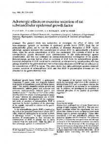
IJOCR 10.5005/jp-journals-10051-0058 Immunohistochemical Expression of Epidermal Growth Factor Receptor in Different Grades of Oral Squamous Cell Carcinomas
Original Article
Immunohistochemical Expression of Epidermal Growth Factor Receptor in Different Grades of Oral Squamous Cell Carcinomas 1
Niraj Patil, 2Harish H, 3Surabhi Sinha, 4Vandana Shah, 5Pratik B Kariya, 6Usha Sharma
ABSTRACT Introduction: Epidermal growth factor receptor (EGFR) is a tyrosine kinase receptor of the ErbB family, which is expressed or highly expressed in a variety of solid tumors, including oral cancers. Aims and objectives: The aim of this study was to evaluate the immunohistochemical (IHC) expression of EGFR in different grades of oral squamous cell carcinoma (OSCC). Materials and methods: A series of 50 paraffin blocks with different grades of previously diagnosed OSCC were analyzed for EGFR, which was determined by standardised histopathology. A total of 32 (64%) cases out of 50 showed positivity for EGFR. Results: A total of 32 cases (64%) showed positivity for EGFR, although statistically, a nonsignificant p-value of 0.814 was obtained for EGFR. Conclusion: We noticed the correlation of increased membrane and cytoplasmic expression of EGFR in poorly differentiated grades of OSCC that can be utilized to predict the prognosis of the patients. Keywords: Epidermal growth factor receptor, Oral cancer, Squamous cell carcinoma. How to cite this article: Patil N, Harish H, Sinha S, Shah V, Kariya PB, Sharma U. Immunohistochemical Expression of Epidermal Growth Factor Receptor in Different Grades of Oral Squamous Cell Carcinomas. Int J Oral Care Res 2016;4(4):259-262. Source of support: Nil Conflict of interest: None
INTRODUCTION Oral squamous cell carcinoma (OSCC) is the commonest head and neck malignancy, representing almost 95% of the
1,3 5,6
Postgraduate Student, 2Reader, 4Professor and Head Senior Lecturer
1-4,6
Department of Oral Pathology, KM Shah Dental College & Hospital: Sumandeep Vidyapeeth, Vadodara, Gujarat, India
head and neck carcinomas. Of all the cancers worldwide, its prevalence rate is 5%.1 Globally, about two-thirds of oral cancers occur in developing countries, and it is a major health hazard. The highest prevalence of oral cancer has been observed in the Indian subcontinent,2 where huge populations are open to the elements, exterior irritants, and carcinogens, such as smoke, tobacco, and betel nut extracts. Usually, cancer develops through rising grades of oral epithelial dysplasia to fatal invasive malignancy.1 Intense research has started a new age of cancer treatment for the duration of the last decade, that of molecular therapeutics. Promising preclinical studies have prompted the expansion of clinical trials testing molecules, such as the epidermal growth factor receptor (EGFR) with conventional cytotoxic therapy.3 The EGFR has a significant role in the differentiation and morphogenesis of numerous organs and proliferation and continued existence of mammalian cells.4 The EGFR is a proto-oncogene, which if activated at the cell surface by transforming growth factor-α serves to advance cellular proliferation.4 It is a transmembrane receptor expressed in an array of human tumors of epithelial origin; higher expressions of EGFR are seen in 80% of squamous cell carcinomas (SCCs), which explains that an unrestrained growth may be aided by abnormal EGFR expression.4 The higher expressions of EGFR have been seen to be of prognostic value in head and neck SCC (HNSCC).5,6 This study may help in indicating that EGFR expressions correlate with the severity of OSCC, and it is also possible that the immunohistochemical (IHC) demonstration of these markers may serve to be a useful prognostic tool for a more precise clinical outcome of OSCC.
Materials and methods A total of 50 cases of OSCC were evaluated immunohistochemically for EGFR expression. Normal buccal mucosa has been taken as a control for EGFR.
5
Department of Paedodontics and Preventive Dentistry, KM Shah Dental College & Hospital: Sumandeep Vidyapeeth Vadodara, Gujarat, India Corresponding Author: Usha Sharma, Senior Lecturer Department of Oral Pathology, KM Shah Dental College & Hospital: Sumandeep Vidyapeeth, Vadodara, Gujarat, India e-mail:
[email protected]
Inclusion Criteria Formalin-fixed paraffin-embedded blocks with adequate tissue size (a minimum of 4–5 mm of lesional tissue) for sectioning and IHC staining with representative histological features.
International Journal of Oral Care and Research, October-December 2016;4(4):259-262
259
Niraj Patil et al Table 1: Ranks of grading for staining evaluation7
Exclusion Criteria The tissue and blocks without representative area of any grade of SCC were excluded. About 4 µm sections were taken, and EGFR was detected by IHC staining using the clone EP38Y (Thermo Scientific rabbit monoclonal antibody #RM-2111-R7, 7 mL). All instances of OSCCs were subjected to histopathological evaluation and sorted as well-differentiated SCC (WDSCC), moderately differentiated SCC (MDSCC), and poorly differentiated SCC (PDSCC) as per Broder’s classification of SCC.
ANALYSIS OF IMUNOHISTOCHEMICAL STAINING Positive immunoreactivity was pointed out by the appearance of brown color as the final result at the antigen target spot. The nonpositive control tissue exhibited lack of staining. Tissue sections of normal oral epithelium were taken as positive control for EGFR. The evaluation of study cases was done subsequently in a similar way, and they were graded as positive or negative. The positive results were assessed further for intensity of staining. The presence of brown-colored end product at the site of target antigen was taken as immunohistochemically positive and the negative control tissue demonstrated absence of staining. Presence of immunostaining in the cell membrane of various layers of epithelium was evaluated in randomized six fields/intensity of positively stained cells as percentage expression at ×40 and graded as 0 (under 10% positively stained cells), 1+ (10–25% positively stained cells: Weak expression), 2+ (25–50% positively stained cells: Mild-to-moderate expression), 3+ (50–75% positive cells: Moderate-to-strong expression) (Table 1). Although judging the depth of staining was looked upon as biased, care was taken to decrease the bias by three autonomous investigators (two other investigators who were senior faculty associates in the branch), which demonstrated intensely positive staining in EGFR.
0
No Labeling
1
Weak labeling
2
Moderate labeling
3
Intense labeling
50% tumor cells, patchy/ homogeneous
Table 2: Age of the OSCC cases considered for this study, with age range being 29–70 years (mean 48 ± standard deviation 12.04 years)
Age (years)
Minimum n (%) age 50 (100) 29.00
Maximum age 70.00
Mean Std. age deviation 48.5200 12.04624
12 (75%) cases out of 16 were diagnosed as WDSCC, 3 (18.8%) as MDSCC, and 1 (6.2%) as PDSCC (Table 2). A total of 32 cases (64%) showed positivity for EGFR, of which 18 (58%) were WDSCC, 4 (66%) cases were MDSCC, and 10 (76%) were PDSCC. Out of the 18 cases of WDSCC that showed positivity for EGFR, 11 (35.5%) showed mild expression and 7 (22.5%) showed moderate expression. Of the 4 cases of MDSCC showing positivity for EGFR, 1 showed mild expression, 2 showed moderate expression, and 1 showed intense expression. Of the 10 cases of PDSCC, 5 showed moderate expression and 5 showed intense expression; p values obtained for EGFR was 0.814, which was nonsignificant (Graph 1, Figs 1 to 4).
DISCUSSION A lot of work is currently focused on the recognition of superior biological and clinical factors that can serve as a predictive and prognostic marker. The perception of the biological initiation of tumor development and its progression in the oral cavity has enhanced as a result of the
Statistical Methods The EGFR expression in different grades of OSCC is compared by using the Pearson correlation test and Student’s t-test. Mann–Whitney and the Kruskal–Wallis tests were employed for comparison of continuous variables.
RESULTS The gender distribution of the cases was, male:female ratio being 2.1:1 (34:16). In males, 19 (55%) cases out of 34 were diagnosed as WDSCC, 12 (35.5%) cases out of 34 as PDSCC, and 3 (8.8%) cases out of 34 as MDSCC. In females,
260
Graph 1: Expression of EGFR in different grades of OSCC
IJOCR Immunohistochemical Expression of Epidermal Growth Factor Receptor in Different Grades of Oral Squamous Cell Carcinomas
Fig. 1: Epidermal growth factor receptor showing intense expression in WDSCC (10×)
Fig. 2: Epidermal growth factor receptor showing intense expression in MDSCC (10×)
Fig. 3: Epidermal growth factor receptor showing intense expression in PDSCC (10×)
Fig. 4: Epithelial growth factor receptor showing moderate expression in WDSCC (10×)
current advances in fundamental research and genomics. As a result, an array of molecular tumor markers evaluated in the laboratory have been recognized in HNSCC for their potential to predict disease result or response to treatment in patients. According to our present study, we expect to gather further proficiency to either anticipate patients at risk for disease development after standard therapy or identify those who can gain from postoperative radiotherapy and those with radioresistant tumors.5 Our study showed moderate-to-intense EGFRexpression in more than 40% of the cases (p-value = 0.814), including all grades of SCC. The status of the lymph node was not recorded for our study. As the grade of the SCC increased, i.e., from WDSCC to PDSCC, the intensity of EGFR increased. It can be summarized that somewhere the expression of EGFR is related to the grade of SCC. Xia et al,8 after studying different family members of the EGFR, conclude that there are no significant
associations between the expression levels of the EGFR family members and the sex, and histological grade (p > 0.05) except that EGFR expression level is significantly associated with age (p