Dorte Glintborg, Kurt Højlund, Nicoline R. Andersen, Bo Falck Hansen, Henning ...... Kim YB, Nikoulina SE, Ciaraldi TP, Henry RR, Kahn BB 1999 Normal in-.
ORIGINAL E n d o c r i n e
ARTICLE R e s e a r c h
Impaired Insulin Activation and Dephosphorylation of Glycogen Synthase in Skeletal Muscle of Women with Polycystic Ovary Syndrome Is Reversed by Pioglitazone Treatment Dorte Glintborg, Kurt Højlund, Nicoline R. Andersen, Bo Falck Hansen, Henning Beck-Nielsen, and Jørgen F. P. Wojtaszewski Department of Endocrinology (D.G., K.H., H.B.-N.), Diabetes Research Centre, Odense University Hospital, DK-5230 Odense, Denmark; Copenhagen Muscle Research Centre (N.R.A., J.F.P.W.), Institute of Exercise and Sport Sciences, Department of Human Physiology, University of Copenhagen, DK-2100 Copenhagen, Denmark; and Department of Diabetes Biology (B.F.H.), Novo Nordisk A/S, DK-2760 Måløv, Denmark
Context: Insulin resistance is a major risk factor for type 2 diabetes in women with polycystic ovary syndrome (PCOS). The molecular mechanisms underlying reduced insulin-mediated glycogen synthesis in skeletal muscle of patients with PCOS have not been established. Subjects and Methods: We investigated protein content, activity, and phosphorylation of glycogen synthase (GS) and its major upstream inhibitor, GS kinase (GSK)-3 in skeletal muscle biopsies from 24 PCOS patients (before treatment) and 14 matched control subjects and 10 PCOS patients after 16 wk treatment with pioglitazone. All were metabolically characterized by euglycemic-hyperinsulinemic clamps and indirect calorimetry. Results: Reduced insulin-mediated glucose disposal (P ⬍ 0.05) was associated with a lower insulinstimulated GS activity in PCOS patients (P ⬍ 0.05), compared with controls. This was, in part, explained by absent insulin-mediated dephosphorylation of GS at the NH2-terminal sites 2⫹2a, whereas dephosphorylation at the COOH-terminal sites 3a⫹3b was intact in PCOS subjects (P ⬍ 0.05). Consistently, multiple linear regression analysis showed that insulin activation of GS was dependent on dephosphorylation of sites 3a⫹3b in women with PCOS. No significant abnormalities in GSK-3␣ or -3 were found in PCOS subjects. Pioglitazone treatment improved insulinstimulated glucose metabolism and GS activity in PCOS (all P ⬍ 0.05) and restored the ability of insulin to dephosphorylate GS at sites 2 and 2a. Conclusions: Impaired insulin activation of GS including absent dephosphorylation at sites 2⫹2a contributes to insulin resistance in skeletal muscle in PCOS. The ability of pioglitazone to enhance insulin sensitivity, in part, involves improved insulin action on GS activity and dephosphorylation at NH2-terminal sites. (J Clin Endocrinol Metab 93: 3618 –3626, 2008)
P
olycystic ovary syndrome (PCOS) is a common endocrine disorder of unknown etiology affecting 5–10% of premenopausal women (1, 2). The disorder is diagnosed by hyperandrogenism, chronic anovulation, and/or polycystic ovaries (1) and frequently characterized by profound peripheral insulin resistance (2, 3). Skeletal muscle is the major site of insulin-stimulated glucose
disposal, and insulin resistance in this tissue represents a major risk factor for type 2 diabetes in women with PCOS (1, 2). Metabolic studies of PCOS patients have shown that impaired insulin-stimulated glucose metabolism is largely accounted for by reduced nonoxidative glucose metabolism (3), similar to the findings in patients with type 2 diabetes and their first-degree relatives (FDR) (4 –11).
0021-972X/08/$15.00/0
Abbreviations: AS160, Akt substrate of 160 kDa; FDR, first degree relatives; FV, fractional velocity; G6P, glucose-6-phosphate; GS, glycogen synthase; GSK, GS kinase; HRP, horseradish peroxidase; IRS, insulin receptor substrate; NOGM, nonoxidative glucose metabolism; PCOS, polycystic ovary syndrome; PI3K, phosphoinositide 3-kinase; Rd, glucose disposal; Ser, serine; TZD, thiazolidinediones.
Printed in U.S.A. Copyright © 2008 by The Endocrine Society doi: 10.1210/jc.2008-0760 Received April 7, 2008. Accepted June 4, 2008. First Published Online June 10, 2008
3618
jcem.endojournals.org
J Clin Endocrinol Metab. September 2008, 93(9):3618 –3626
J Clin Endocrinol Metab, September 2008, 93(9):3618 –3626
Insulin stimulation of glycogen synthesis involves insulin receptor autophosphorylation, activation of insulin receptor substrate (IRS)-1, phosphoinositide 3-kinase (PI3K), and Akt, which in turn leads to inhibition of glycogen synthase kinase (GSK)-3 and hence activation of glycogen synthase (GS) (4). GS is activated by decreased phosphorylation at regulatory sites, of which the COOH-terminal sites 3a [serine (Ser) Ser640] and 3b (Ser644), and the NH2-terminal sites 2 (Ser7) and 2a (Ser10) are probably the most important (4, 12, 13). In patients with type 2 diabetes and their FDR, failure of insulin to activate GS is a hallmark feature of skeletal muscle insulin resistance (4 –11, 14). Recently this defect was linked to an increased phosphorylation of GS at sites 2 and 2a in patients with type 2 diabetes, whereas inhibition of GSK-3 and dephosphorylation at sites 3a and 3b were normal (6). This emphasizes that GS activity and phosphorylation is regulated by other means than insulin-mediated inhibition of GSK-3 (4, 12, 13, 15). The molecular mechanisms underlying skeletal muscle insulin resistance in patients with PCOS in vivo remain to be clarified. Previous studies indicated an impaired insulin-mediated association of PI3K with IRS-1 in vivo (16), and studies of cultured fibroblast and myotubes from PCOS patients suggested a role for enhanced Ser phosphorylation of the insulin receptor and IRS-1 (17–19). Recently we demonstrated that insulin signaling to glucose transport through Akt and the Akt substrate of 160 kDa (AS160) was impaired in muscle of patients with PCOS and that 16 wk treatment with pioglitazone enhanced insulin action on PI3K and normalized Akt and AS160 phosphorylation in parallel with improved insulin-stimulated glucose metabolism (20). However, the latter was not completely normalized, suggesting additional defects. Insulin signaling to GS downstream of Akt, including inhibition of GSK-3, activation of GS by site-specific dephosphorylation, and the effect of treatment with pioglitazone on these signaling elements have not been studied in PCOS. In patients with type 2 diabetes and their FDR, the insulin-sensitizing effect of thiazolidinediones (TZD) has been shown to involve improved insulin signaling through IRS-1, PI3K, and Akt (21–24). In a study of FDR, treatment with TZD did not affect insulin-mediated GS activity despite improved Thr308 phos-
jcem.endojournals.org
3619
phorylation of Akt, suggesting that other factors regulating GS were not normalized by TZD (23, 25). We hypothesized that skeletal muscle insulin resistance in PCOS involves impaired insulin activation of GS and that the lack of near normalization of insulin-stimulated glucose metabolism in response to TZD treatment could be due to an absent effect on GS activity due to phosphorylation of sites regulated by factors other than GSK-3. To test these hypotheses, we studied protein content, activity, and phosphorylation of GS, and its major upstream inhibitor, GSK-3, in skeletal muscle biopsies obtained from metabolically characterized patients with PCOS and matched control women and a subgroup of PCOS patients treated with pioglitazone for 16 wk.
Subjects and Methods Research design Twenty-four obese women of fertile age with PCOS and 14 healthy women, matched according to age and body mass index, participated in the study (Table 1). This cohort represents all the subjects from whom skeletal muscle biopsies were obtained during an euglycemic-hyperinsulinemic clamp before PCOS patients were randomized in a doubleblind manner to 16 wk treatment with either 30 mg pioglitazone or placebo once daily as reported previously (3). In addition to these pretreatment biopsies, another set of muscle biopsies was obtained from 10 of the pioglitazone-treated PCOS patients after treatment. None of these patients experienced side effects related to pioglitazone treatment. No effect on insulin-stimulated glucose metabolism or any other parameters was observed in the placebo group (3), and therefore, the effect of placebo on muscle enzymes was not studied. Two PCOS patients had impaired fasting glucose, but all had hemoglobin A1c within the normal range. Control subjects had normal glucose tolerance, no family history of diabetes, and regular menses. None of the participants were taking medication known to affect hormonal or metabolic parameters. Informed consent was obtained from all subjects before participation. The study was approved by the local ethics committee and the Danish Medicines Agency and was performed in accordance with the Helsinki Declaration II. The trial is registered at www.clinicaltrials.gov (NCT00145340). The euglycemic-hyperinsulinemic clamp studies were performed after an overnight fast as described (3). In brief, a 2-h basal tracer equilibration period was followed by infusion of insulin at a rate of 40
TABLE 1. Clinical and metabolic characteristics of the participants
n Age (yr) Body mass index (kg/m2) Body fat (%) Plasma triglycerides (mmol/liter) Serum free testosterone (mg/liter) Plasma glucose (mmol/liter) Serum insulin (pmol/liter) Plasma FFA basal (mmol/liter)
Controls
PCOS
PCOS before treatment
PCOS after treatment
14 34 ⫾ 2 33.7 ⫾ 1.7 40.5 ⫾ 1.6 0.86 ⫾ 0.11 0.025 ⫾ 0.003 5.6 ⫾ 0.1 51 ⫾ 6 0.47 ⫾ 0.04
24 32 ⫾ 1 33.3 ⫾ 0.9 40.3 ⫾ 1.1 1.66 ⫾ 0.18a 0.048 ⫾ 0.005a 5.9 ⫾ 0.1 104 ⫾ 12b 0.44 ⫾ 0.03
10 30 ⫾ 2 33.2 ⫾ 0.9 39.1 ⫾ 1.3 1.43 ⫾ 0.22 0.053 ⫾ 0.009 5.9 ⫾ 0.2 125 ⫾ 22 0.45 ⫾ 0.05
10 30 ⫾ 2 33.0 ⫾ 1.1 39.8 ⫾ 1.4 1.15 ⫾ 0.16 0.048 ⫾ 0.007 5.6 ⫾ 0.1 69 ⫾ 11c 0.41 ⫾ 0.05
Differences between controls and all PCOS patients before randomization to TZD, and the effect of 16 wk treatment with 30 mg pioglitazone once daily in 10 PCOS patients were tested using one- or two-way ANOVA for repeated measures. Data represent means ⫾ SEM. FFA, Free fatty acids. a
P ⬍ 0.001 vs. PCOS.
b
P ⬍ 0.01 vs. PCOS.
c
P ⬍ 0.05 vs. pretreatment.
3620
Glintborg et al.
Impaired Glycogen Synthase Activity in PCOS Muscle
mU䡠m⫺2䡠min⫺1 for 3 h. The studies were combined with indirect calorimetry, and rates of total glucose disposal (Rd), glucose oxidation, and nonoxidative glucose metabolism (NOGM) were calculated as described (3). Muscle biopsies were obtained from the vastus lateralis muscle immediately before and after the 3-h insulin infusion period using a modified Bergstro¨m needle with suction under local anesthesia. Muscle samples were immediately blotted free of blood, fat, and connective tissue and frozen in liquid nitrogen within 30 sec. Serum levels of insulin, free testosterone, plasma glucose, triglyceride, and nonesterified fatty acid were assayed as described (3). Percent body fat was determined by the bioimpedance method.
Muscle homogenate preparation Homogenates were prepared from 90 mg (wt/wt) muscle, which was freeze dried, dissected free of visible fat, blood, and connective tissue and homogenized as described previously (26) Homogenates rotated end over end at 4 C for 1 h. Total protein content was analyzed by the bicinchoninic acid method (Pierce Chemical Co., Rockford, IL). Unless stated specifically, all chemicals were of analytic grade from Sigma-Aldrich (Brøndby, Denmark).
Muscle glycogen Muscle glycogen content was determined as glycosyl units after acid hydrolysis of freeze-dried muscle tissue by fluorometric methods (27).
SDS-PAGE and Western blotting
J Clin Endocrinol Metab, September 2008, 93(9):3618 –3626
the presence of 0.02 mmol/liter G6P divided by the activity at 8 mmol/ liter G6P (saturated)] or as the percent of fractional velocity (FV) (100 ⫻ activity in the presence of 0.17 mmol/liter G6P divided by the activity at 8 mmol/liter G6P [saturated]).
Statistical analysis Data calculation and statistical analysis were performed using the SPSS for Windows (version 10.0 program; SPSS Inc., Chicago, IL). Variables with skewed distribution (insulin, triglycerides, and free testosterone) were logarithmically transformed before statistical analyses. Results are given as means ⫾ SEM. Statistical evaluation was performed by one- or two-way ANOVA with or without repeated measurements using the Tukey’s post hoc testing. The relationships between continuous variables were examined by calculation of Pearson’s correlation coefficients, and stepwise multiple regression analyses were performed to determine the best predictors of GS I-form activity among the phosphorylation sites measured. Differences between groups were considered statistically significant at P ⬍ 0.05.
Results Clinical and metabolic characteristics As reported for the entire cohort previously (3), the PCOS patients in the present study had increased fasting levels of serum
Muscle homogenate proteins were separated using 10% Tris-HCl gels (Bio-Rad, Herlev, Denmark) and transferred (semidry) to polyvinyl difluoride membranes (Immobilon transfer membrane; Millipore A/S, Copenhagen, Denmark). Standard Western blotting procedures were used for detection of specific proteins as described previously (26). After detection and quantification using a charge-coupled device image sensor and 1D software (Image Station, 2000 mm; Kodak, Ballerup, Denmark), protein content and phosphorylation level were expressed in arbitrary units relative to a standard curve obtained by loading a human skeletal muscle control sample in various amounts on each separate gel.
Antibodies used for Western blotting The following primary antibodies were used in the present study: anti-GSK-3␣ (no. 06 –391) Upstate Biotechnology (Lake Placid, NY); anti-GSK-3 (no. 610202) Transduction Laboratories (Lexington, KY); and anti-GSK-3␣ phos Ser21 (no. 9316) and anti-GSK-3 phosSer9 (no. 9331), Cell signaling Technology (Beverly, MA). GS was detected as a single band at about 90 kDA using a polyclonal GS antibody (kindly provided by Professor Oluf Pedersen, Steno Diabetes Center, Bagsværd, Denmark). For detection of GS site 3a⫹3b (Ser640 and Ser644), site 3a (Ser640), and site 3b (Ser644), phosphorylation antibodies were raised against the peptide RYPRPA(Sx)VPP (Sx)PSLSR (residues 634 –50 of human GS), in which Sx denotes a phosphorylated or nonphosphorylated serine residue, as described (6, 28). The antibodies toward site 2 (Ser7) and site 2 ⫹ 2a (Ser7 and Ser10) were raised against the peptide PLSRSL(Sx)MS(Sx)LPGLED (residues 1–16 of rat GS) as described (6). The antibodies toward site 1a (Ser697) and 1b (Ser710) were raised against the peptides CEWPRRASpSCTSSTG (692–703 residues of human GS) and CSGSKRNSpVDTATS (704 –716 residues of human GS) as described (29). The specificity of these antibodies has previously been investigated (6, 28, 29). The secondary antibodies used were: goat-antirabbit horseradish peroxidase (HRP; P0448), rabbit-antimouse HRP (P0161), and rabbit-antisheep HRP (P0163) (Dako, Glostrup, Denmark).
GS activity GS activity was measured in homogenates by a method described previously (30). GS activity was determined in the presence of 0.02, 0.17, and 8 mmol/liter glucose-6-phosphate (G6P) and given either as the percent of G6P-independent GS activity (I-form activity) [100 ⫻ activity in
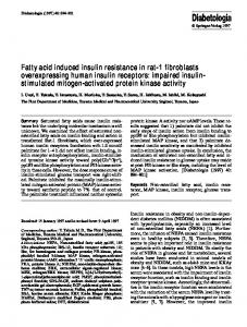
FIG. 1. Glucose metabolism during a 3-h euglycemic-hyperinsulinemic clamp in 14 control subjects and 24 PCOS patients (A) and 10 PCOS patients before (Pre) and after (Post) 16-weeks treatment with pioglitazone (B). Rd rates are displayed as oxidative (black) and nonoxidative glucose metabolism (white) during the basal and insulin-stimulated steady-state periods. Data are means ⫾ SEM. **, P ⬍ 0.001 and *, P ⬍ 0.05 vs. corresponding basal values; ††, P ⬍ 0.001 vs. insulinstimulated values in controls; ‡‡, P ⬍ 0.001 and ‡, P ⬍ 0.05 vs. pretreatment insulin-stimulated values.
J Clin Endocrinol Metab, September 2008, 93(9):3618 –3626
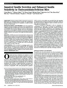
insulin, free testosterone, and plasma triglycerides (Table 1). Insulin-stimulated Rd was 50% lower in PCOS patients than controls (P ⬍ 0.001), and this was primarily accounted for by a 60% reduction in NOGM but also a 39% decrease in glucose oxidation (Fig. 1). Treatment of PCOS subjects with pioglitazone significantly reduced fasting serum insulin (45%) and improved insulin-stimulated Rd (36%) (P ⬍ 0.001), glucose oxidation (26%), and NOGM (50%). Muscle glycogen and GS activity Muscle glycogen content and total GS activity in the basal and insulin-stimulated states were similar in PCOS patients, compared with controls, and were not changed by either insulin or pioglitazone, except for a small decrease in basal glycogen levels in response to pioglitazone (Fig. 2, A and B). Physiological hyperinsulinemia increased GS activity (I-form activity and FV) significantly in both PCOS patients and control subjects. However, in the insulin-stimulated state, GS activity (I-form activity and FV) was significantly reduced in PCOS patients vs. controls (Fig. 2, C and D). In the pioglitazone-treated subgroup of PCOS patients, treatment with pioglitazone significantly increased insulin-stimulated GS activity, compared with pretreatment levels, and induced a near-normal insulin activation of GS. Protein content and site-specific phosphorylation of GS Protein levels of GS were comparable in PCOS patients and controls and were not changed by either insulin or pioglitazone (Fig. 3A). Using phospho-specific antibodies against GS, we observed that physiological hyperinsulinemia caused a significant decrease in the amount of GS phosphorylated at sites 2 ⫹ 2a, site 3a alone, and 3a⫹3b in controls but only at sites 3a⫹3b in PCOS
jcem.endojournals.org
3621
patients (Fig. 3). In control subjects, insulin infusion also tended to decrease GS phosphorylation at site 2 (P ⫽ 0.06), whereas no such effect was seen in PCOS patients. Insulin-induced changes (␦-values) in GS phosphorylation at site 2 alone (P ⫽ 0.027) and sites 2 ⫹ 2a (P ⬍ 0.001) were higher in controls than patients with PCOS (Fig. 3). Treatment with pioglitazone significantly reduced GS phosphorylation at sites 3a alone and sites 3a⫹3b in both the basal and insulin-stimulated state but caused no changes in the response to insulin (␦-values) (Fig. 3). Pioglitazone treatment introduced a significant effect of insulin on site 2 alone and also tended (P ⫽ 0.06) to restore the ability of insulin to dephosphorylate sites 2 ⫹ 2a (Fig. 3). Phosphorylation of GS at sites 3b, 1a, or 1b was not regulated by insulin in either of the groups, insulin regulated, and pioglitazone treatment did not affect the phosphorylation of these sites (Fig. 4). GSK-3␣ and -3 Consistent with GS phosphorylation at sites 3a⫹3b, there was no significant differences in protein content of GSK-3␣ or -3 between the two groups (not shown). Insulin infusion significantly increased phosphorylation of GSK-3␣ at Ser21 in PCOS patients (n ⫽ 24) and phosphorylation of GSK-3 at Ser9 in both groups (Fig. 5). There were no differences in the insulinmediated phosphorylation of GSK-3 between the two groups. In the pioglitazone-treated subgroup of PCOS patients, a significant effect of insulin on the phosphorylation of GSK-3 at Ser9 was seen only after treatment with pioglitazone. Correlation analysis In the total population, Rd and NOGM during insulin stimulation correlated significantly with GS I-form activity (r ⫽ 0.60
FIG. 2. Glycogen content (A) and effect of insulin on GS activity given as total (B), percent I-form activity (C), and percent FV (D) in 14 control subjects and 24 PCOS patients, and (right to the dotted line) in 10 PCOS patients before (Pre) and after (Post) 16-weeks treatment with pioglitazone. Measurements were performed in skeletal muscle biopsies obtained during the basal (white bars) and insulin-stimulated (black bars) steady-state periods of a 3-h euglycemic-hyperinsulinemic clamp. Data are means ⫾ SEM. **, P ⬍ 0.001 and *, P ⬍ 0.01 vs. corresponding basal values; †, P ⬍ 0.001 vs. insulin-stimulated values in controls; ‡, P ⬍ 0.01 vs. pretreatment insulin-stimulated values.
3622
Glintborg et al.
Impaired Glycogen Synthase Activity in PCOS Muscle
J Clin Endocrinol Metab, September 2008, 93(9):3618 –3626
FIG. 3. Protein content (A) and phosphorylation of GS at sites 2 (B), 2 ⫹ 2a (C), 3a (D), and 3a⫹3b (E) in 14 control subjects and 24 PCOS patients, and in 10 PCOS patients before (Pre) and after (Post) 16 wk treatment with pioglitazone (right to the dotted line). F, Representative immunoblots. Measurements were performed in skeletal muscle biopsies obtained during the basal (white bars) and insulin-stimulated (black bars) steady-state periods of a 3-h euglycemic-hyperinsulinemic clamp. Data are means ⫾ SEM. **, P ⬍ 0.001 and *, P ⬍ 0.05 vs. corresponding basal values; †, P ⬍ 0.05 vs. corresponding pretreatment basal or insulin-stimulated values.
and r ⫽ 0.63) and GS FV (r ⫽ 0.59 and r ⫽ 0.59), respectively (all P ⬍ 0.001). In patients with PCOS, NOGM correlated with GS FV (r ⫽ 0.42; P ⫽ 0.04) and tended to correlate with GS I-form activity (r ⫽ 0.39; P ⫽ 0.06), whereas in the control subjects, Rd and NOGM correlated significantly with GS I-form activity (r ⫽ 0.68, P ⫽ 0.008, and r ⫽0.65, P ⫽ 0.013) and tended to correlate with GS FV (r ⫽ 0.47, P ⫽ 0.09, and r ⫽ 0.48, P ⫽ 0.08). In the total population, insulin-stimulated GS FV and GS I-form activity correlated significantly with GS phosphorylation at sites 2 ⫹ 2a, 3a, and 3a⫹3b (r ⫽ ⫺0.39 – 0.48; all P ⬍ 0.02). In the individual groups, GS FV and GS I-form activity correlated significantly with GS phosphorylation at sites 2, 2 ⫹ 2a, 3a, 3a⫹3b, and 1a (r ⫽ ⫺0.40 to 0.54; all P ⬍ 0.05) in PCOS patients but only with sites 2 ⫹ 2a in control subjects (r ⫽ ⫺0.60, P ⫽ 0.024, and r ⫽ ⫺0.70, P ⫽ 0.005). Using stepwise multiple linear regression analysis, we examined the contribution of the different phosphorylation sites to insulin-stimulated GS I-form activity (Table 2). In the total population, a model including phos-
phorylation at sites 2 ⫹ 2a and 3a⫹3b significantly predicted GS-I-form activity. The best model predicting GS I-form activity in patients with PCOS included phosphorylation at sites 3a⫹3b and 1a, whereas in controls the best model included phosphorylation at sites 2 ⫹ 2a (Table 2).
Discussion Insulin resistance in women with PCOS is characterized by impaired insulin-stimulated glucose disposal primarily affecting glycogen synthesis in skeletal muscle (3). Here we demonstrate that impaired insulin activation of GS contributes to insulin resistance at the molecular level in skeletal muscle of women with PCOS. Absent insulin-mediated dephosphorylation of GS at the NH2-terminal sites 2 and 2a, in part, explains impaired insulin activation of GS in PCOS, whereas dephosphorylation at the COOH-terminal sites 3a and 3b was normal consistent with
J Clin Endocrinol Metab, September 2008, 93(9):3618 –3626
jcem.endojournals.org
3623
FIG. 4. Phosphorylation of GS at sites 3b (A), 1a (B), and 1b (C) in 14 control subjects and 24 PCOS patients and in 10 PCOS patients before (Pre) and after (Post) 16 wk treatment with pioglitazone (right to the dotted line). D, Representative immunoblots. Measurements were performed in skeletal muscle biopsies obtained during the basal (white bars) and insulin-stimulated (black bars) steady-state periods of a 3-h euglycemic-hyperinsulinemic clamp. Data are means ⫾ SEM.
adequate inhibition of the major upstream kinase, GSK-3␣/. We provide correlative evidence that insulin activation of GS in human skeletal muscle is dependent on dephosphorylation at sites 2 ⫹ 2a and 3a⫹3b. In women with PCOS the dependency on sites 2 ⫹ 2a seems to be lost and replaced by dependency on sites 3a⫹3b. Moreover, our data suggest that the ability of pioglitazone to improve insulin sensitivity in PCOS involves enhanced insulin-mediated activation of GS and dephosphorylation at sites 2 and 2a. However, the near normalization of insulin activation of muscle GS in PCOS despite less than fully normalization of insulin-stimulated glucose disposal also indicates that other mechanisms than those affecting insulin signaling and GS contribute to insulin resistance in skeletal muscle in PCOS. Insulin-stimulated GS activity is reduced in skeletal muscle of women with PCOS similar to other populations characterized by insulin resistance and impaired insulin-stimulated NOGM such as patients with type 2 diabetes (6 – 8, 14, 31), their FDR (9, 10, 11), patients with HIV-associated lipodystrophy (28), McArdles disease (32), carriers of a mutant IR (33), and obese nondiabetic subjects (8, 14). Consistent with the characterization of this defect in other insulin-resistant conditions (6, 14, 28, 32, 33), protein levels of GS are normal in PCOS patients indicating a major role for altered posttranslational modifications, e.g. phosphorylation. The fact that this defect was observed in normoglycemic, obese women with PCOS, compared with weight-matched healthy women supports the hypothesis that impaired insulin activation of GS is an early defect in the pathogenesis of insulin resistance independent of hyperglycemia and obesity (9 –11). Studies of myotubes established from patients with type 2 diabetes have provided evidence that it may even represent an intrinsic defect (34 –36). However, in a recent study of myotubes established from patients with PCOS, an approximately 60%
increase in basal and insulin-stimulated glucose transport and glycogen synthesis was reported, and the fold-stimulation of glucose transport and glycogen synthesis in response to insulin was normal (18). These findings were, in part, explained by increased GLUT1 abundance (18). Although these data to some extent argue against an intrinsic defect in insulin activation of GS, direct measurements of GS activity in myotubes from women with PCOS are needed to exclude the possibility that elevated intracellular G6P levels caused by a compensatory increase in glucose uptake overrides a potential defect in GS due to enhanced allosteric activation (12, 13). Another important finding of the present study was a significant positive effect of pioglitazone on the activity of GS, the most distal and rate-limiting enzyme in insulin signaling to glycogen synthesis. This has, to our knowledge, not been demonstrated previously in human subjects in vivo. In fact, reports of the effect of TZD on GS activity in muscle of patients with type 2 diabetes are not available. However, in a study of insulin-resistant rhesus monkeys, 6 wk treatment with a TZD increased both insulinstimulated GS fractional activity, total GS activity and G6P content, indicating that TZD enhance both insulin-stimulated dephosphorylation and allosteric activation of GS (37). An improved allosteric activation of GS could explain why 12 wk treatment with troglitazone did not improve GS fractional activity despite improved NOGM during insulin stimulation in obese, glucose tolerant FDR of patients with type 2 diabetes (25). In support of this, 3 months treatment with troglitazone increased im G6P concentrations in type 2 diabetic subjects during hyperglycemic-hyperinsulinemic clamp conditions (38). Nevertheless, we here show that, at least in women with PCOS, pioglitazone also restores a defect insulin action on GS activity by modifying covalent inhibition mediated by phosphorylation.
3624
Glintborg et al.
Impaired Glycogen Synthase Activity in PCOS Muscle
J Clin Endocrinol Metab, September 2008, 93(9):3618 –3626
TABLE 2. Phosphorylation sites predicting insulin-stimulated GS I-form activity Model
Coefficient (B)
SEM
P value
39.641 ⫺0.578 ⫺1.161
2.790 0.167 0.388
0.002 0.005
38.796 ⫺1.543 ⫺0.374
2.949 0.384 0.119
0.001 0.005
41.428 ⫺0.974
3.631 0.357
0.020
a
Total population Constant GS sites 2 ⫹ 2a GS sites 3a ⫹ 3b PCOSb Constant GS sites 3a ⫹ 3b GS site 1a Controlsc Constant GS sites 2 ⫹ 2a
Significant models of GS phosphorylation sites predicting insulin-stimulated GS I-form activity in the total population, PCOS patients, and controls, respectively, determined by stepwise multiple linear regression analyses. All measured GS phosphorylation sites were entered into the models.
FIG. 5. Phosphorylation of GSK-3␣ at Ser21 (A) and GSK-3 at Ser9 (B) in 14 control subjects and 24 PCOS patients and in 10 PCOS patients before (Pre) and after (Post) 16 wk treatment with pioglitazone (right to the dotted line). C, Representative immunoblots. Measurements were performed in skeletal muscle biopsies obtained during the basal (white bars) and insulin-stimulated (black bars) steady-state periods of a 3-h euglycemic-hyperinsulinemic clamp. Data are means ⫾ SEM. **, P ⬍ 0.001 and *, P ⬍ 0.05 vs. corresponding basal values.
The molecular mechanism by which pioglitazone enhances insulin activation of GS and whether this represents a unique response in PCOS remains to be established. In vitro, both NH2- and COOH-terminal sites play a role for the regulation of GS activity (4, 12, 13). Recently we could not demonstrate a significant effect of insulin on sites 2 ⫹ 2a in neither obese nondiabetic nor diabetic male subjects (6), probably because both groups were insulin resistant. However, in later studies of larger cohorts of nondiabetic twins and HIV patients without lipodystrophy (28, 39), we have shown that insulin dephosphorylates GS at sites 2 ⫹ 2a in humans. In the present study, we provide evidence that in healthy obese women, insulin-mediated dephosphorylation of GS at sites 2 ⫹ 2a plays a significant role for the activation of GS during physiological hyperinsulinemia, whereas in women with PCOS an absent effect of insulin on these NH2-terminal sites seems to explain the attenuated insulin activation of GS. In contrast to male patients
a
Adjusted R2 for the model ⫽ 0.38.
b
Adjusted R2 for the model ⫽ 0.54.
c
Adjusted R2 for the model ⫽ 0.35.
with type 2 diabetes (6), we observed no hyperphosphorylation of GS at sites 2 ⫹ 2a during insulin stimulation in women with PCOS. Whether this reflects a unique defect in patient with type 2 diabetes, changes in the metabolic milieu secondary to hyperglycemia or gender-specific differences in conditions with insulin resistance warrants further clarification. However, similarly to that observed in patients with type 2 diabetes, insulin-mediated dephosphorylation of GS at sites 3a⫹3b was normal as also indicated by normal phosphorylation and inhibition of GSK-3␣ and -3. Thus, absent insulin-mediated dephosphorylation of GS at sites 2 ⫹ 2a seems to counteract the dephosphorylation at sites 3a⫹3b mediated by inhibition of GSK-3 in muscle of women with PCOS. Multiple linear regression analysis showed that in the total population of obese women with and without PCOS, insulinmediated dephosphorylation of both sites 2 ⫹ 2a and sites 3a⫹3b plays a major role for activation of GS in response to physiological hyperinsulinemia. This is in accordance with our previous results obtained in larger cohorts of younger and older twins and HIV patients with and without lipodystrophy (28, 39, 40). In healthy obese women, dephosphorylation at sites 2 ⫹ 2a seems to be the best predictor of insulin activation of GS, whereas in women with PCOS, insulin activation of GS seems to be dependent on dephosphorylation at sites 3a⫹3b and perhaps site 1a. These observations are further supported by the finding that normalization of insulin-stimulated GS activity in response to treatment with pioglitazone is accompanied by an enhanced effect of insulin on dephosphorylation of GS at sites 2 and 2a, whereas no significant changes with respect to dephosphorylation of sites 3 ⫹ 3a were seen. In contrast to our working hypothesis, the lack of fully normalization of insulin-stimulated Rd and NOGM in response to pioglitazone was not explained by an absent normalization of insulin-mediated GS activity. This indicates that pioglitazone does not reverse all mechanisms underlying impaired insulin-stimulated NOGM in PCOS. As discussed previously (20), this discrepancy between the near normalization
J Clin Endocrinol Metab, September 2008, 93(9):3618 –3626
of insulin action on PI3K, Akt, AS160, GSK3, and GS and the lack of fully normalization of Rd and NOGM needs to be addressed in future studies. However, there is no reason to believe that this phenomenon is unique to PCOS. It has also been reported in patients with type 2 diabetes (41). In summary, we demonstrate, for the first time, that impaired insulin activation of GS contributes to insulin resistance at the molecular level in skeletal muscle of women with PCOS. In these insulin-resistant women, normal insulin-mediated phosphorylation, and thus likely inhibition, of GSK-3 and subsequent dephosphorylation of GS at sites 3a⫹3b are counteracted by absent dephosphorylation at sites 2 ⫹ 2a. The ability of pioglitazone to improve insulin sensitivity in PCOS clearly involves near-normalized insulin activation of GS and dephosphorylation at sites 2 and 2a. However, other factors controlling insulin-stimulated Rd and NOGM in skeletal muscle need to be investigated to explain the less than fully normalization of these measures in women with PCOS.
Acknowledgments We thank Professor Grahame D. Hardie (Division of Molecular Physiology, University of Dundee, Dundee, Scotland) and Oluf Pedersen (Steno Diabetes Center, Copenhagen, Denmark) for donating materials essential to this study and acknowledge L. Hansen, C. B. Olsen, I. B. Nielsen, and B. Bolmgren for skilled technical assistance. Address all correspondence and requests for reprints to: Kurt Højlund, M.D., Ph.D., Department of Endocrinology, Odense University Hospital, Kloevervaenget 6, DK-5000 Odense C, Denmark. E-mail: k.hojlund@ dadlnet.dk. The trial is registered at www.clinicaltrials.gov (NCT00145340). This work was supported by the Institute of Clinical Research, University of Southern Denmark, The Copenhagen Muscle Research Centre, the Novo Nordisk Research Foundation, The Danish Diabetes Association, an Integrated Project (LSHM-CT-2004 – 005272) funded by the European Commission, and the Danish Medical Research Councils. J.F.P.W. was supported by a Hallas Møller Fellowship from The Novo Nordisk Foundation. Disclosure Statement: The authors have nothing to disclose.
References 1. The Rotterdam ESHRE/ASRM-sponsored PCOS Consensus Workshop Group 2004 Revised 2003 consensus on diagnostic criteria and long-term health risks related to polycystic ovary syndrome. Fertil Steril 81:19 –25 2. Ehrmann DA 2005 Polycystic ovary syndrome. N Engl J Med 352:1223–1236 3. Glintborg D, Hermann AP, Andersen M, Hagen C, Beck-Nielsen H, Veldhuis JD, Henriksen JE 2006 Effect of pioglitazone on glucose metabolism and luteinizing hormone secretion in women with polycystic ovary syndrome. Fertil Steril 86:385–397 4. Højlund K, Beck-Nielsen H 2006 Impaired glycogen synthase activity and mitochondrial dysfunction in skeletal muscle. Markers or mediators of insulin resistance in type 2 diabetes. Curr Diabetes Rev 2:375–395 5. Kim YB, Nikoulina SE, Ciaraldi TP, Henry RR, Kahn BB 1999 Normal insulin-dependent activation of Akt/protein kinase B, with diminished activation of phosphoinositide 3-kinase, in muscle in type 2 diabetes. J Clin Invest 104: 733–741 6. Højlund K, Staehr P, Hansen BF, Green KA, Hardie DG, Richter EA, Beck-Nielsen H, Wojtaszewski JF 2003 Increased phosphorylation of skeletal muscle glycogen synthase at NH2-terminal sites during physiological hyperinsulinemia in type 2 diabetes. Diabetes 52:1393–1402
jcem.endojournals.org
3625
7. Cusi K, Maezono K, Osman A Pendergrass M, Patti ME, Pratipanawatr T, DeFronzo RA, Kahn CR, Mandarino LJ 2000 Insulin resistance differentially affects the PI 3-kinase- and MAP kinase-mediated signaling in human muscle. J Clin Invest 105:311–320 8. Damsbo P, Vaag A, Hother-Nielsen O, Beck-Nielsen H 1991 Reduced glycogen synthase activity in skeletal muscle from obese patients with and without type 2 (non-insulin-dependent) diabetes mellitus. Diabetologia 34:239 –245 9. Vaag A, Henriksen JE, Beck-Nielsen H 1992 Decreased insulin activation of glycogen synthase in skeletal muscles in young nonobese Caucasian first-degree relatives of patients with non-insulin-dependent diabetes mellitus. J Clin Invest 89:782–788 10. Eriksson J, Franssila-Kallunki A, Ekstrand A, Saloranta C, Wide´n E, Schalin C, Groop L 1989 Early metabolic defects in persons at increased risk for non-insulin-dependent diabetes mellitus. N Engl J Med 321:337–343 11. Schalin-Jantti C, Harkonen M, Groop LC 1992 Impaired activation of glycogen synthase in people at increased risk for developing NIDDM. Diabetes 41:598 – 604 12. Lawrence Jr JC, Roach PJ 1997 New insights into the role and mechanism of glycogen synthase activation by insulin. Diabetes 46:541–547 13. Cohen P, Alessi DR, Cross DA 1997 PDK1, one of the missing links in insulin signal transduction? FEBS Lett 410:3–10 14. Nikoulina SE, Ciaraldi TP, Mudaliar S, Mohideen P, Carter L, Henry RR 2000 Potential role of glycogen synthase kinase-3 in skeletal muscle insulin resistance of type 2 diabetes. Diabetes 49:263–271 15. McManus EJ, Sakamoto K, Armit LJ, Ronaldson L, Shpiro N, Marquez R, Alessi DR 2005 Role that phosphorylation of GSK3 plays in insulin and Wnt signalling defined by knockin analysis. EMBO J 24:1571–1583 16. Dunaif A, Wu X, Lee A, Diamanti-Kandarakis E 2001 Defects in insulin receptor signaling in vivo in the polycystic ovary syndrome (PCOS). Am J Physiol Endocrinol Metab 281:E392–E399 17. Dunaif A, Xia J, Book CB, Schenker E, Tang Z 1995 Excessive insulin receptor serine phosphorylation in cultured fibroblasts and in skeletal muscle. A potential mechanism for insulin resistance in the polycystic ovary syndrome. J Clin Invest 96:801– 810 18. Corbould A, Kim YB, Youngren JF, Pender C, Kahn BB, Lee A, Dunaif A 2005 Insulin resistance in the skeletal muscle of women with PCOS involves intrinsic and acquired defects in insulin signaling. Am J Physiol Endocrinol Metab 288:E1047–E1054 19. Corbould A, Zhao H, Mirzoeva S, Aird F, Dunaif A 2006 Enhanced mitogenic signaling in skeletal muscle of women with polycystic ovary syndrome. Diabetes 55:751–759 20. Højlund K, Glintborg D, Andersen NR, Birk JB, Treebak JT, Frøsig C, Beck-Nielsen H, Wojtaszewski JP 2008 Impaired insulin-stimulated phosphorylation of Akt and AS160 in skeletal muscle of women with polycystic ovary syndrome is reversed by pioglitazone treatment. Diabetes 57:357–366 21. Kim YB, Ciaraldi TP, Kong A, Kim D, Chu N, Mohideen P, Mudaliar S, Henry RR, Kahn BB 2002 Troglitazone but not metformin restores insulin-stimulated phosphoinositide 3-kinase activity and increases p110 protein levels in skeletal muscle of type 2 diabetic subjects. Diabetes 51:443– 448 22. Miyazaki Y, He H, Mandarino LJ, DeFronzo RA 2003 Rosiglitazone improves downstream insulin receptor signaling in type 2 diabetic patients. Diabetes 52:1943–1950 23. Meyer MM, Levin K, Grimmsmann T, Perwitz N, Eirich A, Beck-Nielsen H, Klein HH 2002 Troglitazone treatment increases protein kinase B phosphorylation in skeletal muscle of normoglycemic subjects at risk for the development of type 2 diabetes. Diabetes 51:2691–2697 24. Beeson M, Sajan MP, Dizon M, Grebenev D, Gomez-Daspet J, Miura A, Kanoh Y, Powe J, Bandyopadhyay G, Standaert ML, Farese RV 2003 Activation of protein kinase C- by insulin and phosphatidylinositol-3,4,5-(PO4)3 is defective in muscle in type 2 diabetes and impaired glucose tolerance: amelioration by rosiglitazone and exercise. Diabetes 52:1926 –1934 25. Levin K, Hother-Nielsen O, Henriksen JE, Beck-Nielsen H 2004 Effects of troglitazone in young first-degree relatives of patients with type 2 diabetes. Diabetes Care 27:148 –154 26. Birk JB, Wojtaszewski J 2006 Predominant ␣2/2/␥3 AMPK activation during exercise in human skeletal muscle. J Physiol 577:1021–1032 27. Lowry OH, Passonneau JV 1972 A flexible system of enzymatic analysis. New York: Academic Press 28. Haugaard SB, Andersen O, Madsbad S, Frøsig C, Iversen J, Nielsen JO, Wojtaszewski JF 2005 Skeletal muscle insulin signaling defects downstream of phosphatidylinositol 3-kinase at the level of Akt are associated with impaired nonoxidative glucose disposal in HIV lipodystrophy. Diabetes 54:3474 –3483 29. Prats C, Cadefau JA, Cusso R, Qvortrup K, Nielsen JN, Wojtaszewki JFP, Hardie DG, Stewart G, Hansen BF, Ploug T 2005 Phosphorylation-dependent
3626
30.
31.
32.
33.
34.
35.
Glintborg et al.
Impaired Glycogen Synthase Activity in PCOS Muscle
translocation of glycogen synthase to a novel structure during glycogen resynthesis. J Biol Chem 280:23165–23172 Thomas JA, Schlender KK, Larner J 1968 A rapid filter paper assay for UDP glucose-glycogen glucosyltransferase, including an improved biosynthesis of UDP-14C-glucose. Anal Biochem 25:486 – 499 Højlund K, Frystyk J, Levin K, Flyvbjerg A, Wojtaszewski JF, Beck-Nielsen H 2006 Reduced plasma adiponectin concentrations may contribute to impaired insulin activation of glycogen synthase in skeletal muscle of patients with type 2 diabetes. Diabetologia 49:1283–1291 Nielsen JN, Vissing J, Wojtaszewski JF, Haller RG, Begum N, Richter EA 2002 Decreased insulin action in skeletal muscle from patients with McArdle’s disease. Am J Physiol Endocrinol Metab 282:E1267–1275 Højlund K, Wojtaszewski JF, Birk J, Hansen BF, Vestergaard H, Beck-Nielsen H 2006 Partial rescue of in vivo insulin signalling in skeletal muscle by impaired insulin clearance in heterozygous carriers of a mutation in the insulin receptor gene. Diabetologia 49:1827–1837 Gaster M, Petersen I, Højlund K, Poulsen P, Beck-Nielsen H 2002 The diabetic phenotype is conserved in myotubes established from diabetic subjects: evidence for primary defects in glucose transport and glycogen synthase activity. Diabetes 51:921–927 Nikoulina SE, Ciaraldi TP, Abrams-Carter L, Mudaliar S, Park KS, Henry RR 1997 Regulation of glycogen synthase activity in cultured skeletal muscle cells
J Clin Endocrinol Metab, September 2008, 93(9):3618 –3626
36.
37.
38. 39.
40.
41.
from subjects with type II diabetes: role of chronic hyperinsulinemia and hyperglycemia. Diabetes 46:1017–1024 Henry RR, Ciaraldi TP, Abrams-Carter L, Mudaliar S, Park KS, Nikoulina SE 1996 Glycogen synthase activity is reduced in cultured skeletal muscle cells of non-insulin-dependent diabetes mellitus subjects. Biochemical and molecular mechanisms. J Clin Invest 98:1231–1236 Ortmeyer HK, Bodkin NL, Haney J, Yoshioka S, Horikoshi H, Hansen BC 2000 A thiazolidinedione improves in vivo insulin action on skeletal muscle glycogen synthase in insulin-resistant monkeys. Int J Exp Diabetes Res 1:195–202 Petersen KF, Krssak M, Inzucchi S, Cline GW, Dufour S, Shulman GI 2000 Mechanism of troglitazone action in type 2 diabetes. Diabetes 49:827– 831 Poulsen P, Wojtaszewski JF, Petersen I Petersen I, Christensen K, Richter EA, Beck-Nielsen H, Vaag A 2005 Impact of genetic versus environmental factors on the control of muscle glycogen synthase activation in twins. Diabetes 54: 1289 –1296 Poulsen P, Wojtaszewski JF, Richter EA, Beck-Nielsen H, Vaag A 2007 Low birth weight and zygosity status is associated with defective muscle glycogen and glycogen synthase regulation in elderly twins. Diabetes 56:2710 –2714 Bandyopadhyay GK, Yu JG, Ofrecio J, Olefsky JM 2006 Increased malonylCoA levels in muscle from obese and type 2 diabetic subjects lead to decreased fatty acid oxidation and increased lipogenesis; thiazolidinedione treatment reverses these defects. Diabetes 55:2277–2285