INFECTION AND IMMUNITY, Dec. 2005, p. 8393–8396 0019-9567/05/$08.00⫹0 doi:10.1128/IAI.73.12.8393–8396.2005 Copyright © 2005, American Society for Microbiology. All Rights Reserved.
Vol. 73, No. 12
NOTES Impaired Kupffer Cells in Highly Susceptible Mice Infected with Trypanosoma congolense Meiqing Shi, Guojian Wei, Wanling Pan, and Henry Tabel* Department of Veterinary Microbiology, Western College of Veterinary Medicine, University of Saskatchewan, Saskatoon, Canada Received 10 June 2005/Returned for modification 18 July 2005/Accepted 2 September 2005
In highly susceptible BALB/c mice infected with Trypanosoma congolense, the total number of Kupffer cells in the liver remains constant; however, their mean size increases fivefold towards the terminal stage. About 25% of Kupffer cells undergo apoptosis. We suggest that development of an impairment of the macrophage system might be a major mechanism for inefficient elimination of trypanosomes. African trypanosomes are extracellular, single-cell parasites that infect both humans and livestock. The parasites survive in the bloodstream of the mammalian host by escaping the host’s immune responses by sophisticated mechanisms, including antigenic variation of the variant surface glycoprotein, and induction of immunosuppression and polyclonal B-cell activation (1, 17, 23). BALB/c mice are highly susceptible to Trypanosoma congolense and Trypanosoma brucei infections, whereas C57BL/6 mice are relatively resistant, as measured by levels of parasitemia, immunosuppression, and survival time (7, 16, 23). After infection, the parasites multiply in the bloodstream. The infected host will produce antibodies to the variant surface glycoprotein, and the parasitemia is controlled by anti-variant surface glycoprotein-mediated phagocytosis (23). The parasites are predominantly phagocytosed in the liver (2, 12, 20) by Kupffer cells (20, 21). Highly susceptible BALB/c mice are unable to control the first wave of parasitemia and succumb to the infection 7 to 9 days after infection with 103 T. congolense, Trans Mara strain, variant antigenic type (VAT) TC13 (23, 27). Recently, we have shown that in BALB/c mice infected with 103 T. congolense, a small degree of phagocytosis of trypanosomes by Kupffer cells was detectable as early as 5 days after infection. Phagocytosis of trypanosomes increased dramatically from days 5 to 6 (21). Concomitantly, Kupffer cells were greatly enlarged, in some areas almost clogging the sinusoids (20, 21). Presently it is unknown why highly susceptible mice infected with virulent trypanosomes cannot control the first parasitemia. We decided to investigate how the trypanosomes affected the liver, including Kupffer cells. Eight- to 10-week-old BALB/c mice (Animal Resource Center of the University of Saskatchewan, Saskatoon, Canada) were infected intraperitoneally with 103 T. congolense, VAT TC13 (22). Mice were killed on days 0, 5, 6, 7, and 8 postin-
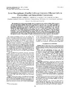
fection. Livers were removed and fixed in 10% neutral buffered formalin for 24 h at 4°C, embedded in paraffin, and cut into 5-m-thick sections. We carried out a quantitative computer analysis of the number and size of the Kupffer cells visualized by immunohistochemical staining for the F4/80 macrophage marker (20), by using software of Northern Eclipse (Empix Imaging, Inc., Mississauga, Canada). The number of Kupffer cells per microscopic field did not change significantly (Fig. 1C), however, the mean size of Kupffer cells per microscopic field increased fivefold towards the terminal stage (Fig. 1 A, B, and D). Sections were stained with hematoxylin and eosin for histological examinations. As shown in Fig. 2B, focal necrosis-like lesions were observed in the livers of mice infected with T. congolense at the terminal stage. In contrast, no such lesions were detected in the livers of normal mice (Fig. 2A). Marked pyknosis of the nuclei of the parenchymal hepatocytes within these focal lesions was observed. These observations indicated that trypanosomal infections caused profound liver injuries at the terminal stages. To demonstrate whether the parenchymal hepatocytes in the lesions were necrotic or apoptotic, terminal deoxynucleotidyltransferase-mediated dUTP-biotin nick end labeling (TUNEL) staining for the liver sections was performed according to Haimovitz-Friedman et al. (9). Briefly, liver tissues were deparaffinized in xylene and rehydrated through a graded series of ethanol. The tissues were incubated with proteinase K (EC 3.4.21.64, Sigma, 20 g/ml in 10 mM Tris/HCl, pH 7.4 to 8.0) for 30 min at 37°C. The tissues were rinsed and incubated with 3% H2O2 in methanol for 10 min, followed by blocking with 2% bovine serum albumin in phosphate-buffered saline. The tissues were incubated with permeabilization solution (0.1% Triton X-100 in 0.1% sodium citrate) on ice for 2 min, rinsed with phosphate-buffered saline, and incubated with TUNEL reaction mixture (Roche Diagnostics Corporation, Indianapolis, IN) for 60 min at 37°C. Finally, they were incubated with antifluorescein antibody conjugated with peroxidase for 30 min at 37°C, followed by development of the color using 3,3⬘-diaminobenzidine (substrate kit, Vector Laborato-
* Corresponding author. Mailing address: Department of Veterinary Microbiology, WCVM, University of Saskatchewan, 52 Campus Drive, Saskatoon, Saskatchewan, S7N 5B4, Canada. Phone: 306-9667221. Fax: 306-966-7244. E-mail:
[email protected]. 8393
8394
NOTES
INFECT. IMMUN.
FIG. 1. In BALB/c mice infected with T. congolense, the number of the Kupffer cells remains fairly constant, but the mean size of Kupffer cells increases fivefold towards the terminal stage. Sizes (A, B, and D) and numbers (C) of Kupffer cells were determined in liver sections of mice. Immunohistochemical stain for macrophage marker F4/80 of liver sections are shown for days 0 (A) and 8 (B). The sizes and numbers of Kupffer cells in livers were determined by microscopic examination of tissue sections and were measured by computer imaging, using software of Northern Eclipse (means ⫾ standard error). The results presented are representative of two experiments.
ries, Burlingame, CA). TUNEL staining detects 3⬘OH DNA ends in apoptotic nuclei. As shown in Fig. 2D and 2E, many nuclei of hepatocytes in the focal lesions were stained by the TUNEL method, suggesting apoptotic death of hepatocytes. We found that there were considerable numbers of single apoptotic cells outside of the focal lesions in the livers of infected mice (Fig. 2D and 2F). Such apoptotic cells were scarcely detected in the livers of normal mice (Fig. 2C). These individual apoptotic cells were found within the sinusoids (Fig. 2F). We speculated that they were Kupffer cells, since Kupffer cells are lining the sinusoids in the liver. Thus, we performed immunofluorescent double staining to identify the phenotype of the apoptotic cells. Liver tissues were deparaffinized, rehydrated, digested, blocked, permeated, and incubated with TUNEL reaction mixture as described above. The tissues were rinsed and incubated with rat anti-mouse F4/80 (to detect macrophages) monoclonal antibody (Serotec Inc., Raleigh, NC) for 30 min. After three washes in PBS, the tissues were incubated with biotin-conjugated goat anti-rat IgG (Cedarlane,
Hornby, Ontario, Canada), followed by washes and incubation with streptavidin Alexa Fluor 488 (Molecular Probes, Eugene, OR) for 30 min. As shown in Fig. 2G and 2H, the apoptotic cells were also F4/80 positive, indicating that Kupffer cells undergo apoptosis in the livers of infected mice at the terminal stage. We further found that 25% ⫾ 2.8% of the total number of Kupffer cells in the livers of infected mice underwent apoptosis. We have provided evidence that a systemic inflammatory response syndrome that is mediated by IFN-␥ is the major cause for the acute death of highly susceptible BALB/c mice infected with T. congolense or T. brucei (20, 27). In this study, we further demonstrate, for the first time, that hepatocytes undergo apoptosis in mice infected with T. congolense. It is noteworthy that apoptosis of hepatocytes developed rapidly and was detected shortly before death, since focal lesions of apoptotic hepatocytes were not observed until day 6 following infection (data not shown). The significance of hepatocyte apoptosis to acute death remains to be elucidated. However, it is
VOL. 73, 2005
NOTES
8395
FIG. 2. Apoptosis of hepatocytes and Kupffer cells of highly susceptible BALB/c mice infected with T. congolense. BALB/c mice were infected with 103 TC13. The mice were killed on day 8 postinfection. Liver sections were subjected to hematoxylin and eosin stain (A and B), immunoperoxidase TUNEL staining (C to F), and immunofluorescent TUNEL staining plus double staining for macrophage marker (anti-F4/80) (G and H). C and D are serial sections of A and B, respectively. (A) Liver section of normal mouse (hematoxylin and eosin) (x100). (B) Liver section of infected mouse (hematoxylin and eosin) (x100). Note: focus of necrosis-like lesion (box). (C) Serial section of A (immunoperoxidase TUNEL stain) (x100). (D) Serial section of B (immunoperoxidase TUNEL stain) (x100). Staining of focal lesion (e) and of individual cells (f). (E) Apoptosis of hepatocytes in focal lesion (TUNEL stain of box e of D) (x400). (F) Apoptosis of individual cells in sinusoids (TUNEL stain of box f of D) (x400). (G) Immunofluorescent double staining (serial section of B) (TUNEL: red; anti-F4/80: green) (x400). (H) Immunofluorescent double staining (box h of G) (TUNEL: red; anti-F4/80: green) (x1000). Note: Individual apoptotic cells are Kupffer cells.
reasonable to suggest that apoptosis of hepatocyte might be associated with the systemic inflammatory response syndrome. The development of the systemic inflammatory response syndrome in T. congolense-infection of highly susceptible mice is associated with significantly enhanced secretion of pro-inflam-
matory cytokines including gamma interferon (IFN-␥) (20, 27). Enhanced plasma levels of IFN-␥ have been reported to be associated with apoptosis of hepatocytes caused by hepatitis B (4, 5), hepatitis C (13, 15) as well as concanavalin A-induced hepatitis (25). It is well established that apoptosis of hepato-
8396
NOTES
INFECT. IMMUN.
cytes caused by concanavalin A is mediated by IFN-␥ through the Fas-Fas ligand pathway (24, 25). We have previously demonstrated that Kupffer cells are loaded with parasite antigen in mice infected with T. congolense (20, 21). Phagocytosis of trypanosomes is mediated by IgM or IgG antibodies specific for the variant surface glycoprotein of the parasites (10, 21). There is ample evidence that phagocytosis of African trypanosomes by macrophages in vitro leads to activation of the pulsed macrophages and that this activation is enhanced by IFN-␥ (8, 10, 21, 23). Antibodymediated phagocytosis of the parasites in vivo also results in significant increase in the size of the activated, trypanosomepulsed Kupffer cells (21). In this study, we show that about 25% of Kupffer cells undergo apoptosis following phagocytosis of trypanosomes at the terminal stage of infection. Other microorganisms, including Mycobacterium tuberculosis (19), Mycobacterium avium (6), Salmonella enterica serovar Typhimurium (14), Shigella flexneri (28), Bordetella pertussis (11), and Entamoeba histolytica (18), have also been shown to induce apoptosis of macrophages. The mechanisms whereby microorganisms promote apoptosis of macrophages still remains poorly understood. An in vitro study has shown that glycoinositolphospholipid ceramide isolated from T. cruzi can induce apoptosis of macrophages in the presence of IFN-␥, suggesting that IFN-␥ contributes to the apoptosis of macrophages (3). We did not directly address this issue. However, our previous results indicate that plasma levels of IFN-␥ are significantly enhanced in T. congolense-infected mice from day 6 onwards (21, 27). It is conceivable that IFN-␥ drives the apoptosis of Kupffer cells which are loaded with trypanosomal antigen. It is possible that trypanosomes might escape phagocytosis by Kupffer cells due to excessive death of Kupffer cells through apoptosis. The conclusion that impairment of Kupffer cells might lead to reduced elimination of the parasites, could be further tested by experimental depletion of Kupffer cells. Relatively resistant C57BL/6 mice infected with T. congolense do control the first parasitemia (23). Treatment of infected C57BL/6 mice with gadolinium chloride, which destroys Kupffer cells (26), might lead to impaired control of parasitemia. In summary, we suggest that trypanosomes might escape elimination in the highly susceptible host due to excessive impairment of the macrophage system.
5. 6. 7. 8. 9.
10.
11. 12.
13. 14. 15. 16. 17. 18. 19.
20. 21. 22. 23. 24.
REFERENCES 1. Cross, G. A. 1990. Cellular and genetic aspects of antigenic variation in trypanosomes. Annu. Rev. Immunol. 8:83–110. 2. Dempsey, W. L., and J. M. Mansfield. 1983. Lymphocyte function in experimental African trypanosomiasis. V. Role of antibody and the mononuclear phagocyte system in variant-specific immunity. J. Immunol. 130:405–411. 3. Freire-de-Lima, C. G., M. P. Nunes, S. Corte-Real, M. P. Soares, J. O. Previato, L. Mendonca-Previato, and G. A. DosReis. 1998. Proapoptotic activity of a Trypanosoma cruzi ceramide-containing glycolipid turned on in host macrophages by IFN-gamma. J. Immunol. 161:4909–4916. 4. Fukuda, R., N. Ishimura, T. X. Nguyen, A. Chowdhury, S. Ishihara, N. Kohge, S. Akagi, M. Watanabe, and S. Fukumoto. 1995. The expression of IL-2, IL-4 and interferon-gamma (IFN-gamma) mRNA using liver biopsies
Editor: W. A. Petri, Jr.
25.
26. 27.
28.
at different phases of acute exacerbation of chronic hepatitis B. Clin. Exp. Immunol. 100:446–451. Galle, P. R., W. J. Hofmann, H. Walczak, H. Schaller, G. Otto, W. Stremmel, P. H. Krammer, and L. Runkel. 1995. Involvement of the CD95 (APO-1/ Fas) receptor and ligand in liver damage. J. Exp. Med. 182:1223–1230. Gan, H., G. W. Newman, and H. G. Remold. 1995. Plasminogen activator inhibitor type 2 prevents programmed cell death of human macrophages infected with Mycobacterium avium, serovar 4. J. Immunol. 155:1304–1315. Greenblatt, H. C., C. L. Diggs, and D. L. Rosenstreich. 1984. Trypanosoma rhodesiense: analysis of the genetic control of resistance among mice. Infect. Immun. 44:107–111. Grosskinsky, C. M., and B. A. Askonas. 1981. Macrophages as primary target cells and mediators of immune dysfunction in African trypanosomiasis. Infect. Immun. 33:149–155. Haimovitz-Friedman, A., C. Cordon-Cardo, S. Bayoumy, M. Garzotto, M. McLoughlin, R. Gallily, C. K. Edwards, 3rd, E. H. Schuchman, Z. Fuks, and R. Kolesnick. 1997. Lipopolysaccharide induces disseminated endothelial apoptosis requiring ceramide generation. J. Exp. Med. 186:1831–1841. Kaushik, R. S., J. E. Uzonna, J. R. Gordon, and H. Tabel. 1999. Innate resistance to Trypanosoma congolense infections: differential production of nitric oxide by macrophages from susceptible BALB/c and resistant C57Bl/6 mice. Exp. Parasitol. 92:131–143. Khelef, N., A. Zychlinsky, and N. Guiso. 1993. Bordetella pertussis induces apoptosis in macrophages: role of adenylate cyclase-hemolysin. Infect. Immun. 61:4064–4071. Macaskill, J. A., P. H. Holmes, D. D. Whitelaw, I. McConnell, F. W. Jennings, and G. M. Urquhart. 1980. Immunological clearance of 75Se-labelled Trypanosoma brucei in mice. II. Mechanisms in immune animals. Immunology 40:629–635. Mita, E., N. Hayashi, S. Iio, T. Takehara, T. Hijioka, A. Kasahara, H. Fusamoto, and T. Kamada. 1994. Role of Fas ligand in apoptosis induced by hepatitis C virus infection. Biochem. Biophys. Res. Commun. 204:468–474. Monack, D. M., B. Raupach, A. E. Hromockyj, and S. Falkow. 1996. Salmonella typhimurium invasion induces apoptosis in infected macrophages. Proc. Natl. Acad. Sci. USA 93:9833–9838. Morshed, S. A., H. Fukuma, Y. Kimura, S. Watanabe, and M. Nishioka. 1993. Interferon-gamma, interleukin (IL)-2 and IL-2 receptor expressions in hepatitis C virus-infected liver. Gastroenterol. Jpn. 28(Suppl. 5):59–66. Ogunremi, O., and H. Tabel. 1995. Genetics of resistance to Trypanosoma congolense in inbred mice: efficiency of apparent clearance of parasites correlates with long-term survival. J. Parasitol. 81:876–881. Oka, M., and Y. Ito. 1987. Polyclonal B-cell-activating factors produced by spleen cells of mice stimulated with a cell homogenate of Trypanosoma gambiense. Infect, Immun. 55:3162–3167. Ragland, B. D., L. S. Ashley, D. L. Vaux, and W. A. Petri, Jr. 1994. Entamoeba histolytica: target cells killed by trophozoites undergo DNA fragmentation which is not blocked by Bcl-2. Exp. Parasitol. 79:460–467. Rojas, M., L. F. Barrera, G. Puzo, and L. F. Garcia. 1997. Differential induction of apoptosis by virulent Mycobacterium tuberculosis in resistant and susceptible murine macrophages: role of nitric oxide and mycobacterial products. J. Immunol. 159:1352–1361. Shi, M., W. Pan, and H. Tabel. 2003. Experimental African trypanosomiasis: IFN-gamma mediates early mortality. Eur. J. Immunol. 33:108–118. Shi, M., G. Wei, W. Pan, and H. Tabel. 2004. Trypanosoma congolense infections: antibody-mediated phagocytosis by Kupffer cells. J. Leukoc. Biol. 76:399–405. Tabel, H. 1982. Activation of the alternative pathway of bovine complement by Trypanosoma congolense. Parasite Immunol. 4:329–335. Tabel, H., R. S. Kaushik, and J. E. Uzonna. 2000. Susceptibility and resistance to Trypanosoma congolense infections. Microbes Infect. 2:1619–1629. Tagawa, Y., S. Kakuta, and Y. Iwakura. 1998. Involvement of Fas/Fas ligand system-mediated apoptosis in the development of concanavalin A-induced hepatitis. Eur. J. Immunol. 28:4105–4113. Tagawa, Y., K. Sekikawa, and Y. Iwakura. 1997. Suppression of concanavalin A-induced hepatitis in IFN-gamma(-/-) mice, but not in TNF-alpha(-/-) mice: role for IFN-gamma in activating apoptosis of hepatocytes. J. Immunol. 159:1418–1428. Thurman, R. G. 1998. II. Alcoholic liver injury involves activation of Kupffer cells by endotoxin. Am. J. Physiol. 275:G605–611. Uzonna, J. E., R. S. Kaushik, J. R. Gordon, and H. Tabel. 1998. Experimental murine Trypanosoma congolense infections. I. Administration of antiIFN-gamma antibodies alters trypanosome-susceptible mice to a resistantlike phenotype. J. Immunol. 161:5507–5515. Zychlinsky, A., M. C. Prevost, and P. J. Sansonetti. 1992. Shigella flexneri induces apoptosis in infected macrophages. Nature 358:167–169.