May 1, 2010 - decreased EGFR internalization and reduced phosphorylation of SHP2, a tyrosine phosphatase required for the full activation of ERK.
NIH Public Access Author Manuscript Cancer Res. Author manuscript; available in PMC 2010 November 1.
NIH-PA Author Manuscript
Published in final edited form as: Cancer Res. 2010 May 1; 70(9): 3843–3850. doi:10.1158/0008-5472.CAN-09-3421.
IMPAIRED SHP2-MEDIATED ERK ACTIVATION CONTRIBUTES TO GEFITINIB SENSITIVITY OF LUNG CANCER CELLS WITH EGFRACTIVATING MUTATIONS Matthew J. Lazzara1,*,†, Keara Lane2,3,*, Richard Chan5, Paul J. Jasper4, Michael B. Yaffe1,2,3, Peter K. Sorger4, Tyler Jacks2,3, Benjamin G. Neel5, and Douglas A. Lauffenburger1,2,3 1 Department of Biological Engineering, Massachusetts Institute of Technology, Cambridge, MA, United States 02139 2
NIH-PA Author Manuscript
Department of Biology, Massachusetts Institute of Technology, Cambridge, MA, United States 02139 3
Koch Institute for Integrative Cancer Research, Massachusetts Institute of Technology, Cambridge, MA, United States 02139 4
Department of Systems Biology, Harvard Medical School, Boston, MA, USA, 02115
5
Cancer Biology Program, Ontario Cancer Institute, Ontario, Canada, M5G 1L7
Abstract
NIH-PA Author Manuscript
Most non-small cell lung cancers (NSCLC) display elevated expression of epidermal growth factor receptor (EGFR), but response to EGFR kinase inhibitors is predominantly limited to NSCLC harboring EGFR-activating mutations. These mutations are associated with increased activity of survival pathways including PI3K/AKT and STAT3/5. We report that EGFR-activating mutations also surprisingly lead to decreased ability to activate ERK compared to wild-type EGFR. In NSCLC cells and mouse embryonic fibroblasts expressing mutant EGFR, this effect on ERK correlates with decreased EGFR internalization and reduced phosphorylation of SHP2, a tyrosine phosphatase required for the full activation of ERK. We further demonstrate that ERK activation levels impact cellular response to gefitinib. NSCLC cells with EGFR mutation display reduced gefitinib sensitivity when ERK activation is augmented by expression of constitutively active mutants of MEK. Conversely, in an NSCLC cell line expressing wild-type EGFR, gefitinib treatment along with or following MEK inhibition increases death response compared to treatment with gefitinib alone. Our results demonstrate that EGFR-activating mutations may promote some survival pathways but simultaneously impair others. This multivariate alteration of the network governing cellular response to gefitinib, which we term “oncogene imbalance”, portends a potentially broader ability to treat gefitinib-resistant NSCLC.
Keywords endocytosis; oncogene addiction; MEK; non-small cell lung cancer Address for correspondence: Douglas A. Lauffenburger, Department of Biological Engineering, Massachusetts Institute of Technology, 77 Massachusetts Avenue, Room 16-343, Cambridge, MA 02139, Telephone: (617) 252-1629, Fax: (617) 258-0204. *Contributed equally to this work. †Present address is Department of Chemical and Biomolecular Engineering, University of Pennsylvania, Philadelphia, PA, USA 19104 DISCLOSURE OF POTENTIAL CONFLICTS OF INTEREST None of the authors declares a conflict of interest.
Lazzara et al.
Page 2
INTRODUCTION NIH-PA Author Manuscript
Up to 80% of non-small-cell lung cancers (NSCLC) display elevated expression of EGFR (1), but response to EGFR kinase inhibitors is predominantly limited to the 10–20% of tumors that harbor somatic EGFR-activating mutations (2–6). Consistent with patient response data, NSCLC cell lines bearing EGFR-activating mutations display enhanced sensitivity to gefitinib and erlotinib in culture (3,6–8). Studies of these cell lines demonstrate increased basal phosphorylation of EGFR and increased activity of survival-associated signaling pathways including PI3K/AKT and STAT3/5 (3,7,9). Exposure of mutant-bearing NSCLC cells to EGF results in more protracted increases in EGFR phosphorylation than for NSCLC cells expressing wild-type EGFR (3), consistent with the reduced endocytosis of EGFR mutants (10,11). Elevated expression of ERBB3 has been observed in some gefitinib-sensitive cells, including those lacking EGFR mutation (7,12). ERBB3 phosphorylation is apparently promoted by EGFR in these cells and is the primary mediator of PI3K/AKT activity (7).
NIH-PA Author Manuscript
EGFR-activating mutations also perturb gefitinib and ATP binding, and promote receptor inhibition at lower gefitinib concentrations (3,13,14). This effect propagates to multiple signaling pathways and in some cases may involve differential inhibition of ERBB3. For example, the phosphorylation of AKT and ERBB3 are equally or more responsive to gefitinib than EGFR phosphorylation in certain gefitinib-sensitive cell lines (7). Activating mutations may result in signaling after EGFR inhibition that favors apoptosis over survival (15). Each of these features may partially explain increased sensitivity to gefitinib of cells with EGFRactivating mutations. In tumors that are initially responsive to gefitinib and erlotinib, resistance mechanisms may arise that abrogate inhibition of survival signaling. In approximately 50% of acquired resistance cases, a secondary T790M mutation disfavors the binding of gefitinib to EGFR (14,16,17). In other cases, MET amplification maintains ERBB3/PI3K/AKT activity after treatment with gefitinib (12). Mutations of BRAF and KRAS also correlate with primary resistance to gefitinib (18–21). Because these mutations increase EGFR kinase activity (22), the finding that the activities of some downstream survival pathways are elevated is not surprising. These alterations apparently lead to cellular dependence on EGFR, or EGFR “oncogene addiction”, as demonstrated by the finding that EGFR inhibition or knockdown leads to apoptosis in NSCLC cells expressing mutant, but not wild-type, EGFR. How elevated survival signaling leads to EGFR dependence, however, remains poorly understood.
NIH-PA Author Manuscript
We report that activation of ERK is impaired by the expression of EGFR mutants compared to wild-type. Reduced EGF-elicited activation of ERK in mutant EGFR-bearing cells correlates with diminished EGFR internalization and reduced phosphorylation of the protein tyrosine phosphatase SHP2, a positive regulator of ERK activity (23). Moreover, the effect on SHP2 phosphorylation is linked to defective EGFR internalization. We further demonstrate that ERK activity impacts cellular sensitivity to gefitinib. NSCLC cells expressing an EGFR mutant exhibit reduced death response to gefitinib when ERK activation is augmented by constitutively active MEK. Conversely, NSCLC cells expressing wild-type EGFR are more sensitive to gefitinib when cotreated or pretreated with the MEK inhibitor U0126. Our results suggest that EGFR-activating mutations are associated with enhancement of some survival signals but impairment of others, with the integrated effects influencing cellular response to gefitinib.
Cancer Res. Author manuscript; available in PMC 2010 November 1.
Lazzara et al.
Page 3
MATERIALS AND METHODS Cells
NIH-PA Author Manuscript
Wild-type Egfr homozygous (EgfrWT/WT), L858R homozygous (EgfrL858R/L858R), and heterozygous (EgfrWT/L858R) primary mouse embryonic fibroblasts (MEFs) were created as described elsewhere (Lane and Jacks, in preparation). 3T3-immortalized MEFs expressing CreER and with one truncated (null) Shp2 allele and one intact allele with a section of exon 11 flanked by LoxP sites (denoted Shp2fl/−) were created as described elsewhere (Chan and Neel, in preparation). H1666 (obtained from American Type Culture Collection) and H3255 (Dr. Passi Janne, functionally characterized by gefitinib sensitivity, as described herein) cells were grown in ACL4. HeLa cells with conditional expression of wild-type or K44A dynamin (Dr. Sandra Schmid, functionally characterized as described herein) were cultured as described elsewhere (24,25). Primary MEFs and Shp2fl/− MEFs were grown in DMEM. All media contained 100 units/mL penicillin, 100 μg/mL streptomycin, 1 mM L-glutamine, and 10% FBS. Shp2fl/− MEFs were cultured in 1 μM 4-hydroxy-tamoxifen (4-OHT) for 24 hrs to induce Shp2 deletion and returned to media without 4-OHT for 36 hrs prior to experiments. Otherwise, cells were plated in six-well dishes and grown for 24–48 hrs prior to serum starving (in media containing 0.1% FBS for 12–16 hrs) or treatment with inhibitors. Egfr expression
NIH-PA Author Manuscript
Wild-type and L858R Egfr cDNA was generated from mRNA isolated from EgfrWT/WT and EgfrL858R/L858R mouse lines, respectively, and inserted into pMSCV expression vectors with puromycin or hygromycin resistance. Plasmids were transfected into ecotropic Phoenix cells (Dr. Gary Nolan, Stanford University), and viral supernatants were used to infect EgfrWT/WT and Shp2fl/− MEFs. Target cells were selected in 2 μg/mL puromycin or 200 μg/mL hygromycin. MEKDD expression VSV-G pseudotyped retrovirus was produced by transfecting HEK 293FT cells with pBABEpuro-MEK1DD (26) or pBABEpuro-MEK2DD (27) and the packaging plasmids pMDG and pMD-g/p. Virus was harvested 48 and 72 hrs after transfection, concentrated by ultracentrifugation, and used to infect H3255 cells. Target cells were selected in 2 μg/mL puromycin. Immunoblotting
NIH-PA Author Manuscript
Lysates were prepared in a standard buffer containing detergents, buffer salts, and protease and phosphatase inhibitors. Lysates were cleared by centrifugation at 4°C and 13,200 rpm for 10 min, and protein concentration was determined by micro-BCA assay. 20 μg of denatured and reduced protein was loaded per lane on 10% polyacrylamide gels and transferred to 0.2 μm nitrocellulose. Membranes were blocked in Odyssey blocking buffer (Licor), and all antibodies were used according to manufacturers’ recommendations. Where needed, blots were stripped with 0.2 N NaOH. Antibodies Antibodies for EGFR, EGFR pY1068, ERK, ERK pT202/Y204, AKT pS473, SHP2 pY542, SHP2 pY580, MEK 1/2, and MEK 1/2 pS217/S221 were purchased from Cell Signaling Technologies. Antibodies for human and mouse Shp2 were purchased from Epitomics and Santa Cruz Biotechnology, respectively. The GAPDH antibody was purchased from Calbiochem. Infrared-dye-conjugated secondary antibodies were purchased from Rockland Immunochemicals.
Cancer Res. Author manuscript; available in PMC 2010 November 1.
Lazzara et al.
Page 4
Other reagents
NIH-PA Author Manuscript
Gefitinib and U0126 were purchased from WuXi Pharmatech and Promega, respectively. Human epidermal growth factor (EGF) and platelet derived growth factor (PGDF) were purchased from Peprotech. Puromycin, propidium iodide, and 4-OHT were purchased from Sigma. Hygromycin B was purchased from Clontech. FBS, penicillin/streptomycin, Lglutamine, geneticin, and all media were purchased from Invitrogen. EGFR internalization assay Rate constants for the endocytosis of 125I-EGF (ke) were measured as described previously (28,29). Propidium iodide permeability Cells were plated in six-well dishes, grown for 24–72 hrs, and switched to complete media containing appropriate concentrations of gefitinib, U0126, and DMSO. After an additional 3– 6 days, adherent and floating cells were pooled and resuspended in PBS-PI. Flow cytometry data was acquired on a Becton Dickinson FACSCalibur and analyzed using FlowJo.
RESULTS NIH-PA Author Manuscript
Previous data demonstrate that ERK phosphorylation may be reduced in cells expressing EGFR mutants compared to counterparts expressing wild-type EGFR (EGFRWT) (3,30), but subsequent studies have focused mainly on the enhancement of pathways including PI3K/AKT and STAT3/5 (3,7,9). To explore the possibility that ERK phosphorylation is reduced in the context of EGFR-activating mutations relative to EGFRWT, we compared the phosphorylation of EGFR, ERK, and AKT in the human NSCLC cell lines H1666 (EGFRWT) and H3255 (EGFRL858R) stimulated with 10 ng/mL EGF for up to 1 hr. We chose H3255 for comparison because it is exquisitely sensitive to gefitinib (3,7) and because it carries EGFRL858R, one of the most common NSCLC-associated EGFR mutations (6,31–33). Western blot analysis is shown in Fig. 1 and Supplementary Fig. S1. As expected, EGFR phosphorylation, monitored at Y1068, was basally elevated and prolonged in response to EGF in H3255 cells compared to H1666. Consistent with previous observations (3,7), the phosphorylation of AKT at S473 was basally elevated in H3255 cells, and AKT phosphorylation actually decreased in response to EGF. In H1666 cells, AKT phosphorylation increased in response to EGF and returned to baseline levels within 60 min. The phosphorylation of ERK at T202/Y204 was uniformly lower in H3255 cells than in H1666 cells, with lower basal levels and delayed increases in response to EGF.
NIH-PA Author Manuscript
H1666 and H3255 lysates were also probed for phosphorylation of the protein tyrosine phosphatase SHP2 at Y542 (Fig 1). SHP2 is required for complete ERK activation (23) and its phosphorylation at Y542 is required for its normal activity downstream of several growth factor receptors (34). SHP2 Y542 phosphorylation was induced in H1666 cells in response to EGF by 5 min, but its phosphorylation was not induced in H3255 cells at any time point. Samples were also probed for SHP2 phosphorylation at Y580, a site of secondary importance for SHP2 regulation (34). An EGF-inducible band at the appropriate molecular weight was observed for H1666 cells but not for H3255 (Supplementary Fig. S2A). EGF-induced SHP2 Y542 phosphorylation was modest in a number of other NSCLC cell lines with EGFRactivating mutations, including PC9, HCC827, and H1975 (Supplementary Fig. S2B). The mutant-bearing, PTEN-null cell line H1650 did exhibit inducible SHP2 Y542 phosphorylation. Experiments using MDA-468 cells, which are PTEN-null, with and without PTEN reconstitution, demonstrated no general effect of PTEN status on SHP2 Y542 phosphorylation (Supplementary Fig. S3).
Cancer Res. Author manuscript; available in PMC 2010 November 1.
Lazzara et al.
Page 5
NIH-PA Author Manuscript
To determine how ERK activity impacts H3255 sensitivity to gefitinib, we created H3255 cells expressing constitutively active mutants of MEK, MEK1DD (26) or MEK2DD (27). Interestingly, MEKDD expression slightly decreased basal ERK phosphorylation below that observed in controls (Supplementary Fig. S4A). Treatment with 1 and 10 μM gefitinib for 10 min, however, resulted in complete inhibition of ERK phosphorylation in control cells but only partial inhibition in MEKDD-transduced cells (Fig 2A), suggesting that both MEKDDs were active. To examine the effect of sustained ERK phosphorylation on H3255 sensitivity to gefitinib, we measured cell permeability to propidium iodide (PI) 72 hrs after treatment with gefitinib (Fig. 2B). At 1 μM gefitinib, the fraction of dead (Fig 2B) and floating (Supplementary Fig. S4B) cells increased significantly above baseline for control cells but not for either MEKDD-expressing line. At higher gefitinib concentrations, PI permeability of control cells increased monotonically but remained at approximately baseline levels for MEKDDexpressing cells. While proliferation was obviously impeded by gefitinib in MEKDDexpressing cells, significant numbers of those cells remained adherent even at 10 μM gefitinib.
NIH-PA Author Manuscript
To assess the potential for increasing cellular sensitivity of NSCLC cells expressing wild-type EGFR to gefitinib through the manipulation of ERK phosphorylation, we measured the response of H1666 cells to treatment with gefitinib alone or in combination with the MEK inhibitor U0126. 30 min exposure of H1666 cells to concentrations of U0126 ≥ 1 μM resulted in complete inhibition of ERK phosphorylation (Supplementary Fig. S5A). Treatment of H1666 cells with gefitinib alone (1 or 10 μM) for 5 and 6 days resulted in little to no increase in the fraction of PI positive cells versus treatment with DMSO. Response to 10 μM U0126 was similarly modest (Fig. 3A and 3B). When gefitinib was combined with U0126, PI permeability 5 and 6 days post-treatment increased beyond that observed for treatment with gefitinib or U0126 alone, especially for 10 μM gefitinib (Fig. 3A and 3B). Microscopy revealed a high percentage of floating cells for combination treatments (Supplementary Fig S5B). To attempt to mimic the condition of chronic low-level ERK phosphorylation encountered in H3255 cells, H1666 cells were cultured in the presence or absence of 10 μM U0126 for 3 weeks and subsequently treated for 72 hrs with DMSO, 1 μM gefitinib, 10 μM U0126, or 1 μM gefitinib and 10 μM U0126 in combination (Fig. 3C and Supplementary Fig. S5C). Control cells that had been maintained in DMSO showed no increase in PI permeability in response to 1μM gefitinib alone and only a modest increase in response to 1μM gefitinib in combination with 10 μM U0126. In contrast, for cells that had been cultured in 10 μM U0126 PI permeability increased more than two-fold above control for treatment with 1 μM gefitinib and more than five-fold above control for treatment with 1 μM gefitinib and 10 μM U0126.
NIH-PA Author Manuscript
We attempted to recapitulate the ERK and SHP2 phosphorylation trends observed in NSCLC lines in primary mouse embryonic fibroblasts (MEFs). EgfrWT/WT MEFs expressing similar levels of murine wild-type and L858R Egfr were stimulated with 10 ng/mL EGF for up to 1 hr and analyzed by Western blot (Figs. 4A and 4B, and Supplementary Fig. S6A). Phosphorylation of Shp2 Y542 and Erk T202/Y204 was more modest in cells expressing EgfrL858R than in cells expressing EgfrWT, but the differences were not as pronounced as in H1666 and H3255 cells. In contrast to what has been observed in NSCLC cells, Akt phosphorylation was not enhanced in cells expressing EgfrL858R and was actually decreased compared to cells expressing EgfrWT at 60 min (Supplementary Fig. S6B). The correlation between SHP2 and ERK phosphorylation levels in NSCLC cells and MEFs expressing EGFRL858R suggests, but does not guarantee, that SHP2 may be less active in these cells (34). We investigated the functional role of Shp2 in promoting Erk phosphorylation by expressing EgfrWT or EgfrL858R in Shp2fl/− 3T3-immortalized MEFs and probing for response to EGF for up to 1 hr (Figs. 4C and 4D, and Supplementary Fig. S7A). Shp2fl/− MEFs constitutively express CreER and contain only one intact Shp2 allele which has a portion of exon 11 flanked by LoxP recombination sites. As described in Materials and Methods,
Cancer Res. Author manuscript; available in PMC 2010 November 1.
Lazzara et al.
Page 6
NIH-PA Author Manuscript
treatment of these cells with 4-hydroxy-tamoxifen (4-OHT) results in loss of Shp2 and its product. For Shp2fl/− cells not treated with 4-OHT (i.e., cells retaining one functional Shp2 allele), Erk phosphorylation was induced more gradually (similar to vector-transduced control cells) in cells expressing EgfrL858R compared to EgfrWT. When this panel of cells was treated with 4-OHT, induction of Erk phosphorylation was decreased for all three cell types, with the largest deviations from baseline (without 4-OHT treatment) observed for cells transduced with vector and EgfrWT. These findings suggest that Shp2 plays a more prominent role in promoting Erk phosphorylation downstream of EgfrWT than EgfrL858R. Interestingly, Shp2fl/− cells became more responsive to gefitinib after treatment with 4-OHT (Supplementary Fig. S7B), suggesting that impairment of Shp2 activity may promote cellular sensitivity to gefitinib.
NIH-PA Author Manuscript
Reduced phosphorylation of ERK downstream of EGFRL858R may be linked to the previously established relationship between EGFR endocytosis and ERK activation (25) given that activating mutations of EGFR are endocytosis-impaired (10,11). Rate constants for the 125IEGF endocytosis rate constant (ke) measured using a well-established technique (28,29) were previously reported for the NSCLC cell lines A549 (EGFRWT, gefitinib-resistant) and PC9 (EGFRdel-E7460-A750, gefitinib-sensitive) as 0.14 and 0.04 min−1, respectively (10). Using the same technique, we measured ke values for H1666 and H3255 of 0.21 and 0.02, respectively (Fig. 5A). We found that this trend was reproduced in a panel of EgfrWT/WT, EgfrWT/L858R, and EgfrL858R/L858R primary mouse embryonic fibroblasts, where ke decreased monotonically with increasing relative abundance of the mutant allele (Fig. 5B). Reductions in ke relative to control were also observed when EgfrWT and EgfrL858R were expressed in the EgfrWT/WT background, with the greatest reduction observed for EgfrL858R expression (Fig. 5C). These results indicate that both receptor expression level and mutational status play a role in determining ke. For Shp2fl/− cells (without 4-OHT treatment), ke for vector control cells was significantly lower than in the EgfrWT/WT background, but increased Egfr expression resulted in further reduced ke values, with the lowest observed ke for EgfrL858R expression (Fig. 5D). No change was observed in the measured ke for Shp2fl/− cells treated with 4-OHT (Supplementary Fig. S7C).
NIH-PA Author Manuscript
To determine if SHP2 Y542 phosphorylation depends upon EGFR endocytosis, we investigated SHP2 phosphorylation in HeLa cells with inducible expression of wild-type and dominant negative K44A dynamin (DynWT and DynK44A, respectively) (25). Dynamin is a GTP-ase involved in clathrin-mediated endocytosis of receptor tyrosine kinases, and its ectopic expression is induced in the HeLa cells we used in the absence of tetracycline. DynK44A expression has been previously shown to reduce rates of EGFR endocytosis compared to DynWT (25), a phenotype we confirmed with the 125I-EGF-based assay (Supplementary Fig. S8A). Western blot analysis of these cells treated with 10 ng/mL EGF for up to 1 hr is shown in Figs. 6A, 6B, and S8B. Phosphorylation of EGFR Y1068 was prolonged (Supplementary Fig. S8B) and that of ERK impaired (Fig. 6B) in cells expressing DynK44A compared to DynWT. Slight reduction in AKT phosphorylation at S473 was also observed with DynK44A expression (Supplementary Fig. S8B). DynK44A expression also resulted in reduced Shp2 Y542 phosphorylation (Fig. 6A), but the effect was more modest than observed in NSCLC lines. Interestingly, a similar SHP2 Y542 phosphorylation trend was observed after stimulation with PDGF (Supplementary Fig. S8C). DynK44A expression also increased cellular sensitivity to gefitinib compared to expression of DynWT (Fig. 6C and 6D). DynWT and DynK44A HeLa cells maintained in tetracycline exhibited no increase in PI permeability when treated with up to 20 μM gefitinib for 72 hrs. In the absence of tetracycline, however, cells expressing DynK44A demonstrated increased death response to gefitinib concentrations ≥ 15 μM.
Cancer Res. Author manuscript; available in PMC 2010 November 1.
Lazzara et al.
Page 7
DISCUSSION NIH-PA Author Manuscript
Our study demonstrates that the EGFRL858R mutant promotes ERK activity less effectively than EGFRWT in human NSCLC cells and MEFs, and that this effect associates with impaired endocytosis of EGFR mutants and concomitant reduction in SHP2 Y542 phosphorylation. Although previous data support the notion that NSCLC-associated EGFR mutants may not promote ERK activity as efficiently as wild-type EGFR (3,30), this effect had not been pursued. Moreover, while connection between EGFR internalization and ERK activation had been established (25), a link between this effect and the impairment of mutant EGFR internalization (10,11) had not been raised. To the best of our knowledge, our present study also represents a first report of differences in the role of SHP2 in promoting ERK activity downstream of wildtype and mutant EGFR.
NIH-PA Author Manuscript
SHP2 is required for complete activation of ERK (23), and its phosphorylation at Y542 is necessary for its full activity downstream of some, but not all, growth factors. For example, Shp2 Y542 phosphorylation has been found to be less important for mediating Erk activation downstream of Egfr than downstream of Fgfr or Pdgfr in mouse fibroblasts (34). Those studies did not explore the effects of receptor expression levels, however, and it has been argued that such effects may alter the occurrence and relevance of SHP2 Y542 phosphorylation in response to EGF (34). In addition to our finding of differential SHP2 phosphorylation, the finding that Shp2 deletion in MEFs leads to less relative impairment of EGF-mediated Erk activity downstream of EgfrL858R than EgfrWT suggests that Shp2 activity may be less efficiently promoted by EgfrL858R. Thus, ERK activity may be reduced downstream of mutant EGFR expression because of defects in receptor trafficking and the functional role (but possibly not phosphatase activity) of SHP2. Data from HeLa cells with inducible DynWT or DynK44A expression indicate that EGFR trafficking and SHP2 phosphorylation are linked. Further studies indicated that, beyond the apparent link to receptor internalization, the mechanism underlying differential SHP2 involvement is beyond the scope of our investigation. Studies of GAB1 phosphorylation at Y627, a site involved in the binding and activation of SHP2, revealed constitutive phosphorylation in H3255 cells compared to ligand-induced phosphorylation in H1666 cells (Supplementary Fig. S9), suggesting that differential SHP2 involvement may not result from an inability to complex with GAB1. Still, another marker of defective SHP2 activity (i.e., impaired phosphorylation of SRC at Y418 (35)) was identified in H3255 cells (Supplementary Fig. S9).
NIH-PA Author Manuscript
We note that we were not able to fully recapitulate the differential phosphorylation of SHP2 and ERK observed in H1666 and H3255 cells in the model systems we explored, for a variety of reasons. Whereas EGF-induced SHP2 Y542 phosphorylation was not detectable in H3255 cells, Shp2 Y542 phosphorylation increased in response to EGF in EgfrWT/WT cells infected with retroviruses encoding EgfrL858R and HeLa cells expressing DynK44A. Those increases were more modest, however, than those observed in EgfrWT/WT cells infected with retroviruses encoding EgfrWT and HeLa cells expressing DynWT, respectively. Similarly, relative reductions or delays in ERK phosphorylation observed in these systems with the expression of EgfrL858R or DynK44A were less substantial than the difference observed between H1666 and H3255. However, H1666 and H3255 are not isogenic, so differences in the expression of proteins other than EGFR may affect SHP2 and ERK phosphorylation. Elevated ERBB3 expression in H3255 (and other gefitinib-sensitive NSCLC cell lines), for example, may play a role in modulating SHP2 and ERK phosphorylation dynamics. H1666 cells also carry an uncommon BRAF mutation (G465V), although implication for altered BRAF or ERK activity in H1666 cells is unclear (36). EGFR expression level can also influence receptor trafficking and signal persistence; we find that EGFR expression in H3255 exceeds that in H1666, in agreement with reports of relative EGFR copy number in these cell lines (30). Finally,
Cancer Res. Author manuscript; available in PMC 2010 November 1.
Lazzara et al.
Page 8
differences in cell background (lung epithelium versus other types) may also affect the extent to which EGFR mutants alter SHP2 and ERK phosphorylation.
NIH-PA Author Manuscript
The failure of MEKDD expression to increase ERK phosphorylation in H3255 cells raises the possibility that additional factors may suppress ERK activity. We explored the possibility that MEKDD expression might increase the activity of MEK phosphatases by probing for MEK phosphorylation at S217/S221. Since both MEKDD isoforms have been mutated at these sites, any reduction in total MEK activity should manifest within the pool of endogenous MEK. We observed no reduction in MEK phosphorylation with MEKDD expression (Supplementary Fig. S10). Interestingly, recent studies demonstrated that transcript levels of MKP3, considered a principal ERK phosphatase, increased more in human bronchial epithelial cells expressing the E746-A750 deletion and L858R EGFR mutants than in those expressing similar levels of wildtype EGFR (37). Thus, the role of MKP3 in determining ERK activity in NSCLC cells with or without EGFR mutation should be explored.
NIH-PA Author Manuscript
Our findings demonstrate an important role for ERK in determining cellular response to gefitinib. The observation that MEKDD expression reduced the death response of H3255 cells to gefitinib suggests that even low ERK activity may promote resistance to EGFR kinase inhibition. This is consistent with reports that primary mutations of BRAF and KRAS that increase ERK activity are associated with gefitinib resistance (18). That cotreatment of H1666 cells with gefitinib and U0126 promoted more cell death than treatment with gefitinib alone also suggests a role for ERK activity levels in determining cellular death response to gefitinib. Our finding that prolonged exposure to U0126 prior to gefitinib administration resulted in further enhanced cell death response may suggest scheduling strategies for the therapeutic combination of MEK and EGFR inhibitors. Our results also raise the notion that the effects of EGFR-activating mutations might be most appropriately characterized as “oncogene imbalance”, since the ERK pathway is altered in converse direction to AKT and STAT. This idea resonates with a proposal holding that EGFR mutant expression results in transient pro-death imbalance of survival and apoptotic signaling in response to EGFR inhibition (15). Whereas NSCLC cells expressing EGFR mutants may have abundant PI3K/AKT and STAT activity, they may be put into especially precarious state by simultaneous impairment of ERK since this pathway can consequently be reduced below critical threshold more easily.
Supplementary Material Refer to Web version on PubMed Central for supplementary material.
NIH-PA Author Manuscript
Acknowledgments Dr. Joan Brugge (Harvard Medical School) for helpful discussions; Dr. Sandra Schmid (Scripps Research Institute) for DynWT/K44A HeLa cells; Dr. William Hahn (Harvard Medical School) for the MEK1DD plasmid; Dr. Sebastian Meloche (University of Montreal) for the MEK2DD plasmid; Dr. Richard Mulligan (Harvard Medical School) for the pMD-G and pMD-g/p plasmids; Dr. Passi Janne (Dana Farber Cancer Institute) for H3255 cells; AstraZeneca (Bristol, United Kingdom) for A549 and PC9 cells; Dr. Eric Haura (Moffitt Cancer Center) for H1650, H1975, and HCC827 cells; Dr. Carlos Arteaga (Vanderbilt Medical School) for MDA-468 cells; Glenn Paradis, Kirsty Smith, Luis Alvarez, Calixte Monast, Alice Macdonald, and Christopher Furcht for technical assistance. MJL was the recipient of an NIH NRSA postdoctoral fellowship. Funding for this project was provided by the NCI Integrative Cancer Biology Program grant U54-CA112967.
Cancer Res. Author manuscript; available in PMC 2010 November 1.
Lazzara et al.
Page 9
References NIH-PA Author Manuscript NIH-PA Author Manuscript NIH-PA Author Manuscript
1. Raben D, Helfrich BA, Chan D, Johnson G, Bunn PA Jr. ZD1839, a selective epidermal growth factor receptor tyrosine kinase inhibitor, alone and in combination with radiation and chemotherapy as a new therapeutic strategy in non-small cell lung cancer. Semin Oncol 2002;29:37–46. [PubMed: 11894012] 2. Lynch TJ, Bell DW, Sordella R, et al. Activating mutations in the epidermal growth factor receptor underlying responsiveness of non-small-cell lung cancer to gefitinib. N Engl J Med 2004;350:2129– 39. [PubMed: 15118073] 3. Sordella R, Bell DW, Haber DA, Settleman J. Gefitinib-sensitizing EGFR mutations in lung cancer activate anti-apoptotic pathways. Science 2004;305:1163–7. [PubMed: 15284455] 4. Mitsudomi T, Yatabe Y. Mutations of the epidermal growth factor receptor gene and related genes as determinants of epidermal growth factor receptor tyrosine kinase inhibitors sensitivity in lung cancer. Cancer Sci 2007;98:1817–24. [PubMed: 17888036] 5. Sequist LV, Bell DW, Lynch TJ, Haber DA. Molecular predictors of response to epidermal growth factor receptor antagonists in non-small-cell lung cancer. J Clin Oncol 2007;25:587–95. [PubMed: 17290067] 6. Pao W, Miller V, Zakowski M, et al. EGF receptor gene mutations are common in lung cancers from “never smokers” and are associated with sensitivity of tumors to gefitinib and erlotinib. Proc Natl Acad Sci U S A 2004;101:13306–11. [PubMed: 15329413] 7. Engelman JA, Janne PA, Mermel C, et al. ErbB-3 mediates phosphoinositide 3-kinase activity in gefitinib-sensitive non-small cell lung cancer cell lines. Proc Natl Acad Sci U S A 2005;102:3788– 93. [PubMed: 15731348] 8. Paez JG, Janne PA, Lee JC, et al. EGFR mutations in lung cancer: correlation with clinical response to gefitinib therapy. Science 2004;304:1497–500. [PubMed: 15118125] 9. Akca H, Tani M, Hishida T, Matsumoto S, Yokota J. Activation of the AKT and STAT3 pathways and prolonged survival by a mutant EGFR in human lung cancer cells. Lung Cancer 2006;54:25–33. [PubMed: 16872715] 10. Hendriks BS, Griffiths GJ, Benson R, et al. Decreased internalisation of erbB1 mutants in lung cancer is linked with a mechanism conferring sensitivity to gefitinib. Syst Biol (Stevenage) 2006;153:457– 66. [PubMed: 17186707] 11. Yang S, Qu S, Perez-Tores M, et al. Association with HSP90 inhibits Cbl-mediated down-regulation of mutant epidermal growth factor receptors. Cancer Res 2006;66:6990–7. [PubMed: 16849543] 12. Engelman JA, Zejnullahu K, Mitsudomi T, et al. MET amplification leads to gefitinib resistance in lung cancer by activating ERBB3 signaling. Science 2007;316:1039–43. [PubMed: 17463250] 13. Carey KD, Garton AJ, Romero MS, et al. Kinetic analysis of epidermal growth factor receptor somatic mutant proteins shows increased sensitivity to the epidermal growth factor receptor tyrosine kinase inhibitor, erlotinib. Cancer Res 2006;66:8163–71. [PubMed: 16912195] 14. Yun CH, Mengwasser KE, Toms AV, et al. The T790M mutation in EGFR kinase causes drug resistance by increasing the affinity for ATP. Proc Natl Acad Sci U S A 2008;105:2070–5. [PubMed: 18227510] 15. Sharma SV, Gajowniczek P, Way IP, et al. A common signaling cascade may underlie “addiction” to the Src, BCR-ABL, and EGF receptor oncogenes. Cancer Cell 2006;10:425–35. [PubMed: 17097564] 16. Pao W, Miller VA, Politi KA, et al. Acquired resistance of lung adenocarcinomas to gefitinib or erlotinib is associated with a second mutation in the EGFR kinase domain. PLoS Med 2005;2:e73. [PubMed: 15737014] 17. Yun CH, Boggon TJ, Li Y, et al. Structures of lung cancer-derived EGFR mutants and inhibitor complexes: mechanism of activation and insights into differential inhibitor sensitivity. Cancer Cell 2007;11:217–27. [PubMed: 17349580] 18. Ji H, Wang Z, Perera SA, et al. Mutations in BRAF and KRAS converge on activation of the mitogenactivated protein kinase pathway in lung cancer mouse models. Cancer Res 2007;67:4933–9. [PubMed: 17510423]
Cancer Res. Author manuscript; available in PMC 2010 November 1.
Lazzara et al.
Page 10
NIH-PA Author Manuscript NIH-PA Author Manuscript NIH-PA Author Manuscript
19. Massarelli E, Varella-Garcia M, Tang X, et al. KRAS mutation is an important predictor of resistance to therapy with epidermal growth factor receptor tyrosine kinase inhibitors in non-small-cell lung cancer. Clin Cancer Res 2007;13:2890–6. [PubMed: 17504988] 20. Pao W, Wang TY, Riely GJ, et al. KRAS mutations and primary resistance of lung adenocarcinomas to gefitinib or erlotinib. PLoS Med 2005;2:e17. [PubMed: 15696205] 21. Riely GJ, Marks J, Pao W. KRAS mutations in non-small cell lung cancer. Proc Am Thorac Soc 2009;6:201–5. [PubMed: 19349489] 22. Zhang X, Gureasko J, Shen K, Cole PA, Kuriyan J. An allosteric mechanism for activation of the kinase domain of epidermal growth factor receptor. Cell 2006;125:1137–49. [PubMed: 16777603] 23. Mohi MG, Neel BG. The role of Shp2 (PTPN11) in cancer. Curr Opin Genet Dev 2007;17:23–30. [PubMed: 17227708] 24. Damke H, Gossen M, Freundlieb S, Bujard H, Schmid SL. Tightly regulated and inducible expression of dominant interfering dynamin mutant in stably transformed HeLa cells. Methods Enzymol 1995;257:209–20. [PubMed: 8583923] 25. Vieira AV, Lamaze C, Schmid SL. Control of EGF receptor signaling by clathrin-mediated endocytosis. Science 1996;274:2086–9. [PubMed: 8953040] 26. Boehm JS, Zhao JJ, Yao J, et al. Integrative genomic approaches identify IKBKE as a breast cancer oncogene. Cell 2007;129:1065–79. [PubMed: 17574021] 27. Gopalbhai K, Jansen G, Beauregard G, et al. Negative regulation of MAPKK by phosphorylation of a conserved serine residue equivalent to Ser212 of MEK1. J Biol Chem 2003;278:8118–25. [PubMed: 12506122] 28. Lund KA, Opresko LK, Starbuck C, Walsh BJ, Wiley HS. Quantitative analysis of the endocytic system involved in hormone-induced receptor internalization. J Biol Chem 1990;265:15713–23. [PubMed: 1975591] 29. Hendriks BS, Opresko LK, Wiley HS, Lauffenburger D. Coregulation of epidermal growth factor receptor/human epidermal growth factor receptor 2 (HER2) levels and locations: quantitative analysis of HER2 overexpression effects. Cancer Res 2003;63:1130–7. [PubMed: 12615732] 30. Tracy S, Mukohara T, Hansen M, Meyerson M, Johnson BE, Janne PA. Gefitinib induces apoptosis in the EGFRL858R non-small-cell lung cancer cell line H3255. Cancer Res 2004;64:7241–4. [PubMed: 15492241] 31. Kosaka T, Yatabe Y, Endoh H, Kuwano H, Takahashi T, Mitsudomi T. Mutations of the epidermal growth factor receptor gene in lung cancer: biological and clinical implications. Cancer Res 2004;64:8919–23. [PubMed: 15604253] 32. Riely GJ, Politi KA, Miller VA, Pao W. Update on epidermal growth factor receptor mutations in non-small cell lung cancer. Clin Cancer Res 2006;12:7232–41. [PubMed: 17189394] 33. Yu J, Kane S, Wu J, et al. Mutation-specific antibodies for the detection of EGFR mutations in nonsmall-cell lung cancer. Clin Cancer Res 2009;15:3023–8. [PubMed: 19366827] 34. Araki T, Nawa H, Neel BG. Tyrosyl phosphorylation of Shp2 is required for normal ERK activation in response to some, but not all, growth factors. J Biol Chem 2003;278:41677–84. [PubMed: 12923167] 35. Zhang SQ, Yang W, Kontaridis MI, et al. Shp2 regulates SRC family kinase activity and Ras/Erk activation by controlling Csk recruitment. Mol Cell 2004;13:341–55. [PubMed: 14967142] 36. Davies H, Bignell GR, Cox C, et al. Mutations of the BRAF gene in human cancer. Nature 2002;417:949–54. [PubMed: 12068308] 37. Sato M, Vaughan MB, Girard L, et al. Multiple oncogenic changes (K-RAS(V12), p53 knockdown, mutant EGFRs, p16 bypass, telomerase) are not sufficient to confer a full malignant phenotype on human bronchial epithelial cells. Cancer Res 2006;66:2116–28. [PubMed: 16489012]
Cancer Res. Author manuscript; available in PMC 2010 November 1.
Lazzara et al.
Page 11
NIH-PA Author Manuscript NIH-PA Author Manuscript
Figure 1.
SHP2 and ERK phosphorylation are reduced, whereas EGFR and AKT phosphorylation are increased, in H3255 cells compared to H1666. Cells were treated with 10 ng/mL EGF for up to 1 hr, and lysates were analyzed by Western blotting with antibodies against indicated proteins. Images are representative of three sets of biological replicates.
NIH-PA Author Manuscript Cancer Res. Author manuscript; available in PMC 2010 November 1.
Lazzara et al.
Page 12
NIH-PA Author Manuscript NIH-PA Author Manuscript NIH-PA Author Manuscript
Figure 2.
Constitutively active MEK mutants reduce H3255 response to gefitinib. A, pBABE-, MEK1DD-, and MEK2DD-transduced H3255 cells were treated with up to 10 μM gefitinib for 10 min and lysates were analyzed by Western blotting with antibodies against indicated proteins. B, Retrovirally-transduced H3255 cells were treated with up to 10 μM gefitinib for 72 hrs and analyzed for propidium iodide permeability. Data are presented as average ± SEM for three sets of biological replicates.
Cancer Res. Author manuscript; available in PMC 2010 November 1.
Lazzara et al.
Page 13
NIH-PA Author Manuscript NIH-PA Author Manuscript NIH-PA Author Manuscript
Figure 3.
Cotreatment with and chronic exposure to U0126 enhances gefitinib-mediated cell death in H1666 cells. A and B, H1666 cells were treated for 5 and 6 days with DMSO (D), 1 or 10 μM gefitinib (G) alone, 10 μM U0126 (U) alone, or gefitinib and U0126 in combination (C) and subsequently analyzed for propidium iodide permeability. C, H1666 cells were cultured in DMSO or 10 μM U0126 for three weeks and then treated for 72 hrs with DMSO, 1 μM gefitinib, 10 μM U0126, or 1 μM gefitinib in combination with 10 μM U0126. For all conditions, data are presented as average ± SEM for three biological replicates.
Cancer Res. Author manuscript; available in PMC 2010 November 1.
Lazzara et al.
Page 14
NIH-PA Author Manuscript NIH-PA Author Manuscript NIH-PA Author Manuscript
Figure 4.
Compared to expression of EgfrWT, expression of EgfrL858R in MEFs results in decreased EGF-mediated Shp2 and Erk phosphorylation and diminished effect of Shp2 deletion on Erk phosphorylation. A and B, EgfrWT/WT primary MEFs and, C and D, Shp2fl/− MEFs transduced with vectors encoding for EgfrWT (WT) or EgfrL858R (L858R) were stimulated with 10 ng/mL EGF, and lysates were analyzed by Western blotting. Densitometry data are presented as average ± SEM.
Cancer Res. Author manuscript; available in PMC 2010 November 1.
Lazzara et al.
Page 15
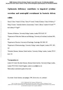
NIH-PA Author Manuscript NIH-PA Author Manuscript Figure 5.
NIH-PA Author Manuscript
EGFR internalization is decreased by activating mutations in NSCLC cells and MEFs. Rate constants for EGF-mediated internalization of EGFR (ke) were determined as described in Materials and Methods for: A, H1666 and H3255 NSCLC cells; B, EgfrWT/WT (WT/WT), EgfrWT/L858R (WT/858), and EgfrL858R/L858R (858/858) MEFs; and C, EgfrWT/WT MEFs and D, Shp2fl/− MEFs transduced with empty vector (EV), EgfrWT (WT) or EgfrL858R (858). Data are presented as average ± SEM for the three sets of biological replicates.
Cancer Res. Author manuscript; available in PMC 2010 November 1.
Lazzara et al.
Page 16
NIH-PA Author Manuscript NIH-PA Author Manuscript Figure 6.
NIH-PA Author Manuscript
Impaired EGFR internalization in HeLa cells decreases SHP2 and ERK phosphorylation and increases gefitinib-mediated death. A and B, HeLa cells with tetracycline-regulated dynamin expression were cultured without tetracycline and stimulated with 10 ng/mL EGF for up to 1 hr, and lysates were analyzed by Western blotting. Densitometry data are presented as average ± SEM for three sets of biological replicates. C and D, DynWT and DynK44A HeLa cells cultured with or without tetracycline were treated with up to 20 μM gefitinib for 72 hrs and analyzed for propidium iodide permeability. Data are presented as average ± SEM for the three sets of biological replicates.
Cancer Res. Author manuscript; available in PMC 2010 November 1.