Jul 19, 2010 - Department of Infectious Diseases, Huashan Hospital, Shanghai Medical College, Fudan University, Shanghai, China3. Received 19 July ...
JOURNAL OF VIROLOGY, Dec. 2010, p. 12850–12861 0022-538X/10/$12.00 doi:10.1128/JVI.01499-10 Copyright © 2010, American Society for Microbiology. All Rights Reserved.
Vol. 84, No. 24
Impairment of Hepatitis B Virus Virion Secretion by Single-Amino-Acid Substitutions in the Small Envelope Protein and Rescue by a Novel Glycosylation Site䌤 Kiyoaki Ito,1† Yanli Qin,1‡§ Michael Guarnieri,1‡¶ Tamako Garcia,1储 Karen Kwei,1# Masashi Mizokami,2 Jiming Zhang,3 Jisu Li,1 Jack R. Wands,1 and Shuping Tong1* Liver Research Center, Rhode Island Hospital, Warren Alpert School of Medicine, Brown University, Providence, Rhode Island1; Research Center for Hepatitis and Immunology, National Center for Global Health and Medicine, Ichikawa, Japan2; and Department of Infectious Diseases, Huashan Hospital, Shanghai Medical College, Fudan University, Shanghai, China3 Received 19 July 2010/Accepted 21 September 2010
Mutations in the S region of the hepatitis B virus (HBV) envelope gene are associated with immune escape, occult infection, and resistance to therapy. We previously identified naturally occurring mutations in the S gene that alter HBV virion secretion. Here we used transcomplementation assay to confirm that the I110M, G119E, and R169P mutations in the S domain of viral envelope proteins impair virion secretion and that an M133T mutation rescues virion secretion of the I110M and G119E mutants. The G119E mutation impaired detection of secreted hepatitis B surface antigen (HBsAg), suggesting immune escape. The R169P mutant protein is defective in HBsAg secretion as well and has a dominant negative effect when it is coexpressed with wild-type envelope proteins. Although the S domain is present in all three envelope proteins, the I110M, G119E, and R169P mutations impair virion secretion through the small envelope protein. Conversely, coexpression of just the small envelope protein of the M133T mutant could rescue virion secretion. The M133T mutation could also overcome the secretion defect caused by the G145R immune-escape mutation or mutation at N146, the site of N-linked glycosylation. In fact, the M133T mutation creates a novel N-linked glycosylation site (131NST133). Destroying this site by N131Q/T mutation or preventing glycosylation by tunicamycin treatment of transfected cells abrogated the effect of the M133T mutation. Our findings demonstrate that N-linked glycosylation of HBV envelope proteins is critical for virion secretion and that the secretion defect caused by mutations in the S protein can be rescued by an extra glycosylation site. The hepatitis B virus (HBV) is an enveloped DNA virus with a tropism for the liver. The 3.2-kb HBV genome harbors genes encoding core protein and its secreted version (called HBeAg), DNA polymerase, the transcriptional transactivator HBx, and envelope proteins. The envelope gene, which is completely overlapped by the polymerase gene, has three in-frame AUG codons that can serve as alternative translation initiation sites. This leads to the expression of three coterminal envelope proteins: large (L), middle (M), and small (S). The sequence unique to the L protein is called the pre-S1 domain, while a downstream sequence shared with the M protein is called the pre-S2 domain. The S domain is present in all three envelope proteins. The S and M proteins are translated from a 2.1-kb subgenomic RNA with a heterogeneous 5⬘ end, while the L
* Corresponding author. Mailing address: Liver Research Center, 4th Floor, 55 Claverick Street, Providence, RI 02906. Phone: (401) 444-7365. Fax: (401) 444-2939. E-mail: Shuping_Tong_MD@Brown .edu. † Present address: Research Center for Hepatitis and Immunology, National Center for Global Health and Medicine, Ichikawa, Japan. ‡ These authors contributed equally to this work. § Present address: Department of Infectious Diseases, Huashan Hospital, Shanghai Medical College, Fudan University, Shanghai, China. ¶ Present address: University of Colorado at Denver, Denver, CO. 储 Present address: Uniformed Services University of the Health Sciences, Washington, DC. # Present address: Boston University School of Medicine, Boston, MA. 䌤 Published ahead of print on 29 September 2010.
protein is expressed from a longer (2.4-kb) subgenomic RNA. The S protein is the most abundantly expressed envelope protein. The L and S proteins exist in nonglycosylated and monoglycosylated forms (L protein, p39 and gp42, respectively; S protein, p24 and gp27, respectively) due to a facultative Nlinked glycosylation site (N-X-S/T) at N146 of the S domain. The M protein contains an extra, constitutive N-linked glycosylation site at position 4 in the pre-S2 domain and consequently exists in monoglycosylated (gp33) and diglycosylated (gp36) forms. HBV genome replication involves a single 3.5-kb terminal redundant transcript, the pregenomic RNA. It serves not only as the messenger for both core protein and DNA polymerase but also as the precursor to the genomic DNA. The newly synthesized core protein self-assembles into the core particle, packaging one molecule each of the pregenomic RNA and DNA polymerase. Inside the core particle the DNA polymerase copies the minus-strand DNA from the RNA template and then degrades the RNA and synthesizes the plus-strand DNA using the minus-strand DNA as the template. Core particles with such a double-stranded DNA genome (with variable degree of plus-strand elongation) are enveloped and secreted as 42-nm virions. The L and S proteins are essential for virion formation, while the M protein is dispensable, although its presence enhances the efficiency of virion secretion (2, 6, 7). The majority of the S and M proteins are secreted alone as the noninfectious subviral particles of 22 nm and detected serologically as hepatitis B surface antigen (HBsAg). The immu-
12850
VOL. 84, 2010
S GENE MUTATIONS MODULATE HBV SECRETION
12851
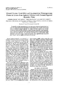
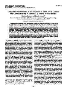
FIG. 1. (A) Impacts of amino acid substitutions in the S domain of viral envelope proteins on virion secretion. The corresponding nucleotide changes and amino acid changes in DNA polymerase, if any, are also shown. The mutations originated from three patient-derived HBV clones. (B) The four amino acid changes of the S domain are located in the immunodominant loops shown in circles (I110M, G119E, and M133T) and a transmembrane segment depicted as boxes (R169P). The secondary structure of the immunodominant loops is maintained by two disulfide bonds: C124OC137 and C139OC147. N146 is the conventional site of N-linked glycosylation. The M133T mutation creates a novel glycosylation site at N131. Shaded circles, cysteine residues. (C) HBV DNA constructs used in this study. The 1.5-mer construct is replication competent but rendered unable to express envelope proteins (L⫺ M⫺ S⫹). The 0.7-mer construct will express all three envelope proteins but can be made to express just the L/M proteins or the S protein. Mutations were introduced into the 0.7-mer construct. replication⫺, replication incompetent.
nodominant loops (residues 107 to 149) within the S domain are exposed on the virion surface, and its ␣ determinant (residues 124 to 147) (Fig. 1B) is the primary target of neutralizing antibodies that arise during natural infection or following vaccination. The recombinant yeast (Saccharomyces cerevisiae)-derived S protein has successfully been used as the HBV vaccine to prevent HBV transmission. However, in 1990 a case of HBV transmission from a carrier mother to her infant, despite a combination of active and passive immunizations, was reported (4). DNA sequencing revealed a G145R mutation within the ␣ determinant which renders the mutant HBsAg poorly recognizable by neutralizing antibodies directed against the wild-type (WT) virus (38). Single-amino-acid substitutions at this and other positions of the ␣ determinant or surrounding sequence that impair HBsAg recognition have since been detected in up to 5% of the vaccine recipients worldwide (5, 13, 14). Such immune-escape mutants have also been detected in liver transplant recipients who were reinfected with HBV, despite prophylaxis with monoclonal antibodies against HBsAg (3, 8, 25, 30, 34), as well as in occult HBV infection, which is defined by the presence of HBV DNA, despite a lack of detectable HBsAg (9, 12, 16, 39). Besides the immune-escape mutants, mutations in the S gene have also been linked to antiviral drug resistance. Nucleos(t)ide analogues currently licensed to treat chronic HBV infection target DNA polymerase, and prolonged therapy is associated with the selection of amino acid changes in the DNA
polymerase that confer drug resistance (22, 42). Due to the overlap between the polymerase and envelope genes, drugresistant mutants can be accompanied by amino acid changes in the envelope proteins as well. So far, only a small fraction of the mutations associated with immune escape or drug resistance has been functionally characterized. Notably, the classic immune-escape mutant with the G145R mutation was reported to be severely impaired in virion secretion (17, 18). We previously performed transfection experiments on 25 full-length genotype A HBV genomes isolated from HBeAgpositive French patients and identified clone 4B to be highly efficient in virion secretion, clone 4C from the same patient to be defective in virion secretion, and clone 3.4 from another patient to have poor virion secretion capacity (27). Through the analysis of chimeric constructs and mutants constructed by site-directed mutagenesis (site-directed mutants), we identified mutations in the envelope gene responsible for impaired virion secretion. As summarized in Fig. 1A, three missense mutations in the S domain of viral envelope proteins could abolish or drastically reduce virion secretion: I110M (clones 4B and 4C), G119E (clone 3.4), and R169P (clone 4C). In contrast, an M133T mutation (clone 4B) could enhance virion secretion when it was present alone and rescue virion secretion when it was combined with the I110M or G119E mutation. All these mutations except R169P are located in the immunodominant loops; R169P is located in a transmembrane segment (Fig. 1B). In the present study, we explored the molecular mechanisms
12852
ITO ET AL.
J. VIROL.
whereby the missense mutations in the envelope gene modulate virion secretion. Our results highlight the importance of N-linked glycosylation of envelope proteins for HBV virion secretion. MATERIALS AND METHODS The 1.5-mer and 0.7-mer HBV constructs and site-directed mutants. The 1.5-mer HBV construct has a 4.8-kb EcoRV-ApaI fragment (nucleotides 1044 to 3221/1 to 2600) of the HBV genome cloned to the pBluescript vector. As already described (7), the envelope-null mutant (L⫺ M⫺ S⫺) has the S gene start codon converted to GCG to prevent S protein expression and an additional G261A nonsense mutation at codon 36 of the S gene to prevent expression of full-length L and M proteins (Fig. 1C). The 0.7-mer construct has a 2.3-kb fragment covering nucleotides 2721 to 3221/1 to 1770 cloned into the pBluescript vector (7). It could express all three envelope proteins (L⫹ M⫹ S⫹) but not any other HBV proteins. Converting the S gene start codon into GCG rendered the 0.7-mer construct unable to express S protein (L⫹ M⫹ S⫺), while converting the 43rd codon of the pre-S2 region into a TAG stop codon truncated both L and M proteins (L⫺ M⫺ S⫹) (Fig. 1C). Missense mutations were introduced into the 0.7-mer construct by overlap-extension PCR (20, 35). Plasmid DNA of the mutant constructs was purified by use of a high-speed plasmid midikit (Qiagen). Transient transfection and analysis of DNA replication and virion secretion. Huh7 human hepatoma cells were seeded in six-well plates and transfected with LT1 (Mirus) as described previously (7, 15, 35). Each well of the six-well plates received 2 g HBV DNA and 5 ng of cDNA encoding secreted alkaline phosphatase (SEAP) to serve as a control for transfection efficiency. Cells and culture supernatant were harvested at day 5 posttransfection. To abolish protein glycosylation, medium was changed 2 days posttransfection, with one well each supplemented with 20 l of dimethyl sulfoxide (DMSO; final concentration, 1%) or tunicamycin dissolved in DMSO (final concentrations, 50 g/ml tunicamycin and 1% DMSO). Cells and culture supernatant were harvested at day 5 posttransfection. Cell lysis, core particle precipitation, and detection of HBV DNA replication by Southern blot analysis have been described previously (7, 20, 35). To concentrate virions from culture supernatant, antibodies were conjugated to protein G agarose beads (Roche) at a ratio of 2.8 l of horse anti-HBs (Ad/Ay; Abcam or Novus), 3 l of mouse anti-pre-S2 antibody (Virogen), and a 10-l bed volume of beads. Next, 10 l of conjugated beads was incubated at 4°C for 2 days with 1.4 ml of precleared culture supernatant. The precipitate was digested with nucleases prior to virion DNA extraction and Southern blot analysis (7, 35). Western blot analysis of cell-associated and secreted envelope proteins. Proteins from the cell lysate were separated by SDS-polyacrylamide gel and transferred to a membrane. Viral envelope proteins were detected by a horse or rabbit polyclonal anti-HBs antibody (Novus) as described previously (7). The blots were stripped and probed again for glyceraldehyde-3-phosphate dehydrogenase (GAPDH) to serve as a loading control (7). For in vitro deglycosylation of envelope proteins, 20 l of cell lysate was boiled with 2 l of denaturing buffer (5% SDS, 0.4 M dithiothreitol) at 100°C. The samples were digested at 37°C with 40 U of endo--N-acetylglucosaminidase H (endo H; New England Biolabs) prior to Western blot analysis. Secreted HBsAg was concentrated from 1.5 ml of culture supernatant by ultracentrifugation at 46,000 rpm for 16 h using an AH65 rotor (Sorvall). The pellet was resuspended in protein loading buffer, followed by Western blot analysis. Measurement of SEAP and secreted HBsAg. SEAP was measured from 5 l of culture supernatant using a Great EscAPe SEAP reporter system (Clontech). HBsAg was detected from 4 l of precleared culture supernatant using an Auszyme monoclonal HBsAg kit (Abbott Laboratories) or 10 to 15 l of culture supernatant using a hepatitis B surface antigen enzyme-linked immunosorbent assay (ELISA) kit (Abazyme) when the Abbott kit was discontinued.
RESULTS Rationale. The goal of the present study was to establish the molecular mechanisms whereby mutations in the envelope protein gene impair or restore virion secretion. The following specific questions were asked. First, do amino acid changes in envelope proteins or DNA polymerase or nucleotide changes in the pregenomic RNA alter virion secretion? In this regard, the I110M, G119E, M133T, and R169P substitutions in the envelope proteins are caused by the T484G, G510A, T552C,
and G660C point mutations in the viral genome, respectively (Fig. 1A), with the T484G mutation further inducing an S466A substitution in the DNA polymerase. Second, if mutations in the envelope proteins impair virion secretion, is the mutant L, M, or S protein responsible? Third, considering that the S protein is the morphogenic factor for the assembly of both virions and subviral particles, is the phenotype of virion secretion linked to the phenotype of HBsAg secretion? Finally, what is the mechanism whereby the M133T mutation rescues virion secretion of the I110M and G119E mutants? To answer these questions, we used a 1.5-mer replication construct with the wild-type sequence at nucleotide positions 484, 510, 552, and 660 as a source of genome replication. Its S gene AUG was mutated to GCG to prevent S protein expression; an additional nonsense mutation at the 5⬘ end of the S gene prevented expression of full-length L and M proteins. By cotransfecting this envelope-null replication construct with a 0.7-mer expression construct for the envelope proteins (wild type or mutant) (Fig. 1C), we could test the effect of mutant envelope proteins on virion secretion without the complication of mutant pregenomic RNA or DNA polymerase. Furthermore, by employing two 0.7-mer constructs for envelope protein expression, one for the L and M proteins and the other for the S protein, we could establish whether mutations in the L/M proteins or the S protein alter virion secretion. The I110M, G119E, and R169P mutations in the envelope gene are sufficient to impair virion secretion. The human hepatoma cell line Huh7 grown in six-well plates was cotransfected with 1.5 g of the 1.5-mer replication construct and 0.5 g of the 0.7-mer expression construct for the WT envelope proteins or the I110M, G119E, or R169P mutant. Cells were harvested at day 5 posttransfection, and core particles were extracted for Southern blot analysis of replicative HBV DNA. Similar levels of HBV DNA were detected, although the R169P mutant displayed little mature (double-stranded) viral DNA (Fig. 2A, lane 10). Considering that naked core particles are also released from transiently transfected Huh7 cells (35), we selectively immunoprecipitated virions from culture supernatant using a combination of monoclonal antibody against the pre-S2 domain and a polyclonal anti-HBs antibody. This approach also minimizes inefficient precipitation of the immune-escape mutants such as G119E (see below). The results shown in Fig. 2B indicate that virion secretion is blocked by the I110M and R169P mutations and severely impaired by the G119E substitution in viral envelope proteins, despite the absence of the T484G, G660C, and G510A mutations in the pregenomic RNA and the S466A substitution in DNA polymerase. The G119 and R169 mutations impair HBsAg detection and secretion, respectively. Besides the naturally occurring I110M, G119E, and R169P mutations, we also tested artificial mutations at these positions to shed light on the secretion defect. The I110D, I110G, and I110T mutations did not impair virion secretion as completely as the naturally occurring I110M sequence did (Fig. 2B, lanes 2 to 5). Among the mutants with mutations at position 119, the G119V mutant displayed more a severe defect in virion secretion than the G119E mutant, whereas the G119A mutant had less impairment (Fig. 2B, lanes 6 to 9). The G119E mutant showed much less secreted HBsAg than the G119T and G119V mutants according to the ELISA method but not according to Western blot analysis of
VOL. 84, 2010
S GENE MUTATIONS MODULATE HBV SECRETION
12853
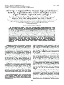
FIG. 2. Impacts of naturally occurring and artificial mutations at S domain positions 110, 119, and 169 on virion secretion and HBsAg secretion and detection. Huh7 cells in six-well plates were cotransfected with 1.5 g of the 1.5-mer replication construct (L⫺ M⫺ S⫺) and 0.5 g of the 0.7-mer envelope protein expression construct (L⫹ M⫹ S⫹). (A and B) Southern blots of replicative HBV DNA from cell lysate (A) and virion DNA from culture supernatant (B). Double-stranded (DS) and single-stranded (SS) DNA forms are indicated. A mixture of 3.2-kb and 1.7/1.5-kb HBV DNAs served as size markers. (C) Western blot analysis of viral envelope proteins in cell lysate using horse and rabbit polyclonal antibodies. The two size forms of the L and S proteins are indicated. (D) Detection of GAPDH from the same blot. (E) Western blot analysis of S protein in culture supernatant following concentration by ultracentrifugation. (F) ELISA of HBsAg from culture supernatant (Abbott kit). Results are expressed as optical density at a wavelength of 450 nm.
HBsAg precipitated by ultracentrifugation (Fig. 2E and F, lanes 6, 8, and 9). This result suggests that G119E is an immune-escape mutant, at least against the monoclonal antibody used in the ELISA. For an unknown reason, the intracellular S protein of all the G119 mutants was poorly recognized by the horse polyclonal antibody, albeit not by the rabbit antibody used in Western blot analysis (Fig. 2C, lanes 6 to 9; compare the upper and lower panels). Among the R169 mutants, the R169G and R169L mutants were similar to the R169P mutant in possessing little doublestranded replicative DNA and in being deficient in the secretion of both virions and HBsAg, whereas the R169H and R169Q mutants showed selective impairment or block of virion secretion (Fig. 2A, B, E, and F, lanes 10 to 14). This result suggests that virion formation or secretion is more sensitive to structural changes in envelope proteins than in subviral particles. These three mutants and the I110M mutant also showed much reduced levels of intracellular L protein (Fig. 2C, lanes 2, 10, 11, and 13).
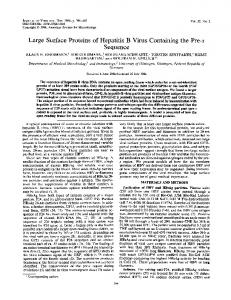
The R169P and R169G mutants manifest dominant negative effects on virion and HBsAg secretion. During natural HBV infection, coexistence of wild-type and secretion-defective mutant genomes may lead to the rescue of the mutant or suppression of the wild-type virus. We therefore conducted cotransfection experiments of the I110M, G119E, G119V, R169P, R169G, and R169Q mutants with the wild-type construct at 1:3, 1:1, and 3:1 ratios. The R169P and R169G mutants showed the strongest dominant negative effects, because they abolished virion secretion even at a 1:1 ratio with the WT construct (Fig. 3B, lane 13; Fig. 3E, lane 9). They also inhibited HBsAg secretion, although to lesser extents (Fig. 3C and F). The other mutants were more efficiently rescued by the wild-type virus (Fig. 3B and E). Mutations in the S protein rather than L/M proteins modulate virion secretion. The I110M, G119E, M133T, and R169P mutations are all located in the S domain, which is present on all three envelope proteins. We generated separate 0.7-mer expression constructs for S protein and L/M proteins (Fig. 1C)
12854
ITO ET AL.
J. VIROL.
FIG. 3. Impact of coexpressing wild-type and mutant envelope proteins on virion secretion. Huh7 cells were transfected with 1.5 g of the 1.5-mer replication construct and 0.5 g of the WT or mutant envelope protein expression construct, or a combination, at the indicated ratios. Lane 16, mock-transfected cells. (A and D) Replicative DNA. (B and E) virion DNA. Lane 1, HBV DNA size markers of 3.2, 1.7, and 1.5 kb. (C and F) ELISA of secreted HBsAg using the Abazyme kit.
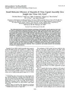
to identify the mutant envelope protein(s) responsible for the virion secretion defect. The L/M proteins harboring the I110M, G119E, or R169P mutation supported efficient virion secretion when they were cotransfected with the wild-type S protein construct, whereas the S protein harboring such mutations failed to reconstitute virion secretion in conjunction with the wild-type L/M proteins (Fig. 4B, compare lanes 4 to 6 with lanes 7 to 9). Consistent with these observations, virion secretion of the I110M mutant (with mutant L, M, and S proteins) could be rescued by supplementing S protein of wildtype origin and could be rescued more efficiently by the S protein of the M133T mutant (Fig. 4E, lanes 8 and 9). In contrast, supplementing the corresponding L/M proteins failed to rescue virion secretion of the I110M mutant (Fig. 4E, lanes 6 and 7). Taken together, the I110M, G119E, R169P, and M133T mutations all exert their effects through the S protein. Moreover, the M133T mutation could work in trans, i.e., from another molecule of the S protein. The M133T mutation also rescues virion secretion of the G145R and R169H mutants and creates a novel N-linked glycosylation site. Introduction of the M133T mutation fully rescued virion secretion of the I110M mutant (Fig. 5B, lane 6) and partially rescued the G119E mutant (Fig. 6B, lane 8). Although it failed to rescue the R169P mutant, it greatly improved the virion secretion of an R169H mutant (Fig. 6B, lanes 11 and 13). Furthermore, M133T could efficiently rescue virion secretion of the classic immune-escape mutant, G145R (Fig. 5B, lane 17). Western blot analysis revealed that the presence of the M133T mutation is associated with additional envelope proteins of higher molecular weights (Fig. 4G, 5C, and 6C). In this regard, M133T is predicted to create a novel N-linked glycosylation site at N131 (131NST133), which should produce additional forms of S (gp30), M (gp39), and L (gp45) proteins. Indeed, endo H treatment of cell lysate simplified the envelope proteins of all the constructs into p24, p30, and p39, corre-
sponding to nonglycosylated S, M, and L proteins, respectively (Fig. 6F). The M133T mutation works through the novel N-linked glycosylation site. A series of experiments were performed to ascertain whether the novel N-linked glycosylation site (N131) or the M133T mutation per se improves the efficiency of virion secretion. We found that the M133I and M133Q mutations failed to significantly improve virion secretion of the I110M mutant (Fig. 5B, lanes 4 and 5) or the G119E mutant (Fig. 6B, lane 6). In contrast, an M133S mutation, which generates the same glycosylation site as M133T, partially rescued the G119E mutant (Fig. 6B and C, lanes 7). Similarly, an artificial G112N mutation creating another novel N-linked glycosylation site (112NST114) could partially rescue the G119E mutant (Fig. 6B and C, lanes 5), although not the I110M or G145R mutant (Fig. 5B, lanes 3 and 19). Importantly, the ability of the M133T mutation to rescue virion secretion of the mutants with the I110M, G119E, G145R, and R169H mutations could be abrogated by an additional N131T or N131Q mutation (Fig. 5B, lanes 7, 8, and 18; Fig. 6B, lanes 9 and 14). As expected, none of the triple mutants expressed extra bands of L (gp45), M (gp39), and S (gp30) proteins (Fig. 5C and 6C). An alternative approach to prevent the M133T-mediated glycosylation at N131 is to treat transfected cells with tunicamycin, which will also block N-linked glycosylation of N146 (for all the constructs) and N112 (for the G112N mutant). Cells were treated with 50 g/ml of tunicamycin at 2 days posttransfection when cells were confluent, because tunicamycin added at earlier time points or a higher concentration was associated with significant cell toxicity. Since tunicamycin was dissolved in DMSO solution, cells treated with same volume of DMSO (final concentration, 1%) served as a control. DMSO is known to maintain the differentiation status of primary duck hepatocytes and trigger differentiation of HepaRG cells, leading to susceptibility to HBV infection (10, 31). DMSO treat-
VOL. 84, 2010
S GENE MUTATIONS MODULATE HBV SECRETION
12855
FIG. 4. Mutations in the S protein impair or rescue virion secretion. (Left panels) Huh7 cells were cotransfected with the replication construct (1.2 g), L and M protein (LM) construct (0.2 g), and S protein construct (0.6 g), with the I110M, G119E, and R169P mutations being present in either the LM or S construct. (Right panels) Huh7 cells were transfected with 1.4 g of the replication construct together with one or two expression constructs, with the DNA amount (in g) indicated. The WT and the I110M and M133T mutants expressing L/M/S proteins are simply designated WT, I110M, and M133T, respectively. (A and D) Replicative DNA from cell lysate; (B and E) virion DNA; (C and F) ELISA for secreted HBsAg (Abbott kit for panel C and Abazyme kit for panel F); (G and H) Western blot analysis of intracellular envelope proteins (G) and GAPDH (H).
ment increased the signals of virion DNA for all the secretioncompetent constructs, including the wild type and the M133T, I110M/M133T, M133T/G145R, M133T/N146Q, M133T/N146S, and M133T/R169H mutants (Fig. 7B and 8B). Considering that DMSO treatment also increased the amount of intracellular replicative DNA for some constructs, albeit to lesser extents (Fig. 7A and 8A), it remains to be determined whether DMSO directly enhances virion secretion efficiency. Treatment of Huh7 cells with 5 g/ml of tunicamycin dissolved in the same volume of DMSO markedly diminished virion secretion for the M133T single mutant or the mutant with the M133T mutation combined with the I110M, G145R, N146Q, N146S, or R169 mutation (Fig. 7B and 8B, lanes 10, 13, and 16). Western blot analysis of cell lysate confirmed complete or nearly complete inhibition of envelope protein glycosylation for the M133T mutants but an incomplete block for the wild-type construct (Fig. 7C and 8C). Since N131 is the only potential site for glycosylation for the M133T/N146S and M133T/N146Q double mutants, the marked effect of tunicamycin treatment on virion secretion of these two con-
structs (Fig. 8B, compare lanes 9, 10, 12, and 13) confirms that the M133T mutation improves virion secretion through glycosylation at N131. For many constructs, especially those with two N-linked glycosylation sites (N131 and N146), tunicamycin treatment markedly increased the amount of secreted HBsAg, according to the ELISA (Fig. 7E, lanes 10 and 13; Fig. 8E, lanes 16 and 19). It also increased the intracellular S protein level of the M133T mutants, according to Western blot analysis (Fig. 7C and 8C). This is probably due to the unmasking of the antigenic site when glycosylation was prevented. Finally, cells treated with tunicamycin released much less SEAP than cells treated with DMSO alone (Fig. 7F and 8F). This is consistent with tunicamycin being a strong inducer of endoplasmic reticulum (ER) stress, which triggers global translational repression. HBV virion secretion requires at least one N-linked glycosylation site and could be enhanced by an extra site. The finding that the M133T mutation rescues virion secretion through the creation of a novel N-linked glycosylation site
12856
ITO ET AL.
J. VIROL.
FIG. 5. Modulation of virion secretion of the I110M and G145R mutants by the G112 and M133 mutations. The 1.5-mer replication construct (1.5 g) was cotransfected with 0.5 g of expression construct of single, double, or triple mutant, as indicated. (A) Replicative DNA; (B) virion DNA; (C) intracellular envelope proteins; (D) GAPDH as a loading control; (E) ELISA for secreted HBsAg (Abazyme kit for lanes 1 to 14 and Abbott kit for lanes 15 to 20).
raised the question of the function of the conventional Nlinked glycosylation site, N146. We therefore generated N146Q and N146S mutants, which as expected expressed only single-size forms of the L (p39), M (gp33), and S (p24) proteins (Fig. 9C, lanes 2 and 3). Both mutants were defective in virion secretion (Fig. 9B). Introduction of the M133T mutation, which restores N-linked glycosylation, albeit at a different position, efficiently rescued virion secretion (Fig. 9B, lanes 4 and 5). To establish whether N146 also contributes to virion secretion of the I110M/M133T, M133T/G145R, and M133T/R169H mutants, we abolished this site by use of an N146Q mutation. The loss of the N146 glycosylation site moderately reduced virion secretion of the I110M/M133T mutant, severely impaired virion secretion of the M133T/R169H mutant, and completely abolished virion secretion of the M133T/G145R mutant (Fig. 9B, lanes 7, 11, and 9). Therefore, the N146 glycosylation site is necessary, although insufficient alone, for efficient virion secretion of the I110M, G119E, and R169H mutants. DISCUSSION Among the three envelope proteins, only L and S are essential for HBV virion formation/secretion (2, 6). Since the S protein but not the L protein is also required for the secretion of subviral particles (HBsAg), it is the critical factor for particle assembly. The L protein suppresses the secretion of subvi-
ral particles in a dose-dependent manner (26, 29), thus channeling a fraction of the S protein toward virion formation. Moreover, the L protein directly interacts with core particles via its pre-S domains. We previously characterized several patient-derived HBV clones and mapped the determinant for virion secretion to the S region of the envelope gene (20, 27). In the present study, we used a transcomplementation assay to confirm that the I110M, G119E, M133T, and R169P amino acid changes in the S domain modulate the efficiency of virion secretion. Triple-transfection experiments established the mutant S protein, rather than the L or M protein, as the determinant for altered virion secretion. Consistent with our findings, mutant S protein was found by other investigators to cause a virion secretion defect of the G145R immune-escape mutant and W172* mutant (conversion of a tryptophan codon into a stop codon) associated with adefovir resistance (17, 36). These findings, together with the HBsAg secretion defect of the R169P mutant, reinforce the S protein as the driving force in particle formation and secretion. The exact mechanisms whereby the I110M, G119E, and R169P mutations impair virion secretion remain to be worked out. Residues 110 and 119 are located inside the ER lumen during virion morphogenesis, while residue 169 is membrane associated (Fig. 1B). Since none of these residues are accessible for interaction with the core particle located in the cytosol, they probably affect a later step of S protein cross-linking
VOL. 84, 2010
S GENE MUTATIONS MODULATE HBV SECRETION
12857
FIG. 6. The M133T mutation rescues virion secretion of the G119E and R169H mutants and creates a novel N-linked glycosylation site. The 1.5-mer replication construct (1.5 g) was cotransfected with 0.5 g of the expression construct of the G119E, R169P, and R169H mutant, with or without additional mutations at positions 112, 131, and 133. (A) Replicative DNA; (B) virion DNA; (C) intracellular envelope proteins; (D) intracellular GAPDH; (E) ELISA for secreted HBsAg using the Abazyme kit; (F) effect of endo H treatment on envelope protein migration. The cell lysate was either treated with endo H (lanes ⫹) or not treated (lanes ⫺) prior to gel electrophoresis. p39, p30, and p24 denote nonglycosylated L, M, and S proteins, respectively.
essential for particle assembly. Indeed, the R169P, R169G, and R169L mutations blocked secretion of both virions and subviral particles. The continued HBsAg secretion for the other mutants with a virion secretion defect (I110M, G119E, G119V, and R169Q) may reflect less dramatic structural changes and a better tolerance of subviral particles, which use a different route for secretion (37). The I110M, G145R, R169P, R169G, and R169L mutants all displayed reduced levels of L protein.
Thus, reduced L/S protein ratios may partly contribute to the impaired virion secretion of these mutants. From the same figures, it is also evident that the G145R and R169L mutants had a lower ratio of gp27/p24, suggesting less efficient S protein glycosylation. HBsAg secreted by the G119E mutant was poorly detected by the ELISA. Therefore, G119E represents an immune-escape mutant. In this regard, G119 is juxtaposed to a cysteine
12858
ITO ET AL.
J. VIROL.
FIG. 7. Impacts of tunicamycin treatment on envelope protein glycosylation and virion secretion of the WT virus and the I110M, M133T, I110M/M133T, M133T/G145R, and N146Q mutants. Huh7 cells in six-well plates were cotransfected with the replication construct (1.5 g), expression construct (0.5 g), and SEAP plasmid (5 ng) in triplicate. Medium was changed 2 days posttransfection, with one well being supplemented with 20 l of DMSO and another well being supplemented with 20 l of tunicamycin (5 mg/ml stock solution in DMSO). Cells and culture supernatant were harvested 3 days later. (A) Replicative DNA; (B) virion DNA; (C) intracellular envelope proteins detected by the rabbit and horse antibodies; (D) GAPDH as a loading control; (E) ELISA for secreted HBsAg (Abazyme kit); (F) SEAP activities as a measurement of ER stress.
residue (C121). The charged glutamic acid probably interferes with the maintenance of a proper intramolecular disulfide bond between C121 and another cysteine residue, thus altering the conformation of the ␣ determinant. Indeed, a G119R mutation has been associated with occult HBV infection (41). Several R169 mutants share similarities with the W172* mutant, which has the C-terminal 55 residues of the S domain deleted (36). These include a block in the secretion of both viral and subviral particles (R169P/G/L), reduced intracellular
levels of L protein (R169P/G/L) and the glycosylated form of S protein (R169L), reduced intracellular levels of the mature (double-stranded) genome (R169P/G/L), and a dominant negative effect on virion secretion (R169P/G). We hypothesize that core particles with a mature genome could be enveloped for R169P/G/L mutants, but such particles are degraded intracellularly, thus depleting both L protein and mature genome. Why the lack of S protein secretion in the case of either the R169P/G/L or W172* mutant (36) fails to cause intracellular
VOL. 84, 2010
S GENE MUTATIONS MODULATE HBV SECRETION
12859
FIG. 8. Impact of tunicamycin treatment on envelope protein glycosylation and virion secretion of the G112N/G119E, M133T/N146Q, M133T/N146S, and M133T/R169P mutants. Huh7 cells in six-well plates were cotransfected with the replication construct (1.5 g), expression construct (0.5 g), and SEAP plasmid (5 ng) in triplicate. Medium was changed 2 days posttransfection, with one well each being supplemented with 20 l of DMSO or tunicamycin (5 mg/ml stock solution in DMSO). Cells and culture supernatant were harvested 3 days later. (A) Replicative DNA; (B) virion DNA; (C) intracellular envelope proteins detected by the rabbit and horse antibodies; (D) GAPDH as a loading control; (E) ELISA for secreted HBsAg (Abazyme); (F) SEAP activities as a measurement of ER stress.
retention remains to be elucidated. The W172* mutant can transform NIH 3T3 cells and has been found in patients with hepatocellular carcinoma (21). It will be interesting to determine whether the R169 mutants, which share many properties
with W172* but which probably do not have the transactivation function of truncated envelope proteins, can promote malignant transformation. The M133T mutants showed reduced levels of both intra-
12860
J. VIROL.
ITO ET AL.
FIG. 9. Requirement of at least one N-linked glycosylation site for virion secretion. The replication construct (1.5 g) was cotransfected with 0.5 g of the expression construct. (A) Replicative DNA; (B) virion DNA; (C) intracellular envelope proteins; (D) ELISA for secreted HBsAg using the Abazyme kit.
cellular S protein (on the basis of Western blot analysis) and secreted HBsAg (on the basis of ELISA). This is most likely due to interference with antibody binding caused by extra polysaccharides, because this phenotype was reversed by either an N131 mutation to destroy the new glycosylation site or tunicamycin treatment of transfected cells to prevent glycosylation at this site. A similar phenotype has been reported for other mutations that create novel N-linked glycosylation sites (40). The M133T mutation efficiently rescues virion secretion of the G145R mutant without altering its immune-escape phenotype (Fig. 5E), thus arguing against a mechanism through restoration of S protein folding or intramolecular interactions mediated by disulfide bonds. Consistent with this finding, the M133T mutation could rescue virion secretion of the I110M mutant when it was present on a different molecule of the S protein. The M133T mutation probably restores correct intermolecular interactions mediated by disulfide bonds (24). Increased virion secretion as a result of the M133T mutation was correlated with moderate reduction of double-stranded DNA inside cells (Fig. 5A, lanes 6 and 13; Fig. 7A, lane 3). Our interpretation is that the M133T mutation increases the envelopment and export of core particles with a mature genome, thus depleting the intracellular pool.
The ability of the M133T mutation to rescue virion secretion of a wide range of mutants (I110M, G145R, N146Q, N146S, R169H, and, to a lesser extent, G119E) argues for a general mechanism of action. Three types of evidence suggest the novel N-linked glycosylation site (most likely in the S protein) as the basis for rescue. First, the M133S mutation but not the M133I or M133Q mutation could achieve a similar effect, and the rescuing capacity of M133T was abrogated by an additional N131T/Q mutation. Second, either an N146Q or an N146S mutation, which prevents N-linked glycosylation of the S and L proteins of wild-type HBV, could block the secretion of HBV virion particles, albeit not subviral particles. Such a secretion defect could be overcome by introduction of the M133T mutation. Third, virion secretion of the M133T/N146S or M133T/ N146Q mutant was markedly reduced by tunicamycin treatment, which nearly completely abolished glycosylated forms of the S protein for these mutants. Secretion of subviral particles was not reduced by tunicamycin treatment. Similarly, others reported that tunicamycin dose-dependently inhibited virion secretion of wild-type HBV from a HepG2 cell line but not the secretion of subviral particles (1, 23, 28, 32). We observed a modest reduction in virion secretion for the wild-type virus (Fig. 8B, lanes 3 and 4), possibly because of an incomplete block of the glycosylation of the wild-type envelope proteins at the concentration used (Fig. 8C, lane 3). Taken together, our findings and those of earlier studies by others demonstrate a critical role of N-linked glycosylation for HBV virion secretion but not for the secretion of subviral particles. In this regard, N-linked glycans mediate interaction with lectin chaperones such as calnexin and calreticulin to guarantee proper folding of the glycoprotein (11). Interestingly, the potential N-linked glycosylation site in the duck hepatitis B virus envelope protein is inefficiently used, suggesting a different mechanism for virion formation (33). In conclusion, immune-escape mutants, such as G119E and G145R, and other mutants, such as I110M and R169P, are impaired in virion secretion. A novel N-linked glycosylation site created by the M133T mutation could rescue virion secretion of the immune-escape mutants without restoring virus recognition by the neutralizing antibodies. Therefore, combination of these mutations will restore viral fitness under the selective pressure of neutralizing antibodies. Indeed, the M133T mutation is found to be highly prevalent in HBsAgnegative patients (12). In patients infected with the T126I or G145K immune-escape mutant, viral persistence correlated with the simultaneous emergence of the M133T mutation (19). This scenario is reminiscent of HBV resistance to nucleotide or nucleoside therapy, where the primary mutation in the P gene reduces viral replication capacity, which is nevertheless compensated for by a second-site mutation that arises later (42). In short, the plasticity of the HBV genome poses a serious challenge to our efforts to prevent and treat HBV infection.
ACKNOWLEDGMENTS We thank Dechun Feng for his help with figure preparation. This work was supported by American Cancer Society Research Scholar Award RSG 06-059-01-MBC to S.T., NIH grant CA109733 to J.L., and NIH grants AA08169 and CA123544 to J.R.W.
VOL. 84, 2010
S GENE MUTATIONS MODULATE HBV SECRETION REFERENCES
1. Block, T. M., X. Lu, F. M. Platt, G. R. Foster, W. H. Gerlich, B. S. Blumberg, and R. A. Dwek. 1994. Secretion of human hepatitis B virus is inhibited by the imino sugar N-butyldeoxynojirimycin. Proc. Natl. Acad. Sci. U. S. A. 91: 2235–2239. 2. Bruss, V., and D. Ganem. 1991. The role of envelope proteins in hepatitis B virus assembly. Proc. Natl. Acad. Sci. U. S. A. 88:1059–1063. 3. Carman, W. F., C. Trautwein, F. J. van Deursen, K. Colman, E. Dornan, G. McIntyre, J. Waters, V. Kliem, R. Muller, H. C. Thomas, and M. P. Manns. 1996. Hepatitis B virus envelope variation after transplantation with and without hepatitis B immune globulin prophylaxis. Hepatology 24:489–493. 4. Carman, W. F., A. R. Zanetti, P. Karayiannis, J. Waters, G. Manzillo, E. Tanzi, A. J. Zuckerman, and H. C. Thomas. 1990. Vaccine-induced escape mutant of hepatitis B virus. Lancet 336:325–329. 5. Cooreman, M. P., M. H. van Roosmalen, R. te Morsche, C. M. Sunnen, E. M. de Ven, J. B. Jansen, G. N. Tytgat, P. L. de Wit, and W. P. Paulij. 1999. Characterization of the reactivity pattern of murine monoclonal antibodies against wild-type hepatitis B surface antigen to G145R and other naturally occurring “a” loop escape mutations. Hepatology 30:1287–1292. 6. Fernholz, D., P. R. Galle, M. Stemler, M. Brunetto, F. Bonino, and H. Will. 1993. Infectious hepatitis B virus variant defective in pre-S2 protein expression in a chronic carrier. Virology 194:137–148. 7. Garcia, T., J. Li, C. Sureau, K. Ito, Y. Qin, J. Wands, and S. Tong. 2009. Drastic reduction in the production of subviral particles does not impair hepatitis B virus virion secretion. J. Virol. 83:11152–11165. 8. Ghany, M. G., B. Ayola, F. G. Villamil, R. G. Gish, S. Rojter, J. M. Vierling, and A. S. Lok. 1998. Hepatitis B virus S mutants in liver transplant recipients who were reinfected despite hepatitis B immune globulin prophylaxis. Hepatology 27:213–222. 9. Grethe, S., M. Monazahian, I. Bohme, and R. Thomssen. 1998. Characterization of unusual escape variants of hepatitis B virus isolated from a hepatitis B surface antigen-negative subject. J. Virol. 72:7692–7696. 10. Gripon, P., S. Rumin, S. Urban, J. Le Seyec, D. Glaise, I. Cannie, C. Guyomard, J. Lucas, C. Trepo, and C. Guguen-Guillouzo. 2002. Infection of a human hepatoma cell line by hepatitis B virus. Proc. Natl. Acad. Sci. U. S. A. 99:15655–15660. 11. Helenius, A., and M. Aebi. 2001. Intracellular functions of N-linked glycans. Science 291:2364–2369. 12. Hou, J., Z. Wang, J. Cheng, Y. Lin, G. K. Lau, J. Sun, F. Zhou, J. Waters, P. Karayiannis, and K. Luo. 2001. Prevalence of naturally occurring surface gene variants of hepatitis B virus in nonimmunized surface antigen-negative Chinese carriers. Hepatology 34:1027–1034. 13. Hsu, H. Y., M. H. Chang, S. H. Liaw, Y. H. Ni, and H. L. Chen. 1999. Changes of hepatitis B surface antigen variants in carrier children before and after universal vaccination in Taiwan. Hepatology 30:1312–1317. 14. Hsu, H. Y., M. H. Chang, Y. H. Ni, H. H. Lin, S. M. Wang, and D. S. Chen. 1997. Surface gene mutants of hepatitis B virus in infants who develop acute or chronic infections despite immunoprophylaxis. Hepatology 26:786–791. 15. Ito, K., K. H. Kim, A. S. Lok, and S. Tong. 2009. Characterization of genotype-specific carboxyl-terminal cleavage sites of hepatitis B virus e antigen precursor and identification of furin as the candidate enzyme. J. Virol. 83:3507–3517. 16. Jeantet, D., I. Chemin, B. Mandrand, A. Tran, F. Zoulim, P. Merle, C. Trepo, and A. Kay. 2004. Cloning and expression of surface antigens from occult chronic hepatitis B virus infections and their recognition by commercial detection assays. J. Med. Virol. 73:508–515. 17. Kalinina, T., A. Iwanski, H. Will, and M. Sterneck. 2003. Deficiency in virion secretion and decreased stability of the hepatitis B virus immune escape mutant G145R. Hepatology 38:1274–1281. 18. Kalinina, T., A. Riu, L. Fischer, H. Will, and M. Sterneck. 2001. A dominant hepatitis B virus population defective in virus secretion because of several S-gene mutations from a patient with fulminant hepatitis. Hepatology 34: 385–394. 19. Kato, J., K. Hasegawa, N. Torii, K. Yamauchi, and N. Hayashi. 1996. A molecular analysis of viral persistence in surface antigen-negative chronic hepatitis B. Hepatology 23:389–395. 20. Khan, N., M. Guarnieri, S. H. Ahn, J. Li, Y. Zhou, G. Bang, K. H. Kim, J. R. Wands, and S. Tong. 2004. Modulation of hepatitis B virus secretion by naturally occurring mutations in the S gene. J. Virol. 78:3262–3270. 21. Lai, M. W., and C. T. Yeh. 2008. The oncogenic potential of hepatitis B virus rtA181T/ surface truncation mutant. Antivir. Ther. 13:875–879. 22. Lok, A. S., F. Zoulim, S. Locarnini, A. Bartholomeusz, M. G. Ghany, J. M. Pawlotsky, Y. F. Liaw, M. Mizokami, and C. Kuiken. 2007. Antiviral drug-
23.
24.
25.
26. 27.
28.
29.
30.
31.
32.
33.
34.
35.
36.
37.
38.
39.
40.
41.
42.
12861
resistant HBV: standardization of nomenclature and assays and recommendations for management. Hepatology 46:254–265. Lu, X., A. Mehta, R. Dwek, T. Butters, and T. Block. 1995. Evidence that N-linked glycosylation is necessary for hepatitis B virus secretion. Virology 213:660–665. Mangold, C. M., F. Unckell, M. Werr, and R. E. Streeck. 1995. Secretion and antigenicity of hepatitis B virus small envelope proteins lacking cysteines in the major antigenic region. Virology 211:535–543. McMahon, G., P. H. Ehrlich, Z. A. Moustafa, L. A. McCarthy, D. Dottavio, M. D. Tolpin, P. I. Nadler, and L. Ostberg. 1992. Genetic alterations in the gene encoding the major HBsAg: DNA and immunological analysis of recurrent HBsAg derived from monoclonal antibody-treated liver transplant patients. Hepatology 15:757–766. Ou, J. H., and W. J. Rutter. 1987. Regulation of secretion of the hepatitis B virus major surface antigen by the preS-1 protein. J. Virol. 61:782–786. Parekh, S., F. Zoulim, S. H. Ahn, A. Tsai, J. Li, S. Kawai, N. Khan, C. Trepo, J. Wands, and S. Tong. 2003. Genome replication, virion secretion, and e antigen expression of naturally occurring hepatitis B virus core promoter mutants. J. Virol. 77:6601–6612. Patzer, E. J., G. R. Nakamura, and A. Yaffe. 1984. Intracellular transport and secretion of hepatitis B surface antigen in mammalian cells. J. Virol. 51:346– 353. Persing, D. H., H. E. Varmus, and D. Ganem. 1986. Inhibition of secretion of hepatitis B surface antigen by a related presurface polypeptide. Science 234:1388–1391. Protzer-Knolle, U., U. Naumann, R. Bartenschlager, T. Berg, U. Hopf, K. H. Meyer zum Buschenfelde, P. Neuhaus, and G. Gerken. 1998. Hepatitis B virus with antigenically altered hepatitis B surface antigen is selected by high-dose hepatitis B immune globulin after liver transplantation. Hepatology 27:254–263. Pugh, J. C., and J. W. Summers. 1989. Infection and uptake of duck hepatitis B virus by duck hepatocytes maintained in the presence of dimethyl sulfoxide. Virology 172:564–572. Sheu, S. Y., and S. J. Lo. 1994. Biogenesis of the hepatitis B viral middle (M) surface protein in a human hepatoma cell line: demonstration of an alternative secretion pathway. J. Gen. Virol. 75(Pt 11):3031–3039. Swameye, I., and H. Schaller. 1997. Dual topology of the large envelope protein of duck hepatitis B virus: determinants preventing pre-S translocation and glycosylation. J. Virol. 71:9434–9441. Terrault, N. A., S. Zhou, R. W. McCory, T. L. Pruett, J. R. Lake, J. P. Roberts, N. L. Ascher, and T. L. Wright. 1998. Incidence and clinical consequences of surface and polymerase gene mutations in liver transplant recipients on hepatitis B immunoglobulin. Hepatology 28:555–561. Tsai, A., S. Kawai, K. Kwei, D. Gewaily, A. Hutter, D. R. Tong, J. Li, J. R. Wands, and S. Tong. 2009. Chimeric constructs between two hepatitis B virus genomes confirm transcriptional impact of core promoter mutations and reveal multiple effects of core gene mutations. Virology 387:364–372. Warner, N., and S. Locarnini. 2008. The antiviral drug selected hepatitis B virus rtA181T/sW172* mutant has a dominant negative secretion defect and alters the typical profile of viral rebound. Hepatology 48:88–98. Watanabe, T., E. M. Sorensen, A. Naito, M. Schott, S. Kim, and P. Ahlquist. 2007. Involvement of host cellular multivesicular body functions in hepatitis B virus budding. Proc. Natl. Acad. Sci. U. S. A. 104:10205–10210. Waters, J. A., M. Kennedy, P. Voet, P. Hauser, J. Petre, W. Carman, and H. C. Thomas. 1992. Loss of the common “A” determinant of hepatitis B surface antigen by a vaccine-induced escape mutant. J. Clin. Invest. 90:2543– 2547. Weinberger, K. M., T. Bauer, S. Bohm, and W. Jilg. 2000. High genetic variability of the group-specific a-determinant of hepatitis B virus surface antigen (HBsAg) and the corresponding fragment of the viral polymerase in chronic virus carriers lacking detectable HBsAg in serum. J. Gen. Virol. 81:1165–1174. Wu, C., X. Zhang, Y. Tian, J. Song, D. Yang, M. Roggendorf, M. Lu, and X. Chen. Biological significance of amino acid substitutions in hepatitis B surface antigen (HBsAg) for glycosylation, secretion, antigenicity and immunogenicity of HBsAg and hepatitis B virus replication. J. Gen. Virol. 91:483– 492. Yuan, Q., S. H. Ou, C. R. Chen, S. X. Ge, B. Pei, Q. R. Chen, Q. Yan, Y. C. Lin, H. Y. Ni, C. H. Huang, A. E. Yeo, J. W. Shih, J. Zhang, and N. S. Xia. Molecular characteristics of occult hepatitis B virus from blood donors in southeast China. J. Clin. Microbiol. 48:357–362. Zoulim, F., and S. Locarnini. 2009. Hepatitis B virus resistance to nucleos(t)ide analogues. Gastroenterology 137:1593–1608.