Implantation of Mouse Embryonic Stem Cell-Derived Cardiac Progenitor Cells Preserves Function of Infarcted Murine Hearts Nicolas Christoforou1*., Behzad N. Oskouei2., Paul Esteso3, Christine M. Hill3, Jeffrey M. Zimmet4, Weining Bian1, Nenad Bursac1, Kam W. Leong1, Joshua M. Hare2, John D. Gearhart3 1 Department of Biomedical Engineering, Pratt School of Engineering, Duke University, Durham, North Carolina, United States of America, 2 Miller School of Medicine, University of Miami, Miami, Florida, United States of America, 3 Institute for Regenerative Medicine, University of Pennsylvania, Philadelphia, Pennsylvania, United States of America, 4 Interventional Cardiology, University of California San Francisco, San Francisco, California, United States of America
Abstract Stem cell transplantation holds great promise for the treatment of myocardial infarction injury. We recently described the embryonic stem cell-derived cardiac progenitor cells (CPCs) capable of differentiating into cardiomyocytes, vascular endothelium, and smooth muscle. In this study, we hypothesized that transplanted CPCs will preserve function of the infarcted heart by participating in both muscle replacement and neovascularization. Differentiated CPCs formed functional electromechanical junctions with cardiomyocytes in vitro and conducted action potentials over cm-scale distances. When transplanted into infarcted mouse hearts, CPCs engrafted long-term in the infarct zone and surrounding myocardium without causing teratomas or arrhythmias. The grafted cells differentiated into cross-striated cardiomyocytes forming gap junctions with the host cells, while also contributing to neovascularization. Serial echocardiography and pressure-volume catheterization demonstrated attenuated ventricular dilatation and preserved left ventricular fractional shortening, systolic and diastolic function. Our results demonstrate that CPCs can engraft, differentiate, and preserve the functional output of the infarcted heart. Citation: Christoforou N, Oskouei BN, Esteso P, Hill CM, Zimmet JM, et al. (2010) Implantation of Mouse Embryonic Stem Cell-Derived Cardiac Progenitor Cells Preserves Function of Infarcted Murine Hearts. PLoS ONE 5(7): e11536. doi:10.1371/journal.pone.0011536 Editor: Arnold Schwartz, University of Cincinnati, United States of America Received February 27, 2010; Accepted June 16, 2010; Published July 12, 2010 Copyright: ß 2010 Christoforou et al. This is an open-access article distributed under the terms of the Creative Commons Attribution License, which permits unrestricted use, distribution, and reproduction in any medium, provided the original author and source are credited. Funding: This work was funded with resources from the Institute for Cell Engineering at Johns Hopkins University. National Heart, Lung and Blood Institute grant HL083342 to N.B. The funders had no role in study design, data collection and analysis, decision to publish, or preparation of the manuscript. Competing Interests: The authors have declared that no competing interests exist. * E-mail:
[email protected] . These authors contributed equally to this work.
cardiac progenitor cells (CPCs) on the basis of Brachyury/Flk1 [11], Isl1/Flk1/Nkx2-5 [12], cKit/Nkx2-5 [13], or Nkx2-5 [14] expression. These cells represent a promising source for heart repair as they have the restricted capacity to differentiate into cardiac muscle, smooth muscle, and vascular endothelium [11,12,13,14]. In this study we hypothesized that mouse ESCderived CPCs will exert functional improvement after myocardial infarction primarily through their multipotential differentiation capacity as well as through the formation of stable and integrated grafts within the host myocardium. We found that when co-cultured with neonatal rat ventricular cardiomyocytes (NRMVs), the CPCs differentiated into cardiomyocytes, formed gap junctions with the rat cells, and supported electrical propagation over a centimeter-scale distance. Temporal assessment performed as long as one month after injection into the infarcted region of the murine myocardium, demonstrated that the CPCs engrafted and differentiated into cardiomyocytes, as well as contributed to neovascularization in the infarcted region. The differentiated cardiomyocytes also formed gap junctions with the host myocardium. The animals that received the CPCs demonstrated significantly improved cardiac function as assessed by echocardiography and pressure/volume (PV) loop analysis. No teratoma formation was observed following cell transplantation.
Introduction Cardiomyocyte loss as a result of myocardial infarction (MI) injury is considered irreversible with the heart lacking sufficient capacity for self-regeneration. Cell-based cardiac therapies are proposed as an attractive therapeutic alternative to reverse cardiomyocyte loss, repair the injured myocardium and ultimately prevent heart failure. To date, a variety of cell sources, both of adult and embryonic origin, have been investigated for use in heart repair with mixed outcomes [1,2]. The use of adult cells is attractive because of their immunocompatible nature, ease of isolation, restricted differentiation potential, and capacity to proliferate rapidly. However, inadequate potential for cardiac differentiation or integration with host cells, limits the benefit of these cells mainly to their paracrine action. On the other hand, embryonic stem cells (ESCs) are able to differentiate into relatively large numbers of early stage cardiomyocytes that functionally integrate with host heart cells [3,4,5]. While ESC-derived cardiomyocytes have been successfully applied for the treatment of myocardial infarction in animal models [6,7,8,9], their clinical application is currently hampered by their neoplastic and immunogenic potential [10]. We and others have recently described the identification, isolation, and characterization of the novel mouse ESC-derived PLoS ONE | www.plosone.org
1
July 2010 | Volume 5 | Issue 7 | e11536
Progenitor Cell Heart Therapy
junctions indicated by punctuate Connexin 43 localization at the site of cell-cell contacts were detected in the differentiated cardiomyocytes (Fig. 2d). However, unlike the NRVMs that expressed Connexin 43 in relatively long plaques at the sites of cell contacts, the CPC-derived cardiomyocytes expressed Connexin 43 sporadically and irregularly. Optical mapping in all co-cultures revealed spontaneous electrical activity at a relatively low rate (1.260.3 Hz) that originated in the CPC islands, suggesting the existence of pacemaking cells amongst the CPC-differentiated cardiomyocytes. Consistent with positive staining for Connexin 43, both spontaneous and pacing-induced action potentials (2 Hz rate) within the CPC island propagated outwards into the surrounding NRVM area (Fig. 2e). Action potentials also propagated into the CPC island when initiated from the surrounding NRVM area (data not shown). The apparent velocity of electrical propagation inside the CPC island was significantly lower as compared to that of the surrounding NRVMs (1.660.2 vs. 16.761.5 cm/s) and dependent on the efficiency of CPC differentiation into cardiomyocytes. Specifically, lower numbers of sparsely distributed cardiomyocytes within the central island yielded significantly lower velocity of propagation (0.460.1 cm/s, Fig. S1g).
Results Isolation and in vitro characterization of mouse ESCderived CPCs The mouse ESC lines D3 [15] and Rosa26 [16] were stably transfected with DNA constructs allowing the expression of the green fluorescent protein (GFP) under the control of the mouse cardiac specific enhancer element of the Nkx2-5 transcription factor as previously described [14]. Following isolation of 50 colonies (clonal) for each cell line, stably transfected clones were identified and further used based on their capacity to express GFP selectively in the spontaneously contracting cardiomyocyte cell clusters. Mouse ESCs were induced to differentiate in suspension forming aggregates termed embryoid bodies (EBs) and initial detection of GFP coincided with initiation of Nkx2-5 expression on differentiation day 5 (Figs. 1a, b). Temporal quantitative RT-PCR analysis performed on differentiating ESCs indicated a time period (days 5–6) during which the CPCs were present but not yet committed into specific cell lineages (Fig. 1g). Prior to initiation of Nkx2-5 expression, coinciding with cardiac progenitor induction, the detection of Brachyury transcripts (day 4) indicated the formation of nascent mesoderm. By day 7, the CPCs underwent differentiationcommitment into cardiac cell lineages including cardiomyocytes as indicated by the initiation of Mhc6 expression along with the detection of spontaneously contracting cell clusters. The initial number and percentage of GFP(+) CPCs decreased temporally between days 5 and 10 (Fig. 1h). This observation, which has been previously reported [13], may be a result of proliferation of other cell types, epigenetic silencing of the specific Nkx2-5 enhancer element used in this set of experiments, and/or differentiation towards the vascular and smooth muscle endothelium cell lineages which downregulate Nkx2-5. To assay the differentiation capacity of the CPCs, following isolation of the GFP(+) progenitor population, the cells were reaggregated and the cell clusters were induced to differentiate for 7–30 days. Several foci of GFP(+) spontaneously contracting cardiomyocytes (a-Actinin expression) were detected in culture (Figs. 1c, d). The multi differentiation potential of the CPCs was determined by demonstrating cell specific expression of cardiac aActinin, endothelial Von-Willebrand factor, and smooth muscle actin in the differentiating clusters (Figs. 1e, f). This was also confirmed by the RT-PCR analysis (previously demonstrated [14]). No colonies of undifferentiated ESCs were detected as confirmed by the absence of Pou5f1 and Nanog expression.
CPC injection in the mouse infracted myocardium The potential of CPCs to treat myocardial infarction injury was assessed by evaluating their ability to form stable grafts in the host myocardium, differentiate into lineage-committed cells, form in vivo gap junctions, and ultimately improve the functional output of the injured hearts. To determine the extent of injury and scar tissue formation in the infarcted hearts, we performed Mason’s trichrome staining on thin heart sections that allowed us to distinguish healthy muscle tissue (red) from scar tissue (blue) (Fig. 3). The control group that underwent sham surgery displayed only minor scarring which could be attributed to injury sustained during the saline injections (Figs. 3a, b). The hearts that underwent LAD ligation and received only saline injections sustained massive infarcts that encompassed almost the entire left ventricle (including apex) and exhibited thin scarred walls and extensive remodeling (Figs. 3c, d). In contrast, the animals that underwent LAD ligations and received CPCs displayed smaller zones of scar tissue formation with decreased cardiac remodeling (Figs 3e–h). While a few hearts displayed minor tissue scarring (Figs. 3e, f) similar to that observed in the sham hearts, others displayed a certain degree of wall thinning and remodeling (Figs. 3g, h). In this case, however, the scar was localized only around the ligation site. To assess the engraftment potential and distribution of the transplanted cells irrespective of their lineage differentiation phenotype within the myocardium we compared parallel serial tissue cross-sections pre-treated either with Mason’s trichrome stain or anti b-Galactosidase, which is constitutively expressed in the Rosa26-derived CPCs (Figs. 3i, l). b-Gal(+) cells were detected within the infarcted region in an area comprising a thin muscle band underneath the scar tissue (Fig. 3i), borders between the scar tissue and healthy muscle (Figs. 3j, k), as well as within the healthy muscle in the vicinity (Fig. 3l) but not remote (not shown) from the scar. These results suggested that the transplanted cells successfully engrafted at all three injection areas including the center of the infarct. Anti b-Galactosidase-specific staining was also used in order to determine the survival rate of the transplanted cells one month post intramyocardial delivery. bGal(+) cells was detected and summed in multiple sections both within and in the surrounding area of the myocardial infarction region. The survival rate was
Determination of the electrocoupling capacity of CPCs The potential of CPC derived cardiomyocytes to form functional gap junctions was determined by utilizing a previously described in vitro NRVM co-culture assay [17,18] which allows the observation of action potential propagation by the means of optical mapping over a macroscopic (3 cm2) culture area [18]. In particular, the CPCs were selectively seeded at high density in the central region of pre-masked coverslips with the NRVMs surrounding them (Fig. 2a). The co-cultures exhibited distinct border between the CPCs and the NRVMs as assayed by GFP and a-Actinin expression (Figs. 2a, b). Immunostaining for cardiomyocyte, smooth muscle and endothelial markers after 14 days of co-culture demonstrated that the majority of the CPCs differentiated into cardiomyocytes (Fig. 2c) with limited smooth muscle or endothelial cell differentiation (data not shown). On the other hand, CPCs plated at low density in the central region of the coverslips formed few patches of cardiomyocytes with the majority of cells differentiating into smooth muscle cells (Fig. S1). Gap PLoS ONE | www.plosone.org
2
July 2010 | Volume 5 | Issue 7 | e11536
Progenitor Cell Heart Therapy
Figure 1. Derivation and in vitro characterization of mouse ESC-derived CPCs. ESCs were induced to differentiate through 3D aggregation and embryoid body formation. Clusters of CPCs, located within the embryoid body, expressed GFP under the control of the Nkx2-5 enhancer element (a–b). GFP(+) CPCs isolated by FACS sorting differentiated exclusively into cardiomyocytes (a-Actinin) while retaining GFP expression (c–d). The CPCs differentiated in vitro into cardiomyocytes (a-Actinin), smooth muscle (Smooth muscle actin), and endothelial cells (Von Willebrand factor) (e–f). Temporal quantitative RT-PCR analysis for the nascent mesoderm marker Brachyury, the cardiac progenitor marker Nkx2-5, and a cardiomyocyte marker Mhc6 (g). The percentage of GFP(+) CPCs was determined temporally following induction of mouse ESC differentiation (h). doi:10.1371/journal.pone.0011536.g001
PLoS ONE | www.plosone.org
3
July 2010 | Volume 5 | Issue 7 | e11536
Progenitor Cell Heart Therapy
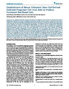
Figure 2. Structural and functional assessment of a NRVM monolayer with a central island comprised of differentiated CPCs. A GFP(+) CPC island (green) within the cardiac network (red) (a–b). Composite image and separate fluorescence channels showing the border between NRVMs and CPCs (c). Note that significant number of GFP positive cells are also a-Actinin positive (cardiomyocytes). Composite image and separate channels showing the Connexin 43 staining within the CPC island. While NRVMs were connected via relatively long Connexin 43(+) gap-junction plaques (data not shown), the gap-junctions in CPC differentiated cardiomyocytes appear small and irregular (d). Electrical propagation initiated inside the CPC island propagated into the surrounding cardiac network (e). Individual hexagonal frames denote 19.5 mm diameter recording area with membrane voltage snapshots shown at given times. Electrical stimulus (pulse sign) was applied in the center of the CPC island at time 0 ms. Membrane voltage is color coded from rest (blue) to peak (red). Circles denote 504 recordings sites. Dashed white line denotes the CPC island. Membrane potentials at selected sites within (1) and outside (2&3) the CPC island are also shown. Electrical stimulus (yellow triangle) yielded action potential propagation that was significantly slower within the island than in the surrounding cardiomyocytes. Isochrones of electrical propagation (white lines) are shown at the bottom right of. Note central isochrone crowding due to slow propagation within the CPC island (black dashed line). doi:10.1371/journal.pone.0011536.g002
PLoS ONE | www.plosone.org
4
July 2010 | Volume 5 | Issue 7 | e11536
Progenitor Cell Heart Therapy
PLoS ONE | www.plosone.org
5
July 2010 | Volume 5 | Issue 7 | e11536
Progenitor Cell Heart Therapy
Figure 3. Scar tissue formation and CPC detection. Histological analysis (Mason’s trichrome staining) and immunohistochemical analysis of the infarcted myocardium demonstrating the extent of scarring (blue) in the myocardium of the three experimental groups at two parallel cross-sections of the myocardium. Sham surgery and saline injections (a–b). Myocardial infarction and saline injections (c–d). Myocardial infarction and CPC injections (e–h). Scale bar: 1 mm. High magnification panel pairs show parallel thin cross-sections stained either with Mason’s trichrome stain or with an antibody against the b-Galactosidase protein which is constitutively expressed in the injected cells (Rosa 26 mouse ESC line). Scar region (i), Interface between the scar and healthy myocardium (j–l). Healthy myocardium (k). Black arrowheads denote b-Galactosidase(+) cells within the myocardium. doi:10.1371/journal.pone.0011536.g003
calculated by extrapolating the number of bGal(+) cells over the entire estimated area of the affected myocardium (