TRAIL (Apo-2L) and TRAIL Receptors in Human Placentas: Implications for Immune Privilege1 Teresa A. Phillips,* Jian Ni,§ Guohua Pan,¶ Steven M. Ruben,§ Ying-Fei Wei,§ Judith L. Pace,‡ and Joan S. Hunt2*† Mechanisms accounting for protection of the fetal semiallograft from maternal immune cells remain incompletely understood. In other contexts, interactions between TRAIL (TNF-related apoptosis-inducing ligand/Apo-2L) and its receptors kill activated lymphocytes. The purpose of this study was therefore to investigate the potential of the TRAIL/TRAIL-R system to protect the placenta against immune cell attack. Analysis by Northern blotting demonstrated mRNAs encoding TRAIL as well as the four TRAIL receptors (DR4, DR5, DcR1/TRID, DcR2/TRUNDD) in human placentas. Immunohistochemical experiments demonstrated that TRAIL protein is prominent in syncytiotrophoblast, an uninterrupted placental cell layer that is continuously exposed to maternal blood, as well as in macrophage-like placental mesenchymal cells (Hofbauer cells). Studies on cell lines representing trophoblasts (Jar, JEG-3 cells) and macrophages (U937, THP-1 cells) showed that both lineages contained TRAIL mRNA and that steady state levels of transcripts were increased 2- to 11-fold by IFN-g. By contrast, cell lineage-specific differences were observed in expression of the TRAIL-R genes. Although all four lines contained mRNA encoding the apoptosis-inducing DR5 receptor, only trophoblast cells contained mRNA encoding the DcR1 decoy receptor and only macrophages contained DcR2 decoy receptor transcripts. DR4 mRNA was present only in THP-1 cells and was the only TRAIL-R transcript increased by IFN-g. Cytotoxicity assays revealed that the two trophoblast cell lines were resistant, whereas the two macrophage lines were partially susceptible to killing by rTRAIL. Collectively, the results are consistent with a role for the TRAIL/TRAIL-R system in the establishment of placental immune privilege. The Journal of Immunology, 1999, 162: 6053– 6059.
T
hroughout pregnancy, the uterus and placenta are sites of immune privilege in which immunologic responses to genetically different fetal tissues are effectively thwarted (reviewed in Refs. 1– 4). The fetal cells that are directly exposed to maternal tissues and blood are trophoblast cells, a unique cell type derived from the trophectoderm layer of the implanted blastocyst. This cell layer completely surrounds and encases the embryo (5, 6). Multiple mechanisms protect trophoblast cells from immunologic rejection (1– 4). Fas ligand (FasL),3 one of the two apoptosisinducing members of the TNF superfamily known to be expressed in trophoblast (7, 8), is believed to be a major contributor to placental immune privilege (8). The TNF-related apoptosis-inducing ligand (TRAIL, also known as Apo-2L) is a newly identified TNF superfamily member with high homology to FasL (9 –12). TRAIL and FasL cooperate in limiting lymphocyte proliferation following activation and use
Departments of *Anatomy and Cell Biology, †Pathology and Laboratory Medicine, and ‡Molecular and Integrative Physiology, University of Kansas Medical Center, Kansas City, KS 66160; §Human Genome Sciences, Rockville, MD 20850; and ¶ Genentech, South San Francisco, CA 94080 Received for publication September 10, 1998. Accepted for publication February 19, 1999. The costs of publication of this article were defrayed in part by the payment of page charges. This article must therefore be hereby marked advertisement in accordance with 18 U.S.C. Section 1734 solely to indicate this fact. 1
This work was supported by grants from the National Institute of Child Health and Human Development to J.S.H. (HD29156 and HD24212), the Kansas Mental Retardation Research Center (HD02528), and the Kansas Reproductive Sciences P30 Center (HD33994). 2 Address correspondence and reprint requests to Dr. Joan S. Hunt, Department of Anatomy and Cell Biology, University of Kansas Medical Center, Kansas City, KS 66160-7400. E-mail address:
[email protected] 3 Abbreviations used in this paper: FasL, Fas ligand; MTT, 3-(4,5-dimethylthiazol2-yl)-2,5-diphenyltetrazolium bromide; hu, human; TRAIL, TNF-related apoptosisinducing ligand.
Copyright © 1999 by The American Association of Immunologists
some common pathways leading to apoptotic cell death (11–18). Unlike FasL and other apoptosis-inducing TNF family members, TRAIL transcripts are detectable in many normal organs and tissues, including human placentas (9, 10). In the TRAIL/TRAIL-R system, control over apoptosis relies on differential display of receptors (TRAIL-R). These include DR4 (TRAIL-R1) and DR5 (TRICK2/TRAIL-R2), which transduce apoptotic signals, and DcR1 (TRID/LIT/TRAIL-R3) and DcR2 (TRUNDD/TRAIL-R4), which lack functional death domains and act as decoys (19 –32). TRAIL-R are widely expressed, and Northern blots have detected messages encoding all four receptors in human placentas (19 –24, 30, 32). Because TRAIL expressed in trophoblast cells might contribute to immune privilege by killing activated lymphocytes and TRAIL-R expression would determine the vulnerability of trophoblast to killing by TRAIL, we undertook evaluation of this system in human placentas and cell lines. Specific transcripts were identified in human placentas, and TRAIL protein was immunolocalized to a restricted number of cell types, which included trophoblast and macrophages. We then documented TRAIL and TRAIL-R mRNAs in trophoblast and macrophage cell lines. Having learned that receptor expression differed in the two lineages, we compared their respective abilities to resist killing by TRAIL.
Materials and Methods Tissues and cell lines Sections of human first trimester (n 5 3) and term placentas (n 5 2) as well as extraplacental membranes (n 5 2) were obtained from elective pregnancy terminations and normal cesarean section deliveries, respectively, in accordance with a protocol approved by the Human Subjects Committee of the University of Kansas Medical Center (Kansas City, KS). The tissues were manually dissected and fixed in 4% paraformaldehyde-PBS overnight at 4°C, then blocked into paraffin at low temperature. The human trophoblast-derived choriocarcinoma cell lines, Jar and JEG-3, and the U937 0022-1767/99/$02.00
6054
TRAIL AND TRAIL RECEPTORS IN HUMAN PLACENTAS
myelomonocytic cells were purchased from the American Type Culture Collection (ATCC, Manassas, VA). The Wilkinson Laboratory for Cancer Research at the University of Kansas Medical Center kindly provided THP-1 monocyte/macrophage cells. The cell lines were cultured at 37°C in RPMI 1640 (Sigma-Aldrich, St. Louis, MO) supplemented with 10% FBS (Atlanta Biologicals, Norcross, GA) and antibiotics (Sigma-Aldrich) (growth medium).
probe for 1 h at 68°C, two 15-min low stringency washes (23 SSC, 0.1% SDS) at room temperature, one 30-min high stringency wash (0.13 SSC, 0.1% SDS) at 60°C, and a second 15-min high stringency wash at the same conditions. The blots were briefly rinsed in 23 SSC at room temperature before being sealed in heat-seal bags (KAPAK, Minneapolis, MN) and exposed to Hyperfilm MP (Amersham Life Science) in the presence of intensifying screens.
Immunoblot analysis
IFN-g treatment of cell lines
Human term placental protein (Human Placenta Protein Medley; Clontech Laboratories, Palo Alto, CA) was fractionated by standard 10% SDSPAGE (33) using a MiniPROTEAN II Dual Slab Cell (Bio-Rad Laboratories, Hercules, CA). The gels were electrophoretically transferred to nitrocellulose membranes (Schleicher & Schuell, Keene, NH) using the BioRad MiniTrans-Blot Cell. TRAIL was identified according to the manufacturer’s instructions using goat anti-human TRAIL (K-18) Ab (1 mg/ml), which recognizes amino acids 233–250 in the C-terminal region of TRAIL, and donkey anti-goat IgG-HRP secondary Ab, both from Santa Cruz Biotechnology (Santa Cruz, CA). Goat IgG from Sigma-Aldrich served as a control for nonspecific binding. The enhanced chemiluminescence substrate was SuperSignal (Pierce, Rockford, IL); detection was by exposure to Hyperfilm MP (Amersham Life Science, Arlington Heights, IL).
Immunohistochemistry The paraformaldehyde-fixed tissues were embedded in paraffin at low temperature, and two 5-micron sections were taken onto glass slides for analysis by immunohistochemistry, as previously described (7), with the following modifications. Following blocking with normal horse serum, the goat IgG anti-TRAIL primary Ab K-18 or control goat IgG (both at 15 mg/ml) was incubated with the tissues overnight at 4°C. Biotinylated horse anti-goat IgG (15 mg/ml; Vector Laboratories, Burlingame, CA) was incubated with the samples for 30 min at room temperature, and endogenous peroxidase was blocked after this step. For peptide inhibitions, primary Ab (10 mg/ml) was incubated with a 19-fold weight excess of peptide K-18 (Santa Cruz Biotechnology) for 1 h at room temperature. The mixtures were centrifuged and tissue sections were incubated with the supernatant as a replacement for the primary Ab. Following incubation with substrate, the tissues were lightly counterstained with hematoxylin, dried, and coverslipped for light microscopy.
Probes cDNAs encoding TRAIL, DR4, DR5, DcR1, and DcR2 were excised from their respective plasmid vectors using appropriate restriction enzymes. The linear fragments were resolved on 1% agarose gels and visualized by UV transillumination, and appropriately sized bands were eluted from the gel by centrifugation on GenElute Agarose Spin Columns (Sigma-Aldrich). Eluted linear ds cDNA was ethanol precipitated and dissolved in 10 mM Tris-HCl, pH 8, 1 mM EDTA. Probes from interior regions of the cDNAs for TRAIL (;550 bp encoding the extracellular soluble region (19)), DR4 (bp 739 –952, 214 bp), DcR1 (bp 217–566, 350 bp), and DcR2 (bp 573– 787, 215 bp) were generated by PCR and agarose gel purified, as described above.
RNA isolation and Northern blot analysis For Northern blot analysis, total RNA was prepared from untreated cultured cell lines (1–2 3 108 cells per preparation) using TRIzol (Life Technologies, Gaithersburg, MD) or TRI reagent (Sigma-Aldrich), according to the manufacturer’s protocol. Total RNA from human term placenta was purchased from Ambion (Austin, TX). RNA samples (8 –10 mg/lane) were resolved on 1% agarose/2 M formaldehyde gels. The gels were examined by UV transillumination and immediately blotted to Nytran using a TurboBlotter, as directed (Schleicher & Schuell). UV cross-linking was performed with a UV Stratalinker 1800 (Stratagene Cloning Systems, La Jolla, CA). Dry blots were either used immediately or sealed in heat-seal bags and stored at 4°C. Twenty-five nanograms of each cDNA or PCRgenerated probe were random-prime labeled with [a-32P]dCTP (3000 Ci/ mmol; ICN Pharmaceuticals, Costa Mesa, CA) using the Random Primers DNA Labeling System (Life Technologies), according to the manufacturer’s instructions. Unincorporated nucleotides were removed from labeled cDNA probes by centrifuging through Micro Bio-Spin 6, SSC Chromatography Columns (Bio-Rad Laboratories). Specific activity of labeled cDNA probes was determined by liquid scintillation, and 5 3 106 cpm/ml (4 ml total hybridization solution) was used for each hybridization. Hybridization was performed in a Hybaid oven using QuikHyb (Stratagene) hybridization solution essentially as recommended by the manufacturer: prehybridization for at least 20 min at 68°C, hybridization with labeled
Four 100-mm tissue culture dishes for each cell line were seeded and incubated at 37°C, 5% CO2 in humid air: the trophoblastic cell lines Jar and JEG-3 at 2 3 106 cells in 10 ml of growth medium 1 day before treatment and the monocytic cell lines U937 and THP-1 at 3 3 106 cells in 10 ml of growth medium 2– 4 h before treatment. A total of 100 ml of 1 3 104 U/ml of human rIFN-g (rhuIFN-g; Genzyme, Cambridge, MA) was added to two of the dishes for each line, and 100 ml of medium was added to the remaining two dishes (controls). Incubation was resumed for 24 h, at which time one medium control and one rhuIFN-g-treated culture dish for each cell line were removed for total RNA preparation using TRIzol, as described above. Total RNA was prepared from the remaining cultures after an additional 24 h (48 h total incubation time). Northern blot analysis was performed as described above. Gels were transilluminated and photographed with Polaroid 665 black and white film to obtain ethidium bromide-stained 28S and 18S bands (loading controls). Northern blot films and the Polaroid negatives were quantitatively analyzed using a Molecular Dynamics (Sunnyvale, CA) Personal Densitometer and ImageQuaNT software.
Cytotoxicity assays JEG-3, Jar, U937, THP-1, and HeLa human endometrial adenocarcinoma cells from ATCC were plated into 96-well microplates (0.1 ml/well, replicates of three wells) in medium containing 10% FBS. U937 and THP-1 cells were plated at 5 3 104 cells/well, and all other cells were plated at 1 3 104 cells/well. After overnight culture at 37°C, rTRAIL (20) was added to a final concentration of 0, 10, or 1000 ng/ml, and cultures were continued for 20 h. Mitochondrial enzyme activity was evaluated using an MTT kit from Promega (Madison, WI), following the manufacturer’s directions. Color intensity was determined spectrophotometrically at A570. Duplicate plates were established, and cytotoxicity was assessed by exclusion of the vital dye, trypan blue.
Nuclear fragmentation assay To determine whether the rTRAIL used in cytotoxicity assays killed by apoptosis, HeLa cells were seeded into eight-chamber Lab-Tek Tissue Culture Chamber Slides (Nunc International, Naperville, IL) at 3 3 105 cells/ well in 0.3 ml of culture medium and cultured overnight at 37°C. On the following day, 30 ml of culture medium (controls) or culture medium containing 1000 ng/ml of rTRAIL was added to each well for a final concentration of 100 ng/ml. The cells were incubated for 2 h at 37°C, then the slides were rinsed briefly in PBS, air dried, and fixed for 30 min in 1% paraformaldehyde at 4°C. The slides were washed in PBS and incubated in the dark at room temperature for 15 min with 0.4 mg/ml DAPI (49, 6-diamidino-2-phenylindole; Sigma) in PBS. The slides were rinsed briefly in PBS and mounted using Slow Fade Light Antifade glycerol mounting medium (Molecular Probes, Eugene, OR) and examined for nuclear fluorescence.
Results Localization of TRAIL protein in human placentas Previous studies have reported that human placentas contain TRAIL mRNA (9, 10). To determine whether the messages were translated and to identify the cells containing TRAIL protein, immunohistochemical experiments were performed. Specificity of a commercially available Ab to a human TRAIL-specific amino acid sequence was first verified by immunoblotting. As shown in Fig. 1, left panel, the goat anti-human TRAIL Ab detected a polypeptide of Mr ; 33,000 –34,000 (major band) as well as less prominent bands at Mr 5 42,000, Mr 5 32,000, and Mr 5 31,000. These results were generally consistent with previous reports on TRAIL protein (17). This same Ab was then used for immunohistochemical experiments.
The Journal of Immunology
6055
FIGURE 1. Immunodetection of TRAIL in human term placenta using a goat Ab to a human TRAILspecific peptide (left) and immunohistochemical localization of TRAIL protein in human placentas and membranes using the same Ab (right). In immunoblots of 60-mg samples of placental protein, the Ab detected a major band at approximately 33–34 kDa and additional bands at approximately 42, 32, and 31 kDa similar to those previously reported (17). In immunohistologic experiments, TRAIL was localized to cells in A, a first trimester placental villus that included syncytiotrophoblast (large arrows) and macrophage-like Hofbauer cells (small arrows and inset); C, first trimester decidual cells (arrowheads), but not leukocytes; and E, term amniochorion. In B, normal goat IgG was substituted for anti-TRAIL, and in D and F, the primary Ab was preincubated with specific peptide. A, amnion membrane; C, chorion membrane; D, decidua; FM, fetal mesenchyme; L, leukocytes. Original magnifications, A–D, 3200; E, F, 3100.
Fig. 1, right panel, shows that TRAIL in first trimester placentas was prominent in syncytiotrophoblast, where it was localized primarily to the apical brush border. Immunoreactive TRAIL was detected in villous stroma and stromal cells, particularly the round, highly vacuolated cells known as Hofbauer cells (placental macrophages), but was low to absent in fibroblastic mesenchymal cells and endothelial cells (Fig. 1A). Normal goat IgG did not bind to any cells in first trimester placentas (Fig. 1B), and these controls were also negative with all other tissues. Fig. 1C shows that large maternal decidual cells in first trimester tissues were TRAIL protein positive. Leukocytic aggregates identified as “L” in Fig. 1C and endothelial cells were TRAIL negative. Staining was essentially abolished by preincubating the anti-TRAIL reagent with specific peptide (Fig. 1D). In term placentas, immunoreactivity with anti-TRAIL was less intense. Positive signals were detected in both syncytiotrophoblast and macrophage-like mesenchymal cells (data not shown). In term extraplacental membranes, TRAIL protein was clearly evident in the amnion membrane (Fig. 1E) as well as in a few macrophagelike stromal cells located between the amnion and chorion membranes and maternal decidual cells. Chorionic cytotrophoblasts contained little or no TRAIL. Staining in the extraplacental membranes was completely abrogated by preincubating the primary Ab with specific peptide (Fig. 1F). These results confirmed translation of TRAIL messages in placentas and indicated that TRAIL is differentially expressed at the maternal-fetal interface, with synthesis probable in syncytiotrophoblast cells, placental macrophages, amnion epithelial cells, and maternal decidual cells.
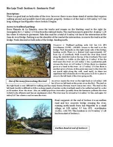
blastic cells, but steady state levels of TRAIL mRNA were reduced in monocytes/macrophages, with lower expression in U937 cells than in THP-1 cells. This and other TRAIL Northern blots used a PCR-generated (approximately 550-bp) DNA probe that identified sequences encoding the external soluble portion of the TRAIL protein. In all Northern analyses, equal loading of lanes and quality of RNA were verified by examining 18S and 28S rRNA content by ethidium bromide staining (Fig. 2, lower panel). These experiments supplied experimental support for the idea that both trophoblasts and placental macrophages transcribe the TRAIL gene. rhuIFN-g enhances TRAIL mRNA in trophoblast and macrophage cell lines Activation with Con A (T cells) and LPS (B cells) increases TRAIL expression in T and B lymphocytes, respectively (17). Macrophages are activated by IFN-g, and trophoblasts have some
Detection of TRAIL mRNA in human placentas and cell lines In accordance with previous reports (9, 10), Northern blots of term placental RNA demonstrated TRAIL transcripts migrating to a position on the gels corresponding to 2 kb (Fig. 2). We then used the same experimental method to test cell lines representing two of the major TRAIL protein-positive cell types identified by immunohistochemistry in human placentas, trophoblasts (Jar, JEG-3 cells), and monocytes/macrophages (U937, THP-1 cells). Fig. 2 shows that TRAIL transcripts were present in all of the cell lines. Transcripts were prominent in Jar and JEG-3 tropho-
FIGURE 2. Upper panel, Detection of 2-kb TRAIL mRNA in human term placenta (Placenta), human trophoblastic cell lines (Jar, JEG-3), and human monocytic cell lines (U937, THP-1) by Northern blot hybridization. A total of 10 mg of total RNA was loaded in each lane, as described in Materials and Methods. Lower panel, The matching ethidium bromidestained agarose gel demonstrates equal loading of samples. In both panels, locations for 28S and 18S rRNA are marked on the right.
6056
TRAIL AND TRAIL RECEPTORS IN HUMAN PLACENTAS
FIGURE 3. Northern blot hybridization experiments showing the effects of IFN-g on steady state levels of TRAIL mRNA in trophoblast cell lines (Jar, JEG-3) and macrophage cell lines (U937, THP-1). Cells were incubated with 100 U/ml of rhuIFN-g for 24 or 48 h. A total of 8 mg of total RNA was loaded in each lane, as described in Materials and Methods. The lower panels show ethidium bromide-stained agarose gels, which verify approximately equal loading of each lane.
macrophage-like characteristics (34). Therefore, we tested rhuIFN-g for effects on TRAIL mRNA in the cell lines. Jar, JEG-3, U937, and THP-1 cells were exposed to 100 U/ml of rhuIFN-g. RNA was harvested at 24 and 48 h, and Northern blot hybridization was used to analyze TRAIL mRNA. Fig. 3 shows that IFN-g efficiently enhanced TRAIL mRNA in all four cell lines, although the kinetics of enhancement varied. Equal loading was verified by examining 28S and 18S bands on ethidium bromide-stained agarose gels. Table I shows the means and SDs of three independent experiments, in which the results were analyzed quantitatively using scanning densitometry. Mean increases in steady state levels of TRAIL mRNA compared with 28S RNA varied from 2–11-fold at 24 h, and 2–3-fold at 48 h. Exposure for 24 h was maximal for Jar, JEG-3, and THP-1 cells, and levels declined at 48 h, whereas 3-fold increases were observed at both 24 and 48 h in U937 cells. No cell lineage-specific patterns were observed in these experiments.
Initially, full-length cDNAs encoding each of the four receptors were used to generate probes. Because the DNA sequences for the four receptors contain sizable regions of identity and similarity, there was some cross-detection, particularly of DR5 by the fulllength probes for DR4, DcR1, and DcR2. (The full-length cDNA probe for DR5 did not detect transcripts for the other three receptors, however.) To reduce this cross-detection, PCR was used to generate smaller probes for DR4, DcR1, and DcR2 from regions of less DNA sequence similarity. Identical specific transcript bands were detected by full-length cDNA and the PCR-generated probes. Cross-detection of DR5 transcript by probes for the other three
Transcripts for TRAIL receptors in placenta and cell lines and effects of IFN-g on steady state levels Potential target cells for placental TRAIL were then investigated by using Northern blot hybridization to detect receptor mRNA. Table I. Effect of IFN-g on TRAIL and TRAIL receptor mRNA levels IFN-g Treatment/Medium Controla mRNA
Cell Line
24 h
48 h
5.6 6 3.0 2.3 6 0.8 3.0 6 0.9 11.1 6 3.8
1.9 6 0.7 1.8 6 0.4 3.3 6 0.9 3.2 6 0.1
TRAIL
Jar JEG-3 U937 THP-1
DR4
THP-1, 5-kb band THP-1, 3-kb band
1.4 6 0.4 1.9 6 0.5
1.3 6 0.8 2.3 6 0.5
DR5
Jar JEG-3 U937 THP-1
1.2 6 0.2 1.0 6 0.2 0.8 6 0.1 1.2 6 0.1
1.1 6 0.1 1.0 6 0.1 1.0 6 0.2 1.4 6 0.3
DcR1
Jar, 5-kb band Jar, 3-kb band Jar, 1.5-kb band JEG-3, 5-kb band JEG-3, 3-kb band JEG-3, 1.5-kb band
1.1 6 0.2 1.0 6 0.2 0.9 6 0.2 1.0 6 0.2 1.0 6 0.1 1.0 6 0.1
1.1 6 0.4 1.1 6 0.4 1.1 6 0.4 1.4 6 0.6 1.2 6 0.5 1.3 6 0.6
U937 THP-1
0.9 6 0.1 0.8 6 0.2
0.7 6 0.1 0.7 6 0.1
DcR2
a The peak area of each transcript band was normalized to the peak area of the 28S ribosomal RNA band for that sample. The resulting value for IFN-g-treated cells at each time point was divided by the value for the medium-treated cells at the same time point. Means 6 SDs for three separate experiments are shown.
FIGURE 4. Detection of apoptosis-inducing TRAIL-R mRNA (DR4/ TRAIL-R1, DR5/TRICK2/TRAIL-R2) (A) and decoy TRAIL-R (DcR1/ TRID/LIT/TRAIL-R3, DcR2/TRUNDD/TRAIL-R4) in human placenta, trophoblastic cell lines (Jar, JEG-3), and human monocytic cell lines (U937, THP-1) by Northern blot hybridization (B). A total of 10 mg of total RNA was loaded in each lane, as described in Materials and Methods. The lower panels show ethidium bromide-stained agarose gels, which verify approximately equal loading of each lane. 28S and 18S rRNA are marked on the right.
The Journal of Immunology
6057
Table II. Results of MTT assays used to assess the cytotoxic effects of rTRAIL on human macrophage cell lines (U937, THP-1), human trophoblast cell lines (JEG-3, Jar), and endometrial adenocarcinoma (HeLa) cellsa TRAIL (ng/ml) Cell Line
Expt. No.
0
10
100
1000
U937
1 2
0 6 5.5b 0 6 2.3
13.6 6 2.4 15.5 6 4.2
30.4 6 4.3 32.0 6 1.7
NDc 34.2 6 0.4
THP-1
1 2
0 6 2.0 0 6 4.3
0.9 6 1.0 3.9 6 1.3
12.3 6 1.0 9.6 6 3.6
ND 24.3 6 2.5
Jar
1 2
0 6 2.9 0 6 1.4
0 6 3.1 2.4 6 2.2
0 6 5.5 2.6 6 1.9
ND 0.1 6 2.0
JEG-3
1 2
0 6 2.7 0 6 2.1
1.5 6 4.6 2.1 6 1.6
0 6 4.6 1.7 6 1.4
ND 1.0 6 1.0
HeLa
1 2
0 6 1.7 0 6 4.6
0 6 7.1 4.2 6 0
27.4 6 1.1 27.3 6 0.6
ND 38.1 6 1.5
a Cells were cultured in 96-well microplates and incubated for 20 h in the presence or absence of rTRAIL as described in Materials and Methods. Mitochondrial enzyme activity was then evaluated using the MTT assay. The results are shown as % cytotoxicity. b Cytotoxicity was calculated using the following formula: ([OD570control 2 OD570experimental]/OD570control) 3 100, where control values were determined from cells cultured in medium. The means 6 SD of cytotoxicity for three replicate wells are shown. c ND, not done.
receptors was greatly reduced or eliminated when the PCR-generated probes were used, as shown in Fig. 4. Approximate sizes of the transcripts detected were 5 and 3 kb for DR4; 4.5 kb for DR5; 5, 3, and 1.5 kb for DcR1; and 4 kb for DcR2. All are consistent with published reports (19 –26, 28 –32). Fig. 4A illustrates mRNAs for the apoptosis-inducing DR4 and DR5 receptors, and Fig. 4B illustrates mRNAs for the decoy receptors, DcR1 and DcR2. Placentas contained specific mRNA encoding each of the four TRAIL-R, although signals were generally weak in comparison with cell lines. This may have been due to the mixed cell types present in tissue and to the comparatively lower quality of the placental RNA available from commercial sources. In the cell lines, transcripts encoding the apoptosis-inducing DR4 receptor were detected only in the THP-1 cells, whereas specific mRNA encoding the second apoptosis-inducing receptor, DR5, were present in all four of the cell lines. The THP-1 cells, which contained mRNA encoding the DR4 receptor, had the least mRNA for the DR5 receptor, and the reverse was true for JEG-3, Jar, and U937 cells. As illustrated in Fig. 4B, only the two cell lines derived from trophoblastic cells, JEG-3 and Jar, contained DcR1 mRNA; specific transcripts were undetectable in the two tumor-derived monocyte/macrophage cell lines, U937 and THP-1. In striking contrast, DcR2 decoy receptor transcripts were present only in macrophage cell lines and not in the trophoblastic cell lines. Matching ethidium bromide-stained agarose gels are shown for each receptor Northern blot to verify equal loading of the lanes. Table I shows the mean fold change in receptor mRNA induced by treating the cell lines with 100 U/ml of rhuIFN-g for 24 or 48 h. Cytokine treatment had essentially no effect on receptor mRNA levels in Jar, JEG-3, and U937 cells. By contrast, DR4 message was doubled in the only cell line that transcribed this gene, the THP-1 cells. Cytotoxicity of TRAIL for trophoblast and macrophage cell lines The results described above regarding expression of specific TRAIL receptors predicted that both trophoblast and macrophage cell lines would be protected against killing by TRAIL, the first by
DcR1 and the second by DcR2. To evaluate this postulate, trophoblast-derived and macrophage cell lines were incubated with rTRAIL and tested for mitochondrial enzyme activity 20 h later using a commercially available MTT assay. Table II shows that trophoblasts were fully protected against TRAIL cytotoxicity. By contrast, U937 and THP-1 tumor macrophages as well as HeLa endometrial adenocarcinoma cells were sensitive to killing by TRAIL and the effects were dose dependent. The MTT results were verified in trypan blue assays for cell viability (results not shown). Nuclear stains were done to determine whether rTRAIL killed cells via apoptosis. Fig. 5 shows nuclear changes induced by exposing HeLa cells to 100 ng/ml of rTRAIL for 2 h. The nuclei of untreated control cells were of fairly uniform size and shape, contained distinct nucleoli, and exhibited comparatively homogenous staining, whereas the nuclei of TRAIL-treated cells varied in size and shape and exhibited areas of intense staining as well as fragmentation characteristic of apoptotic changes.
Discussion The results of this study document for the first time that 1) human placentas transcribe and translate the TRAIL gene, 2) TRAIL protein is differentially distributed among specific cell lineages, and 3) steady state levels of TRAIL mRNA in trophoblast and macrophage cell lines as well as DR4 transcripts in a macrophage cell line are enhanced by IFN-g. We verified transcription of all four TRAIL-R genes in human placentas, and in this study provide evidence that the array of receptor messages in cell types comprising placentas is also cell type specific. The results of a final series of experiments implied that trophoblast is well equipped to participate in TRAIL-mediated killing without sustaining injury, and that this may be due to expression of DcR1. TRAIL is likely to play a major role in maintaining placental immune privilege. Syncytiotrophoblast, which is continuously bathed in maternal blood containing immune cells, was the major cell type containing immunoreactive TRAIL. It is now well established that TRAIL cooperates with FasL in limiting lymphocyte
6058
FIGURE 5. The DAPI (49, 6-diamidino-2-phenylindole) nuclear fluorescence stain reveals alterations in the nuclei of TRAIL-treated HeLa cells consistent with induction of apoptosis. Upper panel, Control HeLa cells. Note that the fluorescent cell nuclei are round and staining is fairly homogenous. Cell cytoplasm is not visible in this stain. Lower panel, TRAILtreated HeLa cells. Note that cell nuclei are of various sizes and shapes, and contain condensations of DNA (arrows). Original magnifications, 31000.
proliferation (17, 35), a process termed activated cell death as opposed to programmed cell death (36). FasL was the first of the apoptosis-inducing TNF family members to be implicated in activated cell death, and the early demise of natural mutant mice deficient in FasL or its receptor, Fas, as a consequence of uncontrolled lymphoproliferation illustrates its biologic importance. Through this ability to kill activated lymphocytes, FasL has been implicated in immune privilege in the testis, eye, and pregnant uterus (8, 37, 38). Our identification of immunoreactive TRAIL in a placental cell layer that is already known to produce FasL strongly suggests cooperativity in this setting, with both TRAIL and FasL potentially involved in killing activated lymphocytes to confer immune privilege to the placenta. Activated lymphocytes express increased TRAIL, and in this study we report that activation of trophoblasts with IFN-g has the same result: steady state levels of TRAIL mRNA are efficiently enhanced. This is probably of importance in vivo; IFN-g is present in placentas and trophoblast has an abundance of IFN-g receptors (39 – 41). In the only reported experiments similar to these, Snell et al. identified IFN-a as an enhancer of TRAIL mRNA in PBMC, but the RT-PCR results were inconsistent among preparations from different individuals (14). TRAIL-mediated self-destruction as well as destruction by paracrine TRAIL may be prevented in trophoblast by the production of
TRAIL AND TRAIL RECEPTORS IN HUMAN PLACENTAS the decoy receptor, DcR1. This idea is offered cautiously; whether DcR1 expression, which was prominent in choriocarcinoma cells (this study), is also a feature of normal trophoblast in situ is unknown. Specific Ab to DcR1 that could be used for protein identification has not been reported. However, trophoblast is the main cell type in placentas, and DcR1 is exceptionally high in this organ, as noted recently by Marsters et al. (30). Trophoblast-derived choriocarcinoma cells containing DcR1 mRNA were entirely resistant to killing by rTRAIL. Although our presumption is that DcR1 provided total protection, it is equally possible that trophoblast cell DR5 apoptosis-inducing receptors are either not produced or are nonfunctional. TRAIL protection is provided differently from both FasL and TNF-a, which are characterized by limited expression of the ligand (Fas/FasL) and soluble receptors that interfere with membrane binding and signal transduction (TNF-a). With its array of four different receptors, TRAIL may have significantly greater flexibility in determining cell life or death than other apoptosis-inducing TNF-related cytokines. Macrophages comprised a second placental cell type that contained TRAIL protein in situ. Further studies on cell lines showed that this lineage expresses the TRAIL gene and contains higher levels of mRNA following activation with IFN-g. This finding was of considerable interest; modulation of TRAIL expression had not been demonstrated in either trophoblasts or macrophages before this study. The functions of TRAIL in villous macrophages are unknown, but it does not seem unreasonable to suggest that these migratory cells, which are phagocytic, produce cytokines, and protect against transport of unwanted substances into the fetus (42), might use their TRAIL for placental modeling and/or killing of neoplastic cells. The studies on macrophage cell lines suggested that at least in this lineage, DR5 delivers a death signal more effectively than DR4. The U937 cells, which were highly susceptible to TRAIL-induced apoptosis, expressed more DR5 and less DR4 mRNA than THP-1 cells, which were less sensitive to killing by TRAIL. Interestingly, the DR4 message was elevated by treating THP-1 cells with IFN-g, whereas a slight decrease in DcR2 mRNA following cytokine treatment was observed, suggesting a reciprocal relationship between an apoptosis-inducing and a protective receptor deserving of further investigation. The inability of extremely high concentrations of rTRAIL to achieve total lysis of macrophage cell lines (Table II) is of interest and implies that, as suggested in other contexts (43), mechanisms other than decoy receptors may be important in protecting cells from destruction by TRAIL. A third type of fetal cell that contained TRAIL protein was the amnion epithelial cell, but no experiments were done to determine whether this lineage produces TRAIL or simply endocytoses the protein, and its potential functions are unclear. Maternal decidual cells in the placental bed residing side by side with migrating trophoblasts were TRAIL positive. It seems unlikely that these TRAIL-expressing cells kill the trophoblast cells; there is little evidence for trophoblast cell death in the placental bed. There has been considerable interest and speculation regarding the role of TRAIL in killing tumor cells (14, 44), suggesting the possibility that destroying neoplastic cells that arise in this dynamic situation of cell growth and proliferation may be a major role for decidual cell TRAIL. In summary, the results of this study strongly suggest that high expression of TRAIL and the decoy receptor, DcR1, in trophoblast cells exposed to maternal blood may contribute importantly to immune tolerance during pregnancy. Moreover, the observations reported in this work are consistent with the postulate that TRAIL has other roles at the maternal-fetal interface that could include
The Journal of Immunology protecting against the development of tumors and contributing to placental modeling and homeostatis.
Acknowledgments We thank J.S. Platt for technical expertise.
References 1. Streilein, J. W. 1995. Unraveling immune privilege. Science 270:1158. 2. Hunt, J. S., and H. Hutter. 1996. Current theories on protection of the fetal semiallograft. In HLA and the Maternal-Fetal Relationship. J. S. Hunt, ed. Landes Pub. Co., Austin, p. 27. 3. Vince, G. S., and P. M. Johnson. 1996. Reproductive immunology: conception, contraception and its consequences. Immunologist 4/5:172. 4. Hunt, J. S., and P. M. Johnson. 1997. Immunology of reproduction. In Encyclopedia of Reproduction, Vol. 2. E. Knobil and J. D. Neill, eds. Academic Press, New York, in press. 5. Fox, H. 1978. Pathology of the Placenta. W. B. Saunders, Philadelphia, p. 1. 6. Benirschke, K., and P. Kaufmann. 1995. Pathology of the Human Placenta, 3rd Ed. Springer-Verlag, New York, p. 1. 7. Chen, H.-L., Y. Yang, X.-L. Hu, K. K. Yelavarthi, J. L. Fishback, and J. S. Hunt. 1991. Tumor necrosis factor-a mRNA and protein are present in human placental and uterine cells at early and late stages of gestation. Am. J. Pathol. 139:327. 8. Runic, R., C. J. Lockwood, Y. Ma, B. Dipasquale, and S. Guller. 1996. Expression of Fas ligand by human cytotrophoblasts: implications in placentation and fetal survival. J. Clin. Endocrinol. 81:3119. 9. Wiley, S. R., K. Schooley, P. J. Smolak, W. S. Din, C.-P. Huang, J. K. Nicholl, G. R. Sutherland, T. Davis Smith, C. Rauch, C. A. Smith, and R. G. Goodwin. 1995. Identification and characterization of a new member of the TNF family that induces apoptosis. Immunity 3:673. 10. Pitti, R. M., S. A. Marsters, S. Ruppert, C. J. Donahue, A. Moore, and A. Ashkenazi. 1996. Induction of apoptosis by Apo-2 ligand, a new member of the tumor necrosis factor cytokine family. J. Biol. Chem. 271:12687. 11. Golstein, P. 1997. Cell death: TRAIL and its receptors. Curr. Biol. 7:R750. 12. Kabelitz, D. 1998. Apoptosis, graft rejection, and transplantation tolerance. Transplantation 65:869. 13. Schulze-Osthoff, K., D. Ferrari, M. Los, S. Wesselborg, and M. E. Peter. 1998. Apoptosis signaling by death receptors. Eur. J. Biol. 254:439. 14. Snell, V., K. Clodi, S. Ahao, R. Goodwin, E. K. Thomas, S. W. Morris, M. E. Kadin, F. Cabanillas, M. Andreeff, and A. Younes. 1997. Activity of TNF-related apoptosis-inducing ligand (TRAIL) in haematological malignancies. Br. J. Haematol. 99:618. 15. Katsikis, P. D., M. E. Garcia-Ojeda, J. F. Torres-Roca, I. M. Tijoe, C. A. Smith, and L. A. Herzenberg. 1997. Interleukin-1b converting enzyme-like protease involvement in Fas-induced and activation-induced peripheral blood T cell apoptosis in HIV infection. J. Exp. Med. 186:1365. 16. Yeh, W.-C., J. L. de la Pompa, M. E. McCurrach, H.-B. Shu, A. J. Elia, A. Shahinian, M. Ng, A. Wakeman, W. Khoo, K. Mitchell, et al. 1998. FADD: essential for embryo development and signaling from some, but not all, inducers of apoptosis. Science 279:1954. 17. Mariani, S. M., and P. H. Krammer. 1998. Differential regulation of TRAIL and CD95 ligand in transformed cells of the T and B lymphocyte lineage. Eur. J. Immunol. 28:973. 18. Jeremias, I., I. Herr, T. Boehler, and K. M. Debatin. 1998. TRAIL/Apo-2-ligandinduced apoptosis in human T cells. Eur. J. Immunol. 28:143. 19. Pan, G., K. O. Rourke, A. M. Chinnaiyan, R. Gentz, R. Ebner, J. Ni, and V. M. Dixit. 1997. The receptor for the cytotoxic ligand TRAIL. Science 276: 111. 20. Pan, G., J. Ni, Y.-F. Wei, G.-l. Yu, R. Gentz, and V. M. Dixit. 1997. An antagonist decoy receptor and a death domain-containing receptor for TRAIL. Science 277:815. 21. Walczak, H., M. A. Degli-Esposti, R. S. Johnson, P. J. Smolak, J. Y. Waugh, N. Boiani, M. S. Timour, M. J. Gerhart, K. A. Schooley, C. A. Smith, et al. 1997. TRAIL-R2: a novel apoptosis-mediating receptor for TRAIL. EMBO J. 16:5386. 22. Screaton, G. R., J. Mongkolsapaya, X.-N. Xu, A. E. Cowper, A. J. McMichael, and J. I. Bell. 1997. TRICK2, a new alternatively spliced receptor that transduces the cytotoxic signal from TRAIL. Curr. Biol. 7:693.
6059 23. MacFarlane, M., M. Ahmad, S. M. Srinivasula, T. Fernandes-Alnemri, G. M. Cohen, and E. S. Alnemri. 1997. Identification and molecular cloning of two novel receptors for the cytotoxic ligand TRAIL. J. Biol. Chem. 272:25417. 24. Sheridan, J. P., S. A. Marsters, R. M. Pitti, A. Gurney, M. Skubatch, D. Baldwin, L. Ramakrishnan, C. L. Gray, K. Baker, W. I. Wood, et al. 1997. Control of TRAIL-induced apoptosis by a family of signaling and decoy receptors. Science 277:818. 25. Wu, G. S., T. F. Burns, E. R. McDonald III, W. Jiang, R. Meng, I. D. Krantz, G. Kao, D.-D. Gan, J.-Y. Zhou, R. Muschel, et al. 1997. KILLER/DR5 is a DNA damage-inducible p53-regulated death receptor gene. Nat. Genet. 17:141. 26. Degli-Esposti, M. A., P. J. Smolak, H. Walczak, J. Waugh, C.-P. Huang, R. F. DuBose, R. G. Goodwin, and C. A. Smith. 1997. Cloning and characterization of TRAIL-R3, a novel member of the emerging TRAIL receptor family. J. Exp. Med. 186:1165. 27. Schneider, P., M. Thome, K. Burns, J.-L. Bodmer, K. Hofmann, T. Kataoka, N. Holler, and J. Tschopp. 1997. TRAIL receptors 1 (DR4) and 2 (DR5) signal FADD-dependent apoptosis and activate NF-kB. Immunity 7:831. 28. Chaudhary, P. M., M. Eby, A. Jasmin, A. Bookwalter, J. Murray, and L. Hood. 1997. Death receptor 5, a new member of the TNFR family, and DR4 induce FADD-dependent apoptosis and activate the NF-kB pathway. Immunity 7:821. 29. Mongkolsapaya, J., A. E. Cowper, X.-N. Xu, A. J. McMichael, J. I. Bell, and G. R. Screaton. 1998. Cutting edge: lymphocyte inhibitor of TRAIL (TNF-related apoptosis-inducing ligand): a new receptor protecting lymphocytes from the death ligand TRAIL. J. Immunol. 159:3. 30. Marsters, S. A., J. P. Sheridan, R. M. Pitti, A. Huang, M. Skubatch, D. Baldwin, J. Yuan, A. Gurney, A. D. Goddard, P. Godowski, and A. Ashkenazi. 1997. A novel receptor for Apo2L/TRAIL contains a truncated death domain. Curr. Biol. 7:1003. 31. Degli-Esposti, M. A., W. C. Dougall, P. J. Smolak, J. Y. Waugh, C. A. Smith, and R. G. Goodwin. 1997. The novel receptor TRAIL-R4 induces NF-kB and protects against TRAIL-mediated apoptosis, yet retains an incomplete death domain. Immunity 7:813. 32. Pan, G., J. Ni, G. Yu, Y.-F. Wei, and V. M. Dixit. 1998. TRUNDD, a new member of the TRAIL receptor family that antagonizes TRAIL signalling. FEBS Lett. 424:41. 33. Laemmli, U. K. 1970. Cleavage of structural proteins during the assembly of the head of bacteriophage T4. Nature 227:680. 34. Guilbert, L., S. A. Robertson, and T. G. Wegmann. 1993. The trophoblast as an integral component of a macrophage-cytokine network. Immunol. Cell Biol. 71: 49. 35. Van Parijs, L., and A. K. Abbas. 1998. Homeostasis and self-tolerance in the immune system: turning lymphocytes off. Science 280:243. 36. Nagata, S. 1997. Apoptosis by death factor. Cell 88:355. 37. Suda, T., T. Okazaki, Y. Naito, T. Yokota, N. Arai, S. Ozaki, K. Nakao, and S. Nagata. 1995. Expression of the Fas ligand in cells of T cell lineage. J. Immunol. 154:3806. 38. Griffith, T. S., T. Brunner, S. M. Fletcher, D. R. Green, and T. A. Ferguson. 1995. Fas ligand-induced apoptosis as a mechanism of immune privilege. Science 270: 1189. 39. Haynes, M. K., K. S. Shepley, L. G. Jackson, R. S. Tuan, and J. B. Smith. 1993. Cytokine production in first trimester chorionic villi: detection of mRNAs and protein products in situ. Cell. Immunol. 151:300. 40. Bulmer, J. N., L. Morrison, P. M. Johnson, and A. Meager. 1990. Immunohistochemical localization of interferons in human placental tissues in normal, ectopic and molar pregnancy. Am. J. Reprod. Immunol. 22:109. 41. Peyman, J. A., and G. L. Hammond. 1992. Localization of IFN-g receptor in first trimester placenta to trophoblasts but lack of stimulation of HLA-DRA, -DRB, or invariant chain mRNA expression by IFN-g. J. Immunol. 149:2675. 42. Vince, G. S., and P. M. Johnson. 1996. Immunobiology of human uteroplacental macrophages: friend and foe? Placenta 17:191. 43. Griffith, T. S., W. A. Chin, G. C. Jackson, D. H. Lynch, and M. Z. Kubin. 1998. Intracellular regulation of TRAIL-induced apoptosis in human melanoma cells. J. Immunol. 161:2833. 44. Rieger, J., U. Naumann, T. Glaser, A. Ashkenazi, and M. Weller. 1998. Apo2 ligand: a novel lethal weapon against malignant glioma? FEBS Lett. 427:124.