Available online at www.sciencedirect.com
ScienceDirect Procedia Chemistry 19 (2016) 259 – 266
5th International Conference on Recent Advances in Materials, Minerals and Environment (RAMM) & 2nd International Postgraduate Conference on Materials, Mineral and Polymer (MAMIP), 4-6 August 2015
In vitro Evaluation of Mesoporous Carbonated Hydroxyapatite in MC3T3-E1 Osteoblast Cells Nur Farahiyah Mohammada,b,Radzali Othmanc, Nurul Asma Abdullahd, Fei Yee YEOHa,* a
School of Materials and Mineral Resources Engineering, Engineering Campus, Universiti Sains Malaysia, 14300 Nibong Tebal, Penang, Malaysia b Biomedical Electronic Engineering Programme, School of Mechatronic Engineering, Pauh Putra Campus, Universiti Malaysia Perlis, 02600 Arau, Perlis, Malaysia c Faculty of Manufacturing Engineering, Universiti Teknikal Malaysia, Melaka, Hang Tuah Jaya, 76100 Durian Tunggal, Melaka dSchool of Health Sciences, Universiti Sains Malaysia Health Campus, 16150 Kubang Kerian, Kelantan Malaysia
Abstract Nanoporous bioceramic recently has gained attention as a drug storage and release host in the therapeutic application. Mesoporous carbonated hydroxyapatite (MCHA) is a nanoporous bioceramic with pore sized ranged between 2-50 nm. In order to use in drug delivery application, this material should demonstrate acceptable cytotoxic activity and osteoinductive response. Therefore, this study aimed to evaluate the effect of mesoporous carbonated hydroxyapatite on viability and alkaline phosphatase (ALP) activity of MC3T3-E1 osteoblasts cell line. These parameters represent cytotoxicity level and osteinductivity capability of the materials. MC3T3-E1 cells were cultured in 25 mg/ml extraction of mesoporous carbonated hydroxyapatite (meso-CHA) and non-porous CHA (np-CHA) for up to 7 days for viability and 14 days for ALP test. Results indicate that meso-CHA shows better cytotoxicity properties compare to np-CHA. Intracellular ALP of cells treated with meso-CHA was higher than np-CHA. © Published by by Elsevier B.V.B.V. This is an open access article under the CC BY-NC-ND license © 2016 2016The TheAuthors. Authors. Published Elsevier (http://creativecommons.org/licenses/by-nc-nd/4.0/). Peer-review under responsibility of School of Materials and Mineral Resources Engineering, Universiti Sains Malaysia. Peer-review under responsibility of School of Materials and Mineral Resources Engineering, Universiti Sains Malaysia Keywords: Mesoporous;Carbonated Hydroxyapatite; Cytotoxicity;Alkaline Phosphatase
* Corresponding author. Tel.: +604-599-6175; fax: +604-594-1011. E-mail address:
[email protected]
1876-6196 © 2016 The Authors. Published by Elsevier B.V. This is an open access article under the CC BY-NC-ND license (http://creativecommons.org/licenses/by-nc-nd/4.0/). Peer-review under responsibility of School of Materials and Mineral Resources Engineering, Universiti Sains Malaysia doi:10.1016/j.proche.2016.03.103
260
Nur Farahiyah Mohammad et al. / Procedia Chemistry 19 (2016) 259 – 266
1. Introduction Porous materials with ordered structure have become an interesting and popular topic for research in adsorptions since the discovery of Mobil Composition of Matter No. 41 (MCM-41) in 19921. According to International Union of Physical and Applied Chemistry (IUPAC) classification, pores can be categorize as micropore (pore size < 2 nm), mesopore ( 2 < pore size < 50 nm) and macropore ( pore size > 50 nm) 2. As these pore sizes fall within the nanoscale range, they can also be described as nanopores. Macroporous hydroxyapatite (HA) which have similar chemical composition to human bone and teeth have been utilized as a vehicle for the delivery of pharmaceuticals molecules such as ibuprofen3-6, alendronate7, zolendronate8, vancomycin9, norfloxacin10 and carvedilol11 to the treated area. Macroporous HA exhibited the ‘burst’ release (the sudden release of a drug in large amounts at the initial stage of the delivery period) profile during drug delivery process 12. This behaviour is considered as a major drawback for its application in drug delivery. It can be attributed to the fact that drug molecules tend to concentrate on the external surface of dense materials rather than attaching to the internal pores13. Consequently, dense and macroporous HA with lower porosity and larger external surface in relative to internal surface area increases the possibility for 'burst' release to occur and thus, less suitable to be used as drug carrier for controlled release. It would be relatively easier for drug molecules to be adsorbed by material with larger surface area and higher adsorption strength. Mesoporous materials have smaller pore size, larger surface area and higher adsorption strength can load more drugs and release in a controllable manner and thus is a much better candidate as a vehicle for drug release. Carbonated hydroxyapatite is a nonstoichiometric version of HA14. The substitution of carbonate in the crystal structure is known to weaken the apatite structure and makes it more soluble 15. Thus, carbonated hydroxyapatite (CHA), which is chemically more similar to human bone constituents, has therefore shown to demonstrated better biocompatibility, bioactivity, and resorbability14, 16 compared to HA. For this reason, a better profile of drug adsorption and release is expected from mesoporous CHA. Biocompatibility is the main concerned for biomaterials; therefore all new developed biomaterials must fulfill specific criteria laid out by the government authorities and international agencies, before receiving approval for clinical application. In practice, standard cellbased toxicity assays are performed in-vitro to evaluate cytotoxic level of the biomaterials. It is important to consider the possible impact of the materials on the process linked to cell proliferation and differentiation. Hence it would be of interest to evaluate the cytotoxicity properties of the mesoporous carbonated hydroxyapatite materials and the effect of this material on the cell differentiations. The aim of this study is to determine the level of toxicity of the in-house synthesise mesoporous CHA and to confirm the effect of mesopore within the CHA nanopowders on cell differentiation compared to non-porous CHA. 2. Methodology 2.1 Sample Preparation Mesoporous carbonated hydroxyapatite (meso-CHA) nanoparticles was synthesise using calcium nitrate tetrahydrate (Ca(NO3)2.4H2O), and diammonium hydrogen phosphate ((NH4)2.HPO4) as calcium and phosphate precursor, respectively. Pluronics® P123 (BASF, USA) was used as non-ionic surfactant to create pore within nanoparticles. As described in our previous paper 17, 18, initially surfactant-calcium ion containing solution was prepared by dissolving 1 g of P123 in 100 mL of deionised water (DI water) followed by the addition of 9.45 g of Ca(NO3)2.4H2O. The solution was then stirred for 30 minutes. The source of phosphate ion was prepared by dissolving 3.17 g of (NH4)2.HPO4 in 60 ml of DI water. Next, the phosphate ion containing solution was mixed with 3.795 g of ammonium hydrogen carbonate (NH4HCO3) as a carbonate ion source. Subsequently, the prior prepared phosphatecarbonate mixture was dripped slowly into the surfactant-calcium containing solution under continuous stirring, producing the milky solution. The alkanity of the milky solution was maintained at pH 11 throughout the mixing process. The milky solutions then poured into a Teflon® bottle and aged at 120°C in an oven for 24 hours. Next, the solution was cooled down and centrifuged at 3000 rpm for 20 minutes to obtain the white precipitate. The excess surfactant was removed from the precipitate by washing and centrifuging for five times with DI water. The white precipitate then dried in an oven at 100°C for 24 hours. The dried precipitate was then ground into fine powders by using mortar and pestle and further calcined in a furnace at 550 °C for 6 hours. In this study, the biocompatibility of synthesise meso-CHA was compared to nonporous carbonated hydroxyapatite (np-CHA). Sample np-CHA was
261
Nur Farahiyah Mohammad et al. / Procedia Chemistry 19 (2016) 259 – 266
prepared using the similar method that mentions previously, but without the addition of P123. 2.2 Sample Characterization The phase and crystallographic structures of synthesised samples were identified by X-ray diffraction (XRD) (Bruker AXS D8) diffractometer using CuKα radiation (λ = 0.15406) over the range of 10° ≤ 2θ ≤ 90. The Fourier Transform Infrared (FTIR) analysis was performed using Perkin Elmer Spectrum One spectrophotometer in the range of wave numbers of 4000-400 cm-1 for the identification of functional group that represent carbonated hydroxyapatite. Morphological and nanoparticle shape of the samples was investigated by using Zeiss SUPRA 35VP field emission scanning electron microscopy (FESEM). High-resolution transmission electron microscope (HRTEM) model Technai™, G2 F20 S-Twin was used to verify pore formation within the sample. Nitrogen adsorption-desorption analysis was conducted using Quantachrome Autosorb® IQ at 77 K to evaluate the surface area and pore characteristic of the samples. The samples were pre-treated by degassing at 573.15 K for 8 hours. Brunauer-Emmett-Teller (BET) equation was used to calculate the specific surface area of the samples. The pore size distribution (PSD) of the samples was calculated from the desorption data of the isotherms using density functional theory (DFT) model. 2.3 Preparation of nanoparticle extract Firstly, all of the sample powders were put under the ultra violet light for 30 minutes for sterilization. Then, 0.25 g of each nanopowders were immersed in 10 ml alpha minimum essential medium (Alpha-MEM, Gibco, USA) in incubators for 24 hours at 37°C to obtained the extract with the concentration of 25 mg/ml. Subsequently, the extract was separated from the powders by centrifuging at 3000 rpm for 10 minutes. 2.4
Cell culture studies
Osteoblast-like cells MC3T3-E1 (Riken, Japan) were used in this study. The Alpha-MEM (Gibco, USA) containing 10% fetal bovine serum (FBS) and 1% penicilin-streptomycin (Gibco, USA) was used as the culture medium. The cells were culture in 75 cm2 flasks (Nunc, Denmark) inside the incubator at 37°C with 5% CO 2 and 95% relative humidity. Cells were cultured until confluenced by changing the culture medium every three days of incubation. 2.4.1
Cytotoxicity study
In vitro cytotoxicity study was conducted using indirect method as per ISO 10993-5 19. Cells cultured in 75 cm2 flasks were trypsinized using TrypLETM Express (Gibco, USA), suspended in a complete Alpha-MEM, and subculture in 96-well culture plates at a cell density of 5000 cells/well with 100 μl of culture medium. The medium was discarded from each well after 24 hours of incubation and 100 μl of 25 mg/ml extracts of the samples were added into the wells. Cells without addition of extracts of the materials served as a negative control. The cells were further incubated for 1, 3, 5 and 7 days. The cytotoxicity of the samples was evaluated by assessing the cell viability using CellTiter 96® AQueous One Solution Reagent (Promega, USA) which contain a novel tetrazolium compound (3-(4,5dimethylthiazol-2-yl)-5-(3-carboxymethoxyphenyl)-2-(4-sulfophenyl)-2H-tetrazolium, MTS) and electron coupling reagent (phenazine ethosulfate, PES). At the end of each incubation period, the assay was added with 20 μl of the reagent and re-incubated in 5% CO2 and 95% relative humidity at 37°C for 4 hours. After 4 hours of incubation, the yellow colour of the reagent changed to brown with different level of concentration. Then, an enzyme-linked immunosorbent assay (ELISA) reader (Sunrise, Tecan, Austria) was used to measure the absorbance of the assay at 492 nm. The cell viability was measured as a ratio of the optical density of the extract medium of treated cell to the optical density of the medium of non-treated cells (control group) using the following formula: Cell viability % = [Absorbance of medium of treated cells/Absorbance of medium controls cells] x 100%.
(1)
If viability is less than 70%, the test sample has a cytotoxic potential. Thus, the lower the cell viability value, the higher the cytotoxicity of the tested item.
262
Nur Farahiyah Mohammad et al. / Procedia Chemistry 19 (2016) 259 – 266
2.5.2
Cell differentiation study by assessment of alkaline phosphatase (ALP) activity
Effect of materials on cell differentiation was investigated using alkaline phosphatase (ALP) test. Bone alkaline phosphatase (ALP) is a bone cell-specific marker that is expressed by the cells during the matrix maturation phase. The expression of ALP by the cells reflect that cell differentiate for bone formation and mineralization20. In this study, ALP activity was studied on 3rd, 7th and 14th day after the addition of meso-CHA and np-CHA in the MC3T3-E1 cells line. ALP activity was detected by the hydrolysis of p-nitrophenyl phosphate (pNPP) to p-nitrophenol using Alkaline Phosphatase Activity Colometric Assay Kit (Biovision, USA) according to manufacturer's protocols. Cells were cultured in triplicate at 5000 cells per well in a 24-well plate (Nunc, Denmark) with 500 μL of culture medium until they reached 80% confluence. Then, medium was discarded from each well and cells were treated with 500 μL of 25 mg/ml of meso-CHA and np-CHA extract. Cells without addition of extracts of the materials served as a negative control. Culture extract medium was changed every three days of incubation. Culture media was discarded from each well at the end of each incubation period and subsequently washed twice with the phosphate buffer saline (PBS). The cells were then lysed with Assay Buffer solutions. The insoluble material was separated from the lysed cells by centrifuging at 13,000 rpm for 3 minutes. About, 80 μl of supernatant obtained from the centrifugation of each sample was pipetted into 96-well plate and added with 50 μl of the pNPP solution, mix well and incubated at 25 °C for 60 minutes. Finally, stop solution (20 μl) was added to stop the ALP reaction and the absorbance was read at 405 nm using ELISA reader (Sunrise, Tecan, Austria). ALP activity of the cells in each extract was calculated using the following equation: ALP activity (U/ml) = A/V/T
(2)
where A is the amount of pNP generated by the samples (in μmol), V is the volume of the sample added in the assay well (in ml), and T is the reaction time (in minutes). 2.5.3
Statistical analysis
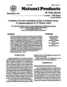
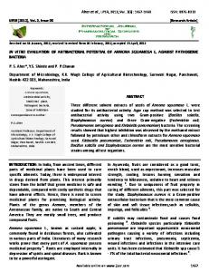
The SPSS software version 16.0 was used for the one-way analysis of variance of the data followed by a multiple comparison Tukey test. Statistical significance was defined as P value < 0.05. 3. Results and discussions 3.1 Characterization of the samples The X-ray diffraction patterns of the synthesised samples are shown in Fig. 1 (a). Sample meso-CHA and npCHA show the pure phase of CHA with the substitution of CO3− ions at the phosphate site, which corresponds to the standard characteristic peaks of carbonate apatite with lattice constant of a = 9.3450 Å and c = 6.920 Å (space group hexagonal P63/m, PDF Number 98-010-1164). The main characteristic peaks of CHA at 2θ = 25.8, 31.8, 32.1° and 33° 21-25 which can be indexed at (002), (121), (112) and (300), were clearly observed in the XRD pattern of sample np-CHA but the peak correspond to (112) were absence in the XRD pattern of meso-CHA due to broadening of the pattern. The main difference between the samples is the level of crystallinity of the materials. The broad diffractions peak of meso-CHA indicates that mesoporous carbonated hydroxyapatite synthesised with the substitution of surfactant was less crystalline than those synthesised without surfactant (np-CHA). Sample synthesise with P123 has the lower fraction of crystallinity (Xc = 0.5196) than non-porous sample (Xc = 0.8403). The crystallinity was taken into consideration in this study because low crystallinity fraction would confer a higher bioresorbability of CHAp26. Highly resorbable materials are favourable in drug delivery application in order to obtain a controlled release of drug delivery from the carrier particles. Fig. 2 exhibits the FTIR spectra of meso-CHA and np-CHA with the bands being labelled accordingly for different functional groups. The bands present at 3568 cm−1 of FTIR spectra are designated for OH −1 27, 28. The absorption peaks at 1050 and 962 attributed to the stretching vibration (v3) of the phosphate (PO43−) groups, and the absorption peaks at 603 cm−1 and 567 cm−1 are corresponding to bending vibration (v4) of the phosphate (PO43−) groups 7, 27. The broad
Nur Farahiyah Mohammad et al. / Procedia Chemistry 19 (2016) 259 – 266
band at around 3435 cm−1 is corresponding to OH group29. The absorption band around 1092 is due to HPO42− ions, indicating that both material is calcium-deficient apatite30. The characteristic bands of B-type CO3−2 substitution are observed at 1472, 1416, and 872 cm−1 31. This finding verified that meso-CHA and np-CHA are B-type carbonated hydroxyapatite, where by the carbonate ions were substituted at the phosphate ions atomic site in the hydroxyapatite lattice32, 33. Therefore, the synthesised carbonate apatite can be expressed as Ca10-x/2[(PO4)6-xCO3)x][(OH2-2y(CO3)y], where x and y are numbers of CO32- ions substituting for PO43- and OH, respectively 34.
Fig.1. XRD diffraction patterns of meso-CHA and np-CHA.
Fig. 2. FTIR spectra of meso-CHA and np-CHA samples.
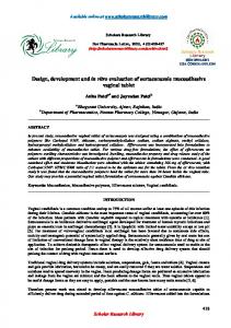
Fig. 3. The (a) FESEM and (b) HRTEM image of nanoparticles of meso-CHA.
FESEM and HRTEM images of sample meso-CHA are shown in Fig. 3. Fig. 3 (a) shows that meso-CHA consists of agglomerated spherical-shaped nanoparticles. However, due to highly agglomerated nanoparticles, the spherical-shaped of meso-CHA was not clearly observed by the HRTEM in Fig. 3 (b). Van der Waals force attraction between the nanoparticles most probably leads to this formation of fine agglomerates. Pores were randomly distributed and not in precisely ordered of alignment. The mesoporous structure of meso-CHA, is confirmed by the nitrogen adsorption-desorption isotherms and the corresponding DFT pore size distribution curve (Fig. 4 (a)). The meso-CHA exhibit the Type IV isotherms with H1 hysteresis loop. The H1 hysteresis loop is often associated with porous materials that consist of agglomerates or compacts of approximately uniform spheres in a fairly regular array35. The isotherms curves of np-CHA is identified
263
264
Nur Farahiyah Mohammad et al. / Procedia Chemistry 19 (2016) 259 – 266
as Type III isotherms with H3 hysteresis loop which indicates that the sample consists of aggregates of plate-like particles, giving rice to slit-shaped pores35. Since the slit-shaped pore due to aggregation of particles, the surface area given is rather low, 4 m2g-1. The inset graphs in Fig. 4 shows the corresponding DFT pore size distribution curve. The pore size for meso-CHA is mainly distributed at 28.4 nm, meanwhile for np-CHA, the pore size is mainly distributed at 11.7 nm. Sample meso-CHA demonstrated the highest BET surface area of 77.5 m2g-1 compared to np-CHA.
Fig. 4. N2 adsorption-desorption isotherms of samples (a) meso-CHA and (b) np-CHA The inset graph shows the corresponding DFT pore size distribution.
3.2 Cytotoxicity study The cytotoxic nature of the samples was assessed with MTS assay after cultured for 1, 3, 5 and 7 days. The MC3T3E1 cells were cultured in 25 mg/ml of meso-CHA and np-CHA extract. The biocompatible nature of meso-CHA was clearly revealed by the viability study using MC3T3-E1 cell line. Cells exhibited more than 70% viability at each incubation period (Fig.5), indicating that meso-CHA and np-CHA are non-toxic materials. The time-dependent result showed that cell viability of meso-CHA is significantly higher than np-CHA on day 1 and day 7 of incubation period (p