EXPERIMENTAL AND THERAPEUTIC MEDICINE 9: 737-743, 2015
Increased adenosine levels contribute to ischemic kidney fibrosis in the unilateral ureteral obstruction model JIN TANG, XIANZHEN JIANG, YIHONG ZHOU, BING XIA and YINGBO DAI Department of Urology, The Third Xiangya Hospital of Central South University, Changsha, Hunan 410013, P.R. China Received December 26, 2013; Accepted June 30, 2014 DOI: 10.3892/etm.2015.2177 Abstract. Renal interstitial fibrosis (RIF) occurs as a result of chronic kidney disease (CKD) and is a common pathway leading to end-stage renal failure. Renal tissue hypoxia and ischemia are present during CKD. Adenosine (ADO) is an important signaling molecule induced under ischemic and hypoxic conditions. In the present study, the association between ADO and RIF was investigated using a mouse model, with the aim of obtaining important information relevant to the prevention and treatment of RIF. A unilateral ureteral obstruction (UUO) model of RIF was established in mice. A total of 44 male mice were randomly divided into sham, model and intervention groups, and samples were collected on days 1, 3, 7, and 14 after modeling. These were collected to detect hypoxia and changes in ADO concentration in obstructed renal tissue as well as to analyze the pathological changes and degree of RIF in the renal tissue. Changes in the levels of collagen deposition and profibrogenic factors in renal tissues were analyzed following intervention with an ADO receptor blocker. Following the UUO procedure, continuous hypoxia was present in the obstructed renal tissue, accompanied by an increased ADO concentration. Tubular injury and interstitial fibrosis progressively increased over time following the UUO procedure. The mRNA expression levels of tissue tumor growth factor β1 (TGF-β1) and α1(I) procollagen were significantly increased. Subsequent to the ADO pathway being blocked by 8-(p-sulfophenyl)‑theophylline, tubular injury and interstitial fibrosis were reduced and the expression of related cytokines was decreased. Increased ADO levels were induced by hypoxia, causing the development of RIF. Following the blocking of the ADO pathway, renal damage was deferred and renal functions were protected.
Correspondence to: Dr Yingbo Dai, Department of Urology, The Third Xiangya Hospital of Central South University, 138 Hexi Yuelu Tongzipo Road, Changsha, Hunan 410013, P.R. China E-mail:
[email protected]
Key words: adenosine, α-smooth muscle actin, cytokines, renal interstitial fibrosis, unilateral ureteral obstruction model
Introduction The most prominent feature of chronic kidney disease (CKD) is renal interstitial fibrosis (RIF). The pathological changes associated with RIF include cellular changes, changes in the extra cellular matrix (ECM) and changes in growth factor interactions (1). Studies have revealed that renal ischemia and hypoxia are caused by microvessel loss during CKD. Adenosine (ADO) is an important signaling molecule that is induced under ischemic and hypoxic conditions (2,3) and which plays a specific biological role following adenosine receptor (AR) binding on the cell surface. In renal tissues, ADO reduces renal blood flow by constricting afferent arterioles, thereby reducing renal transport load (4-8) and protecting short-term renal function. A previous study observed that adenosine deaminase (ADA)‑deficient mice with prolonged exposure to high concentrations of ADO developed RIF lesions and renal damage, indicating that ADO plays an important role in promoting RIF damage (9). Based on these facts, the present study considered the hypothesis that ADO may be an important signaling molecule in the development of RIF. It was hypothesized that under long-term ischemic and hypoxic conditions, intracellular proinflammatory cytokine release is induced by increased ADO concentrations in the renal tissues. Fibroblasts and collagen deposition are activated and regulated, which ultimately results in RIF and leads to kidney damage. Therefore, the present study selected the classic mouse unilateral ureteral obstruction (UUO) model mechanism of RIF. Following modeling, hypoxia of the renal tissue, ADO concentration changes and pathological changes in the obstructed renal tissues were observed at different time points in order to assess the degree of RIF. Changes in the deposition of related profibrogenic factors and interstitial collagen were detected through regulation of the ADO signaling pathway in order to investigate the association between ADO and RIF. Materials and methods Animals. The present study was carried out in strict accordance with the recommendations in the Guide for the Care and Use of Laboratory Animals of the National Institutes of Health, Second Edition (2011). The protocol was approved by the Committee on the Ethics of Animal Experiments of
738
TANG et al: INCREASED ADENOSINE CONTRIBUTES TO ISCHEMIC KIDNEY FIBROSIS
Table I. Primer sequences. Genes α1(I) procollagen-F α1(I) procollagen-R TGF-β1-F TGF-β1-R α-SMA-F α-SMA-R GAPDH (mouse)-F GAPDH (mouse)-R
Upstream and downstream primers (5'-3')
Amplified fragment length (bp)
GTTCTCCTGGCAAAGACGGA 199 CGGCCACCATCTTGAGACTT AGGGCTACCATGCCAACTTC 168 CCACGTAGTAGACGATGGGC GGACTCTGGAGATGGTGTGAC 167 CAATCTCACGCTCGGCAGTA AACTTTGGCATTGTGGAAGG 132 GGATGCAGGGATGATGTTCT
Temperature (˚C) 58 58 58 58/59
TGF-β1, tumor growth factor β1; α-SMA, α-smooth muscle actin; GADPH, glyceraldehyde 3-phosphate dehydrogenase; F, forward; R, reverse.
the Third Xiangya Hospital of Central South University (Changsha, China). All surgery was performed under sodium pentobarbital anesthesia and all efforts were made to minimize suffering. A total of 44 Kunming male mice with an average weight of ~40 g were randomly divided into three groups: Sham group (n=12), model group (UUO group; n=16) and intervention group (PT group; n=16). The mice in the intervention group were intraperitoneally injected with 10 mg/kg/day 8-(p-sulfophenyl)-theophylline (8-PT), a non‑selective AR blocker, once each day following UUO surgery (1 time/day) (10). The mice in the model group were intraperitoneally injected with normal saline. On days 1, 3, 7, and 14 following each surgery, a number of mice were sacrificed, specifically, four each from the UUO and PT groups and three from the sham group. Prior to sacrifice, the mice were placed in metabolic cages for 24 h to enable the collection of urine. A solution of Hypoxyprobe™-1 (pimonidazole HCl; 60 mg/kg) from a Hyproxyprobe‑1 kit (Hypoxyprobe, Burlington, MA, USA) was intravenously injected into the penile region of the mice in order to observe the degree of tissue hypoxia 1.5 h prior to the mice being sacrificed. UUO model. Mice were intravenously injected with 10% sodium pentobarbital (40 mg/kg). A 1.5‑cm left upper quadrant midline incision was made to locate the left ureter. The renal pelvis near the ureter and the middle and upper junctions were ligated with no. 1 silk thread. The ureter between the two ligatures was cut. In the sham group, the left ureter was left free without ligation and the other steps were the same as for the UUO group. Quantitative polymerase chain reaction (qPCR). The mouse cDNA sequence was obtained from GenBank. Primers were designed using Primer Premier software, version 3.0 (Premier Biosoft, Palo Alto, CA, USA) and were synthesized by ProMab (Richmond, CA, USA). Respective primer sequences are shown in Table I. The total RNA in the renal tissue was extracted using TRIzol® reagent (15596-026; Invitrogen Life Technologies, Carlsbad, CA, USA) and RiboLock™ ribonuclease inhibitor (EO0381; Thermo Fisher Scientific, Pittsburgh, PA, USA) was used to eliminate genomic DNA. The reverse transcription (RT) reaction was performed according to the instructions
of the RevertAid™ H Minus First Strand cDNA Synthesis kit (K1631, Thermo Fisher Scientific). The mRNA transcript levels of tumor growth factor β1 (TGF-β1) and α1(I) procollagen were quantified using qPCR according to instructions provided with SYBR® Green PCR Master Mix (4309155; Applied Biosystems, Carlsbad, CA, USA). Pathological analysis. The degree of tubular injury was scored by hematoxylin and eosin (H&E) staining; the degree of RIF was judged by Masson's trichrome staining (11,12). Renal tissue TGF- β1 and α-smooth muscle actin (α-SMA) levels were detected at each time point by immunohistochemistry using anti-TGF-β1 (1:50, Bioss, Ltd., Woburn, MA, USA) and anti‑ α-SMA (1:200, Wuhan Boster Biological Technology, Ltd., Wuhan, China) antibodies. The degree of renal tissue hypoxia was semi-quantitatively determined using a Hypoxyprobe-1 kit (13). A single-blind pathological examination was performed according to a multi‑step procedure. Renal tissue samples were collected from the mice, paraffin‑embedded and sectioned into 5‑µm slices. From each renal sample four slices were randomly selected. Routine H&E and Masson's trichrome staining was performed and the morphology observed using a microscope. Each slice was analyzed by the same individual with five non-overlapping fields randomly selected in each slice. Positive cells or areas were represented by an average optical density, separately calculated in the selected four parts of the renal tissue sample in each mouse. The average value of the four parts was calculated. The average optical density for the positive areas was automatically determined by Image-Pro Plus 6.0 software (Media Cybernetics, Rockville, MD, USA). High performance liquid chromatography (HPLC) assay. Approximately 1/3 of the left kidney was cut immediately upon being extracted from the mouse and preserved in a liquid nitrogen tank to determine the ADO concentration. The HPLC assay was performed on a reversed phase custom ocadecyl‑silica (ODS) column (4.6x254 mm) with a detection wavelength of 260 nm at 30˚C. Following adenine nucleotide extraction, the ADO concentration was designated the abscissa (x) and its corresponding peak area as the vertical axis (y). The regression equation was obtained; the ADO concentration was
EXPERIMENTAL AND THERAPEUTIC MEDICINE 9: 737-743, 2015
739
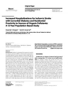
A
B
C
D
Figure 1. (A) Changes in the degree of hydronephrosis at different time periods. (B) The adenosine (ADO) concentration of each group. Data are expressed as the mean ± standard error of the mean (n=3 or 4). **P