Printed in U. S.A.. Indanyloxyacetic Acid-sensitive Chloride Channels from Outer. Membranes of Skeletal Muscleâ. (Received for publication, August 25, 1992).
Vol. 268. No. 1, Issue of January 5, pp. 547-551,1993 Printed in U.S.A.
THEJOURNAL OF BIOLOGICAL CHEMISTRY 0 1993 by The American Society for Biochemistry and Molecular Biology, Inc.
Indanyloxyacetic Acid-sensitive Chloride Channels from Outer Membranes of Skeletal Muscle” (Received for publication, August 25,
1992)
Sabine Weber-SchiirholzS,Erhard WischmeyerS, Monika Laurienl, HaraldJockuschS$, The0 SchurholzT, DonaldW. LandryJI, and Qais Al-AwqatiII From the $.Developmental Biology Unit and nDepartment of Physicochemistry, University of Bielefeld, D- W4800 Bielefeld 1, Federal Republic of Germany and the IICollege of Physicians and Surgeons, Columbia University, New York, New York 10032
In mature skeletal muscle of vertebrates, the level of excitIn maturemammalian muscle, the chlorideconductability, i.e. the current strengths required to cause sufficient ance of the membrane is an important factor in the regulation of excitability. Up to now, no ligand was depolarization to elicit an action potential, is controlled preavailable for thebiochemical characterization of mus- dominantly by chloride conductance which contributes about cle chloride channels. In order tolocalize and charac- 75% of the total conductance at resting potential (Bretag, terize these channels, we have used indanyloxyacetic 1987). Among other causes, elimination ofC1- conductance acid (1AA)-94, a ligand previously used for epithelial can lead to hyperexcitability with spontaneous high frequency C1- channels (Landry, D. W., Reitman, M., Cragoe, E. series of action potentials, the clinical characteristics of the J., Jr., and Al-Awqati, Q. (1987)J. Gen. Physiol. 90, disease myotonia (Rude1 and Lehmann-Horn, 1985). Using 779-798; Landry, D. W., Akabas, M. H., Redhead, C., an indirect approach, the glycerol shock method, it has been Edelman, A., Cragoe, E. J., Jr., and Al-Awqati, Q. inferred that C1- conductance of muscle is associated with the (1989) Science 244, 1469-1472). IAA induced myplasma membrane in frogmuscle or with the transverse otonic responses when microinjected intomature mouse muscle fibers, indicating blockade a of C1- chan- tubular system in mammalian muscle (Dulhunty, 1982). C1nels from the cytoplasmic side. Membrane vesicles channels have been observed by patch-clamping both of frog were prepared from rabbit skeletal muscle and sepa- skeletal muscle (Woll and Neumcke, 1987) and of membrane rated by sucrose gradient centrifugation. Fractions ob- “blebs” from mammalian muscle (Burton et al., 1988), but tained (in the order of increasing density) were sarco- their relation to the macroscopic C1- conductance could not lemma (SL), T-tubules (TT), sarcoplasmaticreticulum be established. Here we show that the channel blocker indanyloxyacetic (SR), and triads and mitochondria (TR/M). The fraction enriched for SL was characterized by high specific acid (IAA-94)’ can be used to assay, enrich for, and to biobinding capacity for [8H]saxitoxin (Na’ channel), chemically characterize C1- channels from mammalian skelwhereasTT-richfractions bound [‘HIPN 200-110 etal muscle. We have localized IAA-sensitive muscular C1(dihydropyridine receptor) withhigh specific activity. channels to the outer sarcolemma and have reconstituted Upon patch-clamping of lipid supplemented vesicles, functional C1- channels from an IAA affinity-enriched IAA-sensitive C1- channels werefound in the SL frac- subfraction of the sarcolemma which contained two specifition but not in the SR. Highest specific activities in cally bound polypeptides. electrical diffusion potential sensitive ‘%l transport and [‘H]IAA-94 binding were found in the SL. SL MATERIALS AND METHODS vesicles were solubilized with 3-[(3-cholamidoproChemi~als-Na~~Cl,NalZ5I, [3H]saxitoxin, and[3H]PN 200-110 pyl)dimethylammonio]-1-propanesulfonateand subwere obtained from Amersham Buchler (Braunschweig, Federal Rejectedto IAA-Sepharose affinitychromatography. Specifically bound protein was eluted with 100 ~ L M public of Germany); ouabain, 3-[(3-cholamidopropyl)dimethylIAA-94 and either analyzedby SDS-gel electrophore- ammoniol-l-propansulfonate(CHAPS), tetrodotoxin, and BHTwere Sigma (Deisenhofen, FRG); and protease inhibitors were from sis or reconstitutedinto phospholipid vesicles. The from Fluka (Neu-Ulm, FRG). Unlabeled PN 200-110was kindly provided eluate contained four polypeptides (specifically bound, by Sandoz AG (Basel, Switzerland). mapp110-120 and 60 kDa; unspecifically bound napp Preparation of Membranes-Membrane fractions were prepared at 6 7 and 50 kDa) and was highly enriched for IAA- 0-4 “C according to a modified pyrophosphate method of Mitchell et sensitive chloride channels as shown by patch-clamp- al. (1983).All buffers contained a mixture of protease inhibitors (0.5 ing after reconstitution. The IAA-sensitive 100/280- mM phenylmethylsulfonyl fluoride, 1 mM benzamidine, 0.5 mM iopicosiemens chloride channels of the sarcolemma are dacetamide, 1 mg/liter pepstatin A, 1 mg/liter leupeptin, 1 mg/liter likely tobe responsible for its major chloride conduct- antipain, 2.8 mg/ml aprotinin). Rabbit hind leg and back muscles ance and thereby for the stabilization of resting poten- (400-450g) were ground in a meat grinder, homogenized in a Waring blender, and centrifuged as described by Mitchell et al. (1983).The tial. supernatant was filtered through cheesecloth, made 0.6 M in KCl,
* This work was supported by the Deutsche Forschungsgemeinschaft (SFB 223/C03). The costs of publication of this article were defrayed in part by the payment of page charges. This article must therefore be hereby marked “advertisement” in accordance with 18 U.S.C. Section 1734 solely to indicate this fact. 3 To whom correspondence should be addressed. Tel.: 49-521-1065618 Fax: 49-521-106-5654.
and stirred for 1 h. After pelleting at 26,400X g for 30 min, the crude membrane fraction was resuspended in 30 ml of 10% (w/v) sucrose, 20 mM Na, pyrophosphate, 20 mM NaH2P04,2 mM MgC12, 1 mM EDTA, pH 7.2, and homogenized in a glass-glass Dounce homogeThe abbreviations used are: IAA, indanyloxyacetic acid SL, sarcolemma; SR, sarcoplasmic reticulum; TT, T-tubule; TR, triad(s); MIT, mitochondria; CHAPS, 3-[(3-cholamidopropyl)dimethylammoniol-l-propanesulfonate; BHT, 2,6-di-tert-butyl-4-methylphenole; TEA, triethylamine hydrochloride; S, siemens.
547
548
Channels
Chloride Sarcolemmal
nizer. The subfractions of membranes were separated by velocity gradient centrifugationusing a combined step andcontinuous sucrose gradient (Mitchell et al., 1983). The following fractions were obtained and characterized by specific marker enzyme activities: SL, 0-14% sucrose; SL/SR/TT, 14-25% sucrose; SR, 25-28% sucrose; TT/SR, 28-36% sucrose; TR/SR/MIT, ~ 3 6 % sucrose. Fractions were diluted in pyrophosphate buffer without sucrose, pelleted at 100,000 X g for 1 h, and resuspended in either 10 mM imidazole, 250 mM sucrose, 1 mM EDTA, pH 7.0 (for [3H]IAA binding or IAA affinity column), or in 130 mM KC1, 10 mM imidazole, 6 mM MgC12,and BHT (1 mg/ml), p H 7.0 (for 36C1-uptakeassays), and stored at -70 "C. Protein concentration was determined by the BCA procedure (Pierce Chemical Co.) using bovine serum albumin as a standard. Enzyme and Ligand Binding Assays-Ouabain-sensitive Na+,K+ATPase, and Ca'+-ATPase activities were measured according to Mitchell et al. (1983), but in the absence of detergent. Succinatecytochrome c reductase activity was measured according to Fleischer and Fleischer (1967). As a marker for Golgi membranes, a-D-mannosidase activity was measured according to Tulsiani et al. (1977). [3H]IAA-94 binding in the presence and absence of detergent was assayed after Landry et al. (1987), but using a newly synthesized ligand with a specific activity of 12 Ci/mmol. [3H]saxitoxin binding was measured according to Brown (1986). For the dihydropyridine receptor assay, membrane fractions (50-100 pg of protein in 125 pl) were suspended in 20 mM Hepes/NaOH, 0.1% bovine serum albumin, p H 6.8, and incubated with [3H]PN 200-110 (830 nCi in 25 pl, 80 Ci/ mmol [3H]PN 200-110) at 4 "C for 1 h in the dark. Two of quadruplicate samples contained unlabeled P N 200-110 (2 p l of a stock concentration 1 mM in EtOH) and the other two 2 p1 of ethanol. A 125-pl aliquot was rapidly filtered on a 2.4-cm fiberglass GFC filter (Whatman, Maidstone,Great Britain).The filters were rapidly washed with 5 ml of 1 M KC1 before and after filtration, and the retained radioactivity was determined in a scintillation counter. Preparation ofVesicles for Patch-clamp Analysis-Vesicles were prepared according to Correa and Agnew (1988) with the exception that soybean phospholipid (Avanti, Birmingham, AL) was used. The S L membrane fragments were washed with 1mM CaC12buffer, filtered through polycarbonate membranes (0.8wm pore size) and pelleted a t 50,000 X g for 50 min. The pellet was resuspended in a lipid vesicle suspension containing 2mg/ml lipid. The amountof lipid added was twice the mass of the protein measured in the membrane fragments. After vortexing and bathsonication (1min), the samples were frozen at -80 "C until use. For patch-clamping, 2 pl for a sample were airdried in a plastic Petri dish and rehydrated with the bath solution. Within a few minutes, large blebs grew out of the protein-lipidfilm which easily sealed to a patch pipette. All measurements were made on excised patches. IAA Affinity Chromatography-The sarcolemmal membrane fraction SL (0.5-1 mgof protein in 1.5 ml of 250 mM sucrose, 10 mM imidazole, 1 mM EDTA, 10% glycerol, pH 7.0) was solubilized by 1% (w/v) CHAPS and shaken for 2 h at 4 "C. Insoluble material was removed by centrifugation at 143,100 X g for 1h, and the supernatant was mixed with IAA-23 affinity resin (1 ml), prepared according to Landry et al. (1989). After agitation overnight, the slurry was transferred to a 10-ml column and washed at 0.3 ml/min with 40 ml of 250 mM sucrose, 10 mM imidazole, 10% glycerol, 0.6% CHAPS, 100 p M benzoic acid, pH 6.0. Specifically bound proteins were eluted with 5 ml of an elution buffer composed of 250mM sucrose, 10 mM imidazole, 10% glycerol, 0.6% CHAPS, and 100 NM IAA-94, pH 6.0. Iodination and SDS-Polyacrylamide Gel Electrophoresis of Proteins-Proteins from the last 1.5 ml of the wash and from the IAA eluate were concentrated by methanol precipitation and dissolved in 0.1 ml SDS sample buffer (50 mM Tris-C1, 2.5% SDS, 0.2 mg/ml bromphenol blue, pH 7.0). Iodination was performed according to the IODO-GEN method (Pierce Chemical Co.), using 200 pCi of NaIz5I (specific activity, 588 MBq/pg) in 100 p1 of SDS sample buffer. The reaction was stopped after 10 min at room temperature by decantation. dithiothreitol (final concentration 30 mM) was added and iodinated proteinsseparated by conventionalelectrophoresison 10% SDS-polyacrylamide gels. Hyperfilm p-max (Amersham-Buchler, Braunschweig, FRG) was exposed to the dried gels. Reconstitution into Phospholipid Vesicles-The proteinseluted from the affinity column were concentrated from 2 to 0.5 ml on a Centricon 10 (Amicon, Witten, FRG) membrane (30 min, 3,000 rpm Sorvall SS 34-rotor). The concentrated proteins (approximately 1pg) were mixed with 30 pl of phospholipid mixture (containing 27 mg of Avanti phospholipid, 3 mg of cholesterol, 40 mg of CHAPS, 0.1% (w/ v) BHT in 1 ml of HzO) and dialyzed overnight against 3 X 650 ml
of buffer (135 mM NaCI, 5 mM Hepes-NaOH, 0.5 mM CaC12, 1 mM MgC12, pH 7.4). RESULTS
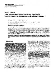
IAA Sensitiuity of the Excitability of the Muscle Fiber-A myotonic reaction of a muscle fiber may serve as an indicator fora reduced chloride conductance (Rude1 and LehmannHorn, 1985). When IAA (100 p ~ was ) added to the bath solution, single action potentials were elicited by single stimuli, as in control fibers. In contrast, 16 of 36 fibers microinjected withIAA showed high frequency seriesof action potentials ( p < 0.01, chi Square test; Fig. 1). These results are compatible with the notion that themajor chloride channels of mature muscle are IAA-sensitive and that IAA acts from the cytoplasmic side. Identification of Membrane Fractions fromMuscle-In order to preparatively enrich for the IAA-sensitive C1- channels, membrane fractions were prepared from rabbit muscle. Fractionation of rabbit muscle membranes by sucrose gradient centrifugation was monitored by the distribution of marker enzymes. Na+,K+-ATPasewas used as a marker for sarcolemma, ea2+-ATPase for sarcoplasmic reticulum, a-D-mannosidase for Golgi membranes, and succinate-cytochrome c reductase for mitochondria.The specific enzyme activities are summarized in Table I. In addition, electronmicroscopy was employed to testfor the presence of triad structures. Fractions were furthercharacterized by assaying binding of several ligands and the uptake of 36Cl-, driven by an electrical diffu-
>
IAA microinjection
IAA bath
I
0.3 s
FIG. 1. Effect of the modeof application of indanyloxyacetic acid (IAA) to muscle fibers on excitability. Intracellular recordings of membranepotentialsaftera single hyperpolarizing stimulus (1 s, 100 nA). Mouse sternocostalis muscle in standard solution (140 mM NaCl, 2 mM CaC12, 1 mM MgC12,5 mM KCH3S03, 7 mM NaCH3S03, 12 mM glucose, 2 mM Hepes-NaOH, pH 7.2) at 36 "C wasused. Left, effect of IAA applied from the cytoplasmic side. About 5 pl of 1 mM IAA were microinjected into each fiber (approximate volume, 4nl).When tested 5-10 min later,16out of 39 microinjected fibers showed typical myotonic runs. Controls, of twenty fibers (two preparations) microinjected with buffer and 70 fibers (seven preparations) microinjected with10 p~ IAA, none showed repetitive firing. Right, bath application of 1 mM IAA was ineffective; all 26 tested fibers showed a single action potential upon stimulation.
TABLE I Marker enzyme activities in membrane fractions from rabbit muscle Results of three to five independent preparations. Measurements on each preparation were done in triplicate. Membrane fraction
Ouabain-sensitive Ca2+-ATPase Succinate-cytochrome Na', K+-ATPase c reductase
pmol PJmg.h
Homogenate SL SL/TT/SR
SR TT/SR TR/SR/MIT
pmol Pi/mg.h nmol cytochrome reduced per mg. min
0.8 (0.38-1.6) 6.1 (5-7) 12.0 (11-13) 4.5 (3.6-5.4) 4.5 (3.8-5.2) 9.8 (6.9-11.7) 88.0 (69-107) 1.5 (1-2) 6.5 (5.5-7.5) 63.4 (60-68) (3.9-5.2) 4.54.5 (2-10) (60-69) 64.0
2.1 (1.8-2.4)