Jul 20, 1981 - Hynie & Zbarsky (1973) found that up to 39% of intestinal alkaline ...... mucosal cells isolated from the same level of the villus-crypt axis (Young ...
Biochem. J. (1981) 200, 645-654 Printed in Great Britain
645
Independent biosynthesis of soluble and membrane-bound alkaline phosphatases in the suckling rat ileum Graeme P. YOUNG,* Steven T. YEDLIN and David H. ALPERSt Division ofGastroenterology, Department of Internal Medicine, Washington University School of Medicine, 660 S. Euclid Avenue, St. Louis, MO 63110, U.S.A.
(Received 20 July 1981/Accepted 31 July 1981) Enzymically active intestinal alkaline phosphatase exists in both soluble and membrane-bound forms in the suckling rat. Antiserum prepared against purified soluble alkaline phosphatase (anti-AlP) was shown to be monospecific when assessed by Ouchterlony double-diffusion analysis and immunoelectrophoresis. The two forms of alkaline phosphatase were antigenically identical and possessed similar affinities for anti-AlP. To study the biosynthesis of the two forms, 14-day-old rats were injected intraperitoneally with [ 3Hlleucine. The labelling kinetics of alkaline phosphatase, extracted from supernatant and brush-border membrane fractions with anti-AlP, was followed over 20h. Incorporation of [3Hlleucine into membrane-bound alkaline phosphatase was rapid, reaching a plateau at 6h. The soluble enzyme showed slower incorporation of label and maximal radioactivity was not reached until 12h after labelling, a lag of 6 h behind the membrane-bound enzyme. Soluble alkaline phosphatase could not have been a precursor of the membrane form, as there was no early peak of radioactivity in the soluble form. To determine if the soluble enzyme was irreversibly derived from the membrane enzyme, a newly developed technique of labelling brush-border membrane proteins in vivo by intraluminal injection of diazotized f1251liodosulphanilic acid was used. The appearance of 1251 in soluble and membrane alkaline phosphatase was then monitored over a 7h period, encompassing the lag between maximal leucine labelling of the two forms. The results failed to show either a proportional transfer of radioactivity from membrane to soluble alkaline phosphatase or an absolute increase in radioactivity of the soluble form during degradation of brush-border alkaline phosphatase. Therefore there does not appear to be a serial precursor/product relationship between the soluble and membrane-bound forms of suckling-rat intestinal alkaline phosphatase. Certain hydrolases of the intestinal brush border can occur in the supernatant as well as the membrane fraction after homogenization of mucosa. Hynie & Zbarsky (1973) found that up to 39% of intestinal alkaline phosphatase was in a soluble form in suckling-rat small bowel. Similarly, Galand & Forstner (1974) found 44% of maltase activity in the supernatant fraction of suckling rat gut homogenates. The soluble proportion fell to 7% after Abbreviation used: anti-AIP, antiserum monospecific for intestinal alkaline phosphatase. * Present address: Department of Medicine, University of Melbourne, Royal Melbourne Hospital, Melbourne, Vic. 3050, Australia. t To whom reprint requests should be addressed.
Vol. 200
weaning, and after treatment of suckling rats with cortisol. Seetharam et al. (1977) confirmed these findings and showed that this phenomenon was more marked in the ileum, that it persisted to a lesser degree in adult rats, and that the soluble enzymes corresponded to their brush-border but not their lysosomal counterparts. The cellular location of these soluble enzymes was not determined. Forstner & Forstner (1979) have shown that developmental changes in soluble maltase are not coincident with those of lysosomal marker enzymes, indicating the lack of dependence between the soluble enzyme phenomenon and the endocytic-lysosomal apparatus of the villous cell. The genetic relationship of these two intestinal alkaline phosphatases in uncertain. They may arise 0306-3283/81/120645-10$01.50/1 ©) 1981 The Biochemical Society
646
from two different gene codes, and the changes which occur with maturation could represent a change in gene expression. To clarify their relationship, we purified the two forms from the suckling-rat ileum (Yedlin et al., 1981). Comparison of the purified hydrolases revealed similar kinetics for multiple substrates and similar amino acid compositions, suggesting that they originated from the same gene code. However, the membrane enzyme differed from the soluble enzyme in a number of ways. First, the membrane enzyme was incorporated readily into synthetic liposomes. Second, it possessed a different carbohydrate composition, with more mannose and less fucose than the soluble enzyme. Third, the principal isoelectric points of membrane alkaline phosphatase were more acidic that those of the soluble form. The soluble enzyme was not an artefact of proteolysis during enzyme purification. It was concluded that the soluble form lacked the hydrophobic anchor-piece necessary for membrane incorporation. If soluble and membrane-bound alkaline phosphatase were derived from the same gene code, they may be biosynthetically related in one of two different ways: synthesis could proceed in parallel from a common precursor (e.g. nascent peptide), or alternatively they could have a serial precursor/ product relationship. The different carbohydrate compositions of the two forms suggests that the latter is unlikely (Yedlin et al., 1981), but such evidence is not conclusive. Therefore we have further studied the relationship between the two forms of intestinal alkaline phosphatase in the suckling rat by using highly specific antibody prepared against soluble alkaline phosphatase. The kinetics of incorporation of 1I3H1leucine in vivo into each form of alkaline phosphatase have been elucidated. We have also examined the possibility that soluble alkaline phosphatase is derived from the membrane-bound form, by using a newly developed method of labelling with diazotized [125lliodosulphanilic acid. We conclude that soluble alkaline phosphatase is neither a precursor nor a product of the membrane enzyme. Materials and methods Animals Pregnant albino Wistar rats were obtained from National Laboratory Animal Co., O'Fallon, MO, U.S.A. After delivery, all litters were made equal in size, to ensure uniform growth rates.
Preparation of brush borders and soluble fraction At the desired age, suckling rats were stunned and decapitated, and the small bowel was immediately removed and rinsed with cold iso-osmotic saline (0.9% NaCI). The distal half of the small bowel (i.e.
G. P. Young, S. T. Yedlin and D. H. Alpers
the ileum) was cut into small pieces. This was homogenized at 40C in 2mM-Tris/HCl, pH7.4, containing 50mM-mannitol, with five up-and-down strokes of a Potter-Elvehjem-type size B tissue grinder (A. H. Thomas Co., Philadelphia, PA, U.S.A.) with Teflon pestle rotating at 900rev./min. The method of Schmitz et al. (1973) was used to isolate brush-border fragments, which were then resuspended in 10mM-Tris/HCl, pH7.4, containing 0.2 mM-MgCl2 (hereafter called Tris/Mg buffer) before further treatment. The supernatant fraction remaining after isolation of brush borders was then spun at 105 0OOg for 60min and the resultant supernatant fraction used as the source of soluble alkaline phosphatase. Enzyme and protein assays Alkaline phosphatase was assayed by the'method of Forstner et al. (1968) at pH9.2 and acid phosphatase was assayed by the method of Henning & Plattner (1974) at pH4.8, with p-nitrophenyl phosphate (Sigma Chemical Co., St. Louis, MO, U.S.A.) as substrate in both cases. One unit of activity is the amount of enzyme that hydrolyses 1,umol of substrate in min at 37°C. Samples'for alkaline phosphatase and acid phosphatase assays were always pretreated with 0.1% Nonidet P40 (BDH Chemicals, Poole, Dorset, U.K.). Protein was assayed by the method of Lowry et al. (1951), with bovine serum albumin (97-99% pure; Sigma) as standard.
Polyacrylamide-gel electrophoresis Disc gel electrophoresis was performed by the method of Davis (1964), with or without 0.1% Triton X-100 (Fisher Scientific Co., Pittsburgh, PA, U.S.A.). Total acrylamide concentration was 5% (acrylamide:bisacrylamide 38:1). On each gel 0.1 unit of soluble or solubilized membrane alkaline phosphatase was loaded and electrophoresed at 2 mA per gel. After electrophoresis, gels were stained for alkaline phosphatase activity at room temperature with a solution of 2mM-fi-naphthyl acid phosphate, 1 mM-tetrazotized o-dianisidine, and 0.4 mM-MgCl2 in 7.5 mM-sodium barbital buffer, pH 9.4. The o-dianisidine was added last. All chemicals were obtained from Sigma. Detergent solubilization of membrane fractions Membrane alkaline phosphatase was solubilized from either brush borders or total membrane preparations (i.e. the 105 OOOg pellet of intestinal homogenates). In each case solubilization was performed at 40C for 16 h in Tris/Mg buffer. The following non-ionic detergents were used in various concentrations: Nonidet P40, Triton X-100, Emulphogen and Empigen BB (Marchon, White-
1981
Intestinal alkaline phosphatases
647
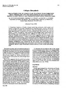
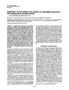
haven, Cumbria, U.K.). Solubilized material was collected after centrifugation at 45 0OOg for 15 min. Ethanol precipitation ofproteins Both detergent-solubilized membrane proteins and soluble tissue proteins were precipitated with 70% (v/v) ethanol precooled to -15°C. Precipitation was performed in precooled containers in a freezer room at -15 0C, for 30min. The precipitated proteins were collected by centrifugation at 45 0OOg for 15 min. The pellet was redissolved in Tris/Mg buffer containing 0.1% Nonidet P40 and denatured protein removed by repeat centrifugation. Preparation and characterization of antisera Antiserum was prepared against purified soluble intestinal alkaline phosphatase in rabbits as described in detail by Yedlin et al. (1981). Immunization was performed over a 6-week period by using Freund's complete adjuvant. Antiserum was adsorbed with the supernatant fraction from the sucklingrat duodenum before use. Anti-AlP was monospecific when assessed by Ouchterlony doublediffusion analysis (Fig. la) and immunoelectrophoresis (Fig. lb). Titration of anti-AlP against the two forms of ileal alkaline phosphatase (Fig. 2) revealed a similar titre and affinity for each form. Anti-AlP did not cross-react with rat bone or liver alkaline phosphatase or with rat acid phosphatase. There was no evidence of precipitation of other brush-border hydrolases (sucrase, maltase and lactase) during the titration studies.
[3HlLeucine-labelling studies For this, 47 14-day-old non-starved suckling rats from five litters were each' injected intraperitoneally with 50,uCi of [3H]leucine (specific radioactivity 45.7 Ci/mmol; New England Nuclear, Boston, MA, U.S.A.) contained in 0.25ml of 50mM phosphatebuffered iso-osmotic saline, pH7.4. Animals were returned to their mothers and then killed at the following time points: 10, 20, 30 and 60min, 2, 3, 4, 6, 8, 12, 16 and 20h. Ileal homogenates were prepared in 18ml of buffer as described above. A sample (0.5ml) of homogenate was centrifuged at 105OOOg for 60min to allow determination of the total amount of enzyme in the soluble and membrane fraction of each animal's ileum. The remaining 17.5 ml was used to make brush-border and soluble fractions. After solubilization of brushborder proteins in Nonidet P40, both the soluble fraction and detergent-solubilized brush-border proteins were precipitated by ethanol as described above. Precipitated proteins were resolubilized in 0.1% Nonidet P40 in Tris/Mg buffer. Alkaline phosphatase was then isolated by immunoprecipitation. Vol. 200
Fig. 1. (a) Ouchterlony double-diffusion analysis of anti-AIP against various forms of intestinal alkaline phosphatase, and (b) immunoelectrophoresis of ileal supernatant fraction by the method of Grabar & Williams (1953) (a) The central well contains anti-AlP. Commencing from the top well and moving clockwise, the peripheral wells contain: ileal supernatant fraction, purified soluble alkaline phosphatase, purified membrane alkaline phosphatase, Nonidet-P40-solubilized ileal brush-border proteins, ileal supernatant fraction and Nonidet-P40-solubilized ileal total membrane fraction. Immunodiffusion was performed in 0.8%-agarose containing 50mM-sodium barbital, pH 8.6, and 0.1% Nonidet P40. (b) Supernatant fraction (105OO0g) from the ileum containing 0.9 unit of alkaline phosphatase activity was placed in the top well. The same amount of protein from a duodenal supernatant fraction was placed in the other well. Electrophoresis was performed towards the anode for 2h. A 1:4 dilution of anti-AlP was then placed in the trough. Histochemical staining for alkaline phosphatase (see the Materials and methods section) showed concentration of enzymic activity in the precipitin line. The support medium was the same as in Fig. 1 (a).
Immunoprecipitation of alkaline phosphatase Alkaline phosphatase was isolated from brushborder and soluble fractions by immunoprecipitation from 1 ml samples containing a known
G. P. Young, S. T. Yedlin and D. H. Alpers
648
100
\.
80
60
.
40
-
-
20
0
32
64
128
512
256
512
1024 1024
1 /Titre
Fig. 2. Titration curve of anti-AlP againsitvarious forms of ileal alkaline phosphatase V, Purified soluble alkaline phosphata supernatant fractions; O, Nonidet-P44
ise;*, ileal O-solubilized entration of
ileal brush-border proteins. The conc( alkaline phosphatase in each case was I1.4 units/ml. For further details see the Materials a nd methods
section.
amount of alkaline phosphatase acitivity, in the range 5-10 units. To determine thie amount of
antibody required to
precipitate at
Ileast 98% of
were pe -rformed with of dilu 1: 16 Nwas required. Sample/anti-AlP mixtures were then incubated at for 2h and maintained at 4c Visible immune pellets were collectc by centrifugation at 1200g for 10min, and th e supernatant
activity, pilot studies 50,ul of sample and 50,ul Generally a titre of 1:8 or
ted anti-AlP.
37°C
~C ed
enzyme
overnight.
fraction was assayed to ensure that greater than 98% enzyme activity had been precipit ;ated. Immune pellets were washed three times in T ris/Mg buffer containing 0.1% Nonidet P40. Non-sp4 ecific trapping in the immune pellet of radioactive I proteins other than alkaline phosphatase was determiined by adding non-radioactive concentrated alkaline phosphatase to the immune-supernatant fractioni an amount equal to the original enzyme activity.. Precipitation was repeated with the same amount of and the pellets were washed as described ab )ove. To determine the incorporation 3I of into alkaline phosphatase, washed immune pelletE were resuspended in 50,ul of water and then transferred to scintillation vials containing 0.5ml of NCS Tissue Solubilizer (0.6M; Amersham/Searlle, Arlington Heights, IL, U.S.A.) After solubiliza tion at room temperature for 16h, lOml of a toluene-based of which scintillation fluid was added, each i contained 6g of 2,5-diphenyloxazole and 75 mg of
in anti-AlP
HI
itre
1,4-bis-(5-phenyloxazol-2-yl)benzene (New England Nuclear). Radioactivity was determined in a Packard Tri-Carb liquid-scintillation spectrophotometer (model 3320) until 10000 counts were accumulated or 50 min had elapsed. Background radioactivity was 15.7c.p.m. Samples with the lowest radioactivity gave at least twice this number of counts. Counting efficiency determined by the channels-ratio method was uniform, in the range 40.5-42.5%. For soluble alkaline phosphatase, non-specific trapping of radioactivity was always less than 4.6% of total radioactivity. When alkaline phosphatase was immunoprecipitated from detergent-solubilized brush borders, non-specific trapping was fairly constant, being no higher than twice background. This represented 2-27% of radioactivity in alkaline phosphatase, depending on the time point. Preliminary studies showed that if ethanol precipitation of fractions was omitted before immuno-
precipitation, non-specific trapping could reach 5 [3H]Leucine incorporation into soluble or membrane alkaline phosphatase was calculated for each
times background.
animal, after correction for non-specific trapping and background. Incorporation was calculated in two ways: (a) c.p.m. of 3H per unit of enzyme activity; and (b) c.p.m. in the total mass (i.e. c.p.m. per unit of activity x total units in each fraction) of ileal soluble or brush-border-bound alkaline phosphatase. The first method fails to account for variations in the total mass of enzyme and for a possible difference in catalytic efficiency (units of enzyme activity per mol of enzyme protein) between the two forms. Labelling studies
with[[25lIiodosulphanilic
[125Ilodosulphanilic acid (>1OOOCi/mmol; New England Nuclear) was converted into the diazonium salt according to the instructions provided by New England Nuclear and used immediately. The use of this agent in vivo to label brush-border membrane proteins of adult rats has been described in detail elsewhere (Young & Alpers, 1981). Nine 16-day-old rats from a single litter were operated on, under ether anaesthesia; 65,uCi of diazotized [i25J]iodosulphanilic acid in 0.2 ml of 10 mM-potassium phosphate-buffered iso-osmotic saline, pH7.4, was injected via a 27-gauge needle into the distal bowel at the jejuno-ileal junction. No obvious peritoneal leak was seen through the small puncture hole. All animals recovered rapidly from the procedure after closure of the abdominal wound with acontinuous silk suture. Animals were returned to their mothers and killed after 1, 3 or 7 h, three at each time point. After extensive rinsing of the intestine with phosphate-buffered saline, ileal homogenates were prepared as described above by using eight strokes of the homogenizer. Homogenates were then im1981
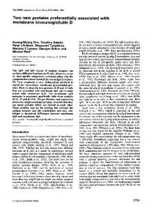
Intestinal alkaline phosphatases mediately treated with 70% ethanol as described and precipitated for 10min. This extracted over 90% of non-protein-bound diazotized [1251]iodosulphanilic acid (assessed by precipitation with 10% trichloroacetic acid). This made multiple washing of membranes unnecessary and achieved separation of released soluble intracellular proteins from free label, thus stopping the reaction. Ethanol-precipitated homogenate proteins were redissolved in Tris/Mg buffer, and soluble alkaline phosphatase was separated from membrane-bound alkaline phosphatase by centrifugation at 105000g for 60min. Supernatant fractions were dialysed overnight at 4°C against Tris/Mg buffer. Membrane proteins were solubilized, precipitated with ethanol and resolubilized in 0.1% Nonidet P40 as described for brush-border membranes. These treatments resulted in over 95% of diazotized [ '25lliodosulphanilic acid being bound to protein, as assessed by trichloroacetic acid precipitation. Alkaline phosphatase was immunoprecipitated from each fraction as described above and incorporation of 125I measured in a Nuclear-Chicago gamma counter for 10min. Background was 14 c.p.m., counting efficiency was 71%. Results were expressed as c.p.m. in total mass of soluble or membrane alkaline phosphatase. Uptake of 125I into the liver was determined by counting radioactivity of ml samples of a 10% (w/v) liver homogenate prepared in 50 mM-mannitol/ lOmM-Tris/HCI (pH 7.4). Results In the 14-day-old rat, 55% of small-bowel alkaline phosphatase activity was found in the ileum. Of this ileal enzyme, 64.9 + 6.7% (mean + S.D.) was found in the 105 OOOg supernatant fraction. That is, in the ileum the pool of soluble enzyme is twice the size of that of the membrane-bound enzyme, based on enzyme activity. In the jejunum, only 10.9 + 4.3% was soluble. Fig. 3 shows the results of polyacrylamide-gel electrophoresis of ileal supernatant and membrane-bound alkaline phosphatase. The membrane (i.e. brush-border) form migrated slowly, with a mean RF of 0.23, and would not enter the gel in the absence of non-ionic detergent. The supernatant form entered the gel whether or not detergent was present, with a mean RF of 0.44 in the presence of detergent. The slow form was not seen in the ileal supernatant fraction (105 000g), nor was the fast form seen in washed pellets. Hence centrifugation at high speed for 1 h was a satisfactory means of separating the two. Acid phosphatase activity was distributed differently along the intestine from alkaline phosphatase; 75% of activity was found in the distal half of the gut. The amount of activity released into the supernatant fraction by homogenization was Vol. 200
649 (a)
(b)
:.
NW.
(c)
-:X:
.w.... .1
Fig. 3. Polyacrylamide-disc-gel electrophoresis of membrane (a) and soluble (b and c) fractions of ileal homogenates, stainedfor alkaline phosphatase activity Enzyme activity was detected in these Tris/glycine gels as described in the Materials and methods section. Gels and samples for (a) and (b) contained 0.1% Triton X- 100. No detergent was present in (c). The RF values for alkaline phosphatase are: (a), 0.23 (b) 0.044, (c) 0.50. The slightly dense area which is seen behind the dye front in gels (a) and (b) is due to interaction between Triton X- 100 and a component of Bromophenol Blue.
33.6 + 1.6% (mean + S.E.M.) in the ileum and 31.9 ± 3.6% in the jejunum. When untreated ileal supernatant proteins were fractionated on Sephadex G-200, alkaline phosphatase activity was eluted with a molecular weight of 97000, in close agreement with the findings of Yedlin et al. (1981) for the purified enzyme.
owl
650
G. P. Young, S. T. Yedlin and D. H. Alpers
Immunoprecipitation
of
these
fractions
showed
radioactivity to be proportional to the amount of enzymically active alkaline phosphatase present (Fig. 4). No other peaks of radioactivity were observed, indicating that anti-AlP did not precipitate enzymically inactive fragments of the alkaline phosphatase molecule or other radioactive proteins.
. ~64;o
,f