NEUROENDOCRINOLOGY
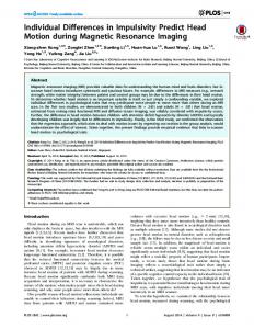
Individual Differences in Reactivity to Social Stress Predict Susceptibility and Resilience to a Depressive Phenotype: Role of Corticotropin-Releasing Factor Susan K. Wood, Hayley E. Walker, Rita J. Valentino, and Seema Bhatnagar Department of Anesthesiology and Critical Care Medicine, The Children’s Hospital of Philadelphia, University of Pennsylvania School of Medicine, Philadelphia, Pennsylvania 19104
Previous social stress exposure is a common risk factor for affective disorders. However, factors that determine vulnerability or resiliency to social stress-induced psychopathologies remain unclear. Using a rodent model of social stress, the present study was designed to identify putative neurobiological substrates that contribute to social stress-induced psychopathology and factors that influence or predict vulnerability. The resident-intruder model of defeat was used as a social stressor in adult male Sprague Dawley rats. The average latency to assume a subordinate posture (signaling defeat) over seven daily defeat exposures was calculated and examined with respect to endpoints of hypothalamic-pituitary-adrenal activity, components of the corticotropin-releasing factor (CRF) system, and behaviors that are relevant to human depression. In the present studies, a bimodal distribution emerged in an otherwise homogeneous population of Sprague Dawley rats such that 42% of rats exhibited short defeat latencies (⬍300 sec), whereas 58% of rats resisted defeat and exhibited longer latencies (⬎300 sec). These two phenotypes were associated with distinct endocrine and behavioral profiles as well as differences in components of the CRF system. Notably, the short-latency subpopulation exhibited hypothalamic-pituitary-adrenal dysregulation and behavior similar to that observed in melancholic depression. Examination of components of the CRF system suggested that proactive behavior in resisting defeat exhibited by long-latency rats was associated with decreased efficacy of CRF. Together, these data suggest that inherent differences in stress reactivity, perhaps as a result of differences in CRF regulation, may predict long-term consequences of social stress and vulnerability to depressive-like symptoms. (Endocrinology 151: 1795–1805, 2010)
tress exposure, often of a social nature (1–3), is known to precipitate psychopathological disorders. However, because stressful events are part of everyday life and only a select population of individuals develops stressinduced pathologies, elucidating the biological basis of individual differences in stress vulnerability or resiliency has been at the forefront of clinical and preclinical research (4). An ethologically relevant animal model of social stress useful for studying the link between stress and psychopathologies is the resident-intruder paradigm. This model involves threats and antagonistic encounters by a large, aggressive male rat (resident) toward a smaller male rat
S
(intruder). Rodents exposed to this type of social defeat stress exhibit decreased motivation, increased behavioral despair, anhedonia, and decreased social interactions (5– 7). Social stress also produces long lasting changes in hypothalamic-pituitary-adrenal (HPA) axis function in rodents (8 –11). The potent repercussions of social defeat are well documented; however, less is understood regarding the individual differences in vulnerability to these effects. One characteristic that may help elucidate the underlying basis for individual differences in stress-related pathology is the type of strategy used to cope with stress. Human studies suggest that passive coping during a stressful life event is
ISSN Print 0013-7227 ISSN Online 1945-7170 Printed in U.S.A. Copyright © 2010 by The Endocrine Society doi: 10.1210/en.2009-1026 Received August 28, 2009. Accepted January 7, 2010. First Published Online February 16, 2010
Abbreviations: CRF, Corticotropin-releasing factor; FST, forced swim test; HPA, hypothalamic-pituitary-adrenal; LL, long latency; PVN, paraventricular nucleus of the hypothalamus; SL, short latency.
Endocrinology, April 2010, 151(4):1795–1805
endo.endojournals.org
1795
1796
Wood et al.
Individual Stress Vulnerability
associated with the development of stress-induced depression, whereas proactive coping is correlated with resiliency (12, 13). In addition, differing coping styles result in opposing physiological and neuroendocrine responses to stress (14, 15). For example, proactive coping during stress is correlated with high sympathetic activity, whereas passive coping is associated with greater activation of the HPA stress axis (16). Therefore, passively coping during stress may increase the risk of developing not only depression but also HPA dysfunction. To gain insight into the neurobiological factors conferring vulnerability or resiliency to stress-induced psychiatric disease, we evaluated the acute behavioral response to social stress, indicated by the latency to be defeated, and examined resultant effects on HPA reactivity and behavior in rats. Corticotropin-releasing factor (CRF) initiates the endocrine limb of the stress response by stimulating synthesis and release of ACTH from the anterior pituitary, which leads to corticosteroid release by the adrenal cortex (17). CRF receptor activation in extrahypothalamic regions also results in autonomic and behavioral aspects of the stress response (18 –20). Elevated CRF function occurs in depressed subjects and contributes to the HPA dysregulation that characterizes melancholic depression (reviewed in Ref. 21). Moreover, CRF levels normalize after antidepressant treatment (reviewed in Refs. 22 and 23). Given the central effects of CRF in the brain, overactivity of this system can contribute to the neuroendocrine, cognitive, and behavioral aspects of depression. Thus, individual differences in components of the CRF system may contribute to vulnerability to depression. We hypothesized that individual differences in the response to social defeat are related to differences in central CRF activity and will be associated with distinct neural and behavioral repercussions. Moreover, we proposed that rats exhibiting an exaggerated submissive response, indicated by a short latency (SL) to exhibit the submissive defeat posture, during repeated social defeat would be more susceptible to HPA and depressive-like dysfunction.
Materials and Methods Animals Male Sprague Dawley rats (275–300 g on experiment d 1) were used as controls or intruders, and male Long-Evans retired breeders (650 – 850 g) served as residents (Charles River, Wilmington, MA). Rats were singly housed with a 12-h light, 12-h dark cycle (lights on at 0700 h) in a climate-controlled room with ad libitum food and water. Studies were approved by the Children’s Hospital of Philadelphia Institutional Animal Care and Use Committee and conformed to the National Institutes of Health Guide for the Use of Laboratory Animals. All experimentation was conducted between 0900 and 1200 h.
Endocrinology, April 2010, 151(4):1795–1805
Social defeat The social defeat paradigm used in these studies was modified from the resident-intruder model originally developed by Miczek (24). Rats were randomly assigned to either a social defeat or control group for a consecutive 7 d (based on Refs. 8 and 9). During each episode of social stress, a rat was placed into the home cage territory of an unfamiliar Long-Evans resident previously screened for high aggression (8, 9). A typical agonistic encounter resulted in intruder subordination or defeat, signaled by the intruder assuming a supine position for approximately 3 sec. After defeat, a wire mesh enclosure was placed in the cage to prevent physical contact between the resident and intruder but allowing visual, auditory, and olfactory contact for the remainder of the 30-min defeat session. Latency to assume a submissive posture (defeat) was recorded and averaged over the seven daily defeat exposures. If an intruder resisted defeat for 15 min, rats were separated with the wire partition for the remainder of the session. Controls were placed behind a wire partition in a novel cage for 30 min daily. Rats were returned to their home cage after each session, and body weight was recorded on d 1 and 8.
Experimental design All rats were assigned into either a control or defeat stress group. The stress protocol was kept constant in all experiments such that rats were exposed to a consecutive 7 d of a 30-min social defeat or control novel cage exposure except for rats in the cFos study, in which brain tissue was harvested on d 5 of stress exposure. Adrenal and thymus gland weights were also recorded after most experiments.
Study 1: individual differences in the HPA response to repeated daily defeats In this study, we examined whether rats with differing behavioral responses to social defeat stress would also exhibit variations in HPA axis habituation to the defeat itself. Five to 6 d before experimentation, intruders (n ⫽ 24) and controls (n ⫽ 12) were anesthetized using ketamine (ip; 80 mg/kg) and xylazine (ip; 10 mg/kg). Micro-Renathane catheter tubing (MRE-40; Braintree Scientific Inc., Braintree, MA) was inserted 3 cm into the right jugular vein. Intravenous blood (280 –300 l) was collected from indwelling venous catheters on d 1, 4, and 7 of control or social defeat exposure immediately preceding social stress or control (0 min), during defeat or novel cage stress (15 and 30 min) and 30 min after termination of stress (60 min). Catheters were flushed daily with 100 U/ml heparinized saline. Plasma samples were later assayed for ACTH and corticosterone levels using commercially available RIA kits (MP Biomedicals, Orangeburg, NY) as previously described (25). In a separate population of rats (n ⫽ 15 intruders; n ⫽ 8 controls), brains were transcardially perfused with 4% paraformaldehyde 90 min after the start of d 5 of stress or control exposure. Using a previously published protocol (26), free-floating brain slices (30 m) containing the parvocellular division of the paraventricular nucleus of the hypothalamus (PVN) were processed for immunohistochemical detection of cFos expression. The cFos antibody used in these studies (1:25,000) was generously donated by Dr. Paul E. Sawchenko (The Salk Institute, La Jolla, CA).
Endocrinology, April 2010, 151(4):1795–1805
endo.endojournals.org
1797
Study 2: individual differences in defeat-induced changes in hypothalamic CRF and AVP mRNA and CRF1 and V1b receptor expression within the pituitary
via tail vein 0, 15, 30, and 60 min after the initiation of restraint and later assayed for ACTH and corticosterone.
In this study, we examined whether differing behavioral responses during social defeat were related to changes within the central components of the HPA axis. Therefore, we evaluated CRF and AVP mRNA density within the PVN and CRF1 and V1b receptor expression in the pituitary. Rats (n ⫽ 13 intruders; n ⫽ 9 controls) were decapitated 24 h after the seventh defeat or control exposure under resting conditions, and brain and pituitary tissue was stored at ⫺80 C until processing. Brains were sliced in coronal serial sections (14 m thick) and mounted onto slides (Fisherbrand ProbeOn Plus; Fisher Scientific, Pittsburgh, PA) before in situ hybridization. Riboprobes generously donated by Drs. Audrey Seasholtz and Robert Thompson (University of Michigan, Ann Arbor, MI) were used to localize CRF and AVP mRNA within the parvocellular division of the PVN and was performed using a previously published protocol (27). Briefly, tissue was fixed for 60 min at room temperature with 10% formalin before being acetylated (0.25% acetic anhydride), and dehydrated in ascending ethanol concentrations (50 –100%). The sections were hybridized, in situ, with a 35S-labeled antisense riboprobe to CRF or AVP. After hybridization, slides were exposed to Kodak Biomax film for 4 and 72 h for AVP and CRF, respectively. Films were scanned into Photoshop using Adobe Photoshop Elements (version 4.0). Using Image J software, the background density was subtracted from the integrated OD of each region of interest. Two sections expressing CRF or AVP mRNA were averaged for each rat. To determine differences in CRF1 and V1b protein expression, pituitary tissue was homogenized in a RIPA buffer and protease inhibitor cocktail and centrifuged (14,000 ⫻ g, 15 min) before Western blot analysis. Western blotting was conducted using a previously published protocol with slight modifications to accommodate the use of the Odyssey infrared imaging system (LI-COR Biosciences, Lincoln, NE) (28). Pituitary protein extracts (100 g) were subjected to SDS-PAGE and transferred to a polyvinylidene difluoride membrane. Membranes were blocked with Odyssey buffer (diluted 1:1 with PBS; LI-COR) and incubated for 90 min at room temperature in the presence of goat anti-CRF1 (1:400; Santa Cruz Biochemicals, Santa Cruz, CA) or overnight at 4 C in rabbit anti-AVPR1B (1:2000; Lifespan Biosciences, Seattle, WA). All membranes were also incubated with mouse anti--actin (1:5000, Sigma, St. Louis, MO) for 90 min at room temperature. Membranes were incubated with corresponding infrared secondary antibodies for 60 min; IR Dye 800 donkey antigoat (LI-COR IRDye 800-labeled) or IR Dye 680 donkey antirabbit, and donkey antimouse (Alexa Fluor 680labeled). Membranes were scanned using the Odyssey infrared imaging system (LI-COR). The Odyssey application software (version 2.1) was used to determine the molecular weight and integrated intensity of each band. Ratios of the CRF1 and V1b receptors to -actin were calculated for each sample.
Study 4: individual differences in defeat-induced functional changes within the HPA axis
Study 3: individual differences in the HPA response to novel restraint stress In this study, we determined whether the behavioral response during social defeat differentially impacted the ability to respond to a novel stressor. Rats (n ⫽ 15 intruders; n ⫽ 9 controls) were exposed to 30 min restraint stress in a Plexiglas tube 24 h after the seventh defeat or control manipulation. Blood was collected
In this study, we examined whether the behavioral response during social defeat was associated with functional changes that occurred in the HPA axis as a result of repeated social defeat. Rats implanted with venous catheters from study 1 were used in this study and exposed to a pharmacological challenge of vehicle or CRF (0.3 g/kg) 24 h after d 7 of defeat. Pilot studies quantifying ACTH release in response to 0, 0.3, and 1.0 g/kg established that 0.3 g/kg is on the linear portion of the dose-response curve. This dose also produces moderate ACTH release comparable to that elicited by restraint stress (300 – 400 pg/ml) (10, 29). Ovine CRF was obtained from Dr. Jean Rivier (Clayton Foundation Laboratories for Peptide Biology, The Salk Institute, La Jolla, CA) and dissolved in sterile saline (0.3 mg/ml) the day of the test, and blood was collected after vehicle (saline) or CRF was administered via an indwelling iv catheter in the home cage under resting conditions. Twenty-four hours later, a dexamethasone suppression test was conducted (48 h after d 7 of defeat or control exposure). Dexamethasone was dissolved in ethanol and brought up to concentration (0.03 mg/ml) with sterile saline. Either vehicle or dexamethasone (0.03 mg/kg, sc; Sigma Chemical Co., St. Louis, MO) was injected 90 min before a 30-min restraint stress. A similar dose of dexamethasone was previously reported from our laboratory to produce half-maximal suppression of restraint-induced ACTH release (30). In both studies, blood was collected via iv catheter immediately before (0 min) and 15, 30, and 60 min after the initiation of drug infusion (CRF challenge) or restraint (dexamethasone suppression test). No single rat received an injection of both CRF and dexamethasone. In the event that the venous catheter was no longer patent for blood withdrawal, tail vein blood was collected. ACTH concentrations in plasma were determined using the same methods as in study 1.
Study 5: individual differences in vulnerability to depressive-like behavior In this study, we determined whether rats with different behavioral responses during social defeat were differentially vulnerable to developing depressive-like behaviors. Rats (n ⫽ 15 intruders; n ⫽ 12 controls) were exposed to the Porsolt forced swim test (FST) 24 and 48 h after d 7 of stress. The FST was conducted as previously published (31), and the water depth was modified to 40 cm.
Statistics A two-way repeated-measures ANOVA was used to identify significant differences in daily defeat latencies between SL and long-latency (LL) rats 关day (1–7) ⫻ group (SL vs. LL)兴.
Study 1 Integrated (picograms per milliliter per minute) ACTH and corticosterone release was calculated using time points 0, 15, 30, and 60 min. Significant differences in defeat-induced integrated HPA reactivity over d 1, 4, and 7 were identified using a one-way ANOVA within each group (control, SL, and LL). To identify whether previous defeat experience altered resting corticoste-
1798
Wood et al.
Individual Stress Vulnerability
Endocrinology, April 2010, 151(4):1795–1805
rone levels on d 1, 4, and 7, one-way ANOVA (control vs. SL vs. LL) were used to compare 0-min corticosterone concentrations at each day. Differences in cFos expression within the PVN of control, SL, and LL rats were identified using a oneway ANOVA.
Study 2 One-way ANOVA were used to identify differences in CRF and AVP mRNA in the PVN and CRF1 and V1b receptor expression in the pituitary between control, SL, and LL rats.
Study 3 The HPA response to restraint was also evaluated between groups using a three- by four-way ANOVA 关group (control, SL, and LL) ⫻ time point (0, 15, 30, or 60)兴.
Study 4 Differences in integrated ACTH release induced by the CRF challenge and dexamethasone suppression tests were compared between groups using two- by three-way ANOVA 关treatment (vehicle or drug) ⫻ group (control, SL, or LL)兴.
Study 5 Differences in behavior during the FST and defensive burying tests between control, SL, and LL rats were identified using oneway ANOVA. Group differences in body weight and gland weight were evaluated using one-way ANOVA. To determine whether certain endpoints were related, defeat latency was correlated with adrenal weight, CRF1 pituitary protein concentrations, and behavior in the FST. In addition, CRF mRNA densities in the PVN were correlated with adrenal weight. Post hoc analyses were conducted using Bonferroni comparisons. All data are presented as mean ⫾ SE.
Results Behavioral reactivity during social defeat (average defeat latency) The average latency to be defeated over seven consecutive defeats was calculated for each intruder rat. Although all intruders were identical with respect to strain, vendor, housing, handling conditions, and exposure to residents, the population was bimodally distributed with respect to average defeat latency (Fig. 1A). Based on this distribution, we chose an average defeat latency of 300 sec as a cutoff to define rats’ behavioral response to social defeat. Across all experiments described herein, a subpopulation of 42% assumed a subordinate posture (n ⫽ 34) in a relatively short time (⬍300 sec, SL) and the other subpopulation (58%) resisted defeat (n ⫽ 48) resulting in longer latencies (⬎300 sec, LL) to assume this posture. Average defeat latencies per study for SL and LL rats can be found in the figure legends. Overall, the SL and LL populations had mean (⫾SEM) latencies to assume a subordinate posture of 191 ⫾ 12 sec and 478 ⫾ 15 sec across all studies described here, respectively 关t(80) ⫽ 14.05; P ⬍
FIG. 1. Individual differences in the latency to be defeated. A, A frequency distribution indicated that the average latency to exhibit a supine position (signaling defeat) over seven consecutive social defeat exposures was bimodally distributed among all rats used in these studies (n ⫽ 82). Based on this distribution, we used 300 sec as a cutoff to further subdivide rats. Rats with an average defeat latency of less than or greater than 300 sec were termed SL and LL, respectively. B, These data represent a subset of SL (n ⫽ 18) and LL (n ⫽ 21) rats from A that were exposed to the same colony of residents. Two distinct populations emerged by d 3 of stress as indicated by the average daily latency to assume a submissive posture. One-way ANOVA indicated an effect of group.
0.0001兴. Average defeat latencies of SL rats were consistent between studies 关F(10,52) ⫽ 1.05; P ⫽ 0.42兴. LL rats average defeat latencies, however, were somewhat variable between studies 关F(10,81) ⫽ 3.6; P ⫽ 0.0006兴. To determine whether these subpopulations of rats were initially different in their response to social defeat or whether differences developed over repeated exposures, we examined latency to be defeated on a day-to-day basis (Fig. 1B). Figure 1B represents only a subset of rats (SL, n ⫽ 18; LL, n ⫽ 21) included in Fig. 1A that were exposed to the same residents over four separate experiments to reduce the variability that is observed by comparing rats’ defeat latencies exposed to many different sets of residents. In this subset of rats, the average latency to be defeated on a daily basis revealed that the bimodal distribution was a result of increasing duration of onset for LL rats to assume a submissive posture as the number of exposures to residents increased, whereas the latency to be defeated for SL remained unchanged over days 关Fig. 1B; group effect: F(1,216) ⫽ 36.7; P ⬍ 0.0001兴. To identify potential defeat latency-dependent changes in behavior, the first four defeat episodes were recorded from a subset of rats (n ⫽ 22) and later reviewed by two experimenters blind to each rats’ classification. Behavioral analysis revealed that although all rats experienced similar frequency of attacks, by the fourth defeat episode, LL rats spent 73 ⫾ 44 sec in an upright posture (forepaws raised standing only on hind
Endocrinology, April 2010, 151(4):1795–1805
endo.endojournals.org
1799
legs), whereas SL rats spent only 6 ⫾ 2.0 sec in an upright posture (P ⫽ 0.07). These data suggest that the emergence of active behaviors may contribute to resistance to defeat and increased defeat latency in LL rats. These differences in active behavior could not be explained by underlying differences in plasma testosterone concentrations because testosterone levels were comparable in control, SL, and LL rats 24 h before the start of experimentation (2.6 ⫾ 0.5, 2.5 ⫾ 0.8, and 2.2 ⫾ 0.5, respectively). Study 1: SL and LL rats exhibit distinct differences in HPA habituation to social defeat To determine whether the divergence in defeat latency was associated with a differential habituation to the social defeat itself, integrated ACTH and corticosterone release were evaluated within groups on d 1, 4, and 7 during the 7-d social defeat exposure. Control manipulation produced modest ACTH release that did not significantly change between d 1, 4, and 7 (P ⫽ 0.17). Robust defeatinduced ACTH release was evident in SL and LL rats on the first defeat exposure. However, in SL rats, ACTH release did not display habituation until d 7; however, this decrease was not significant (P ⫽ 0.22). LL rats, on the other hand, exhibited ACTH habituation as early as d 4 关F(2,17) ⫽ 3.6; P ⫽ 0.05兴. Therefore, differences in defeat latency emerged at the same time that HPA axis habituation occurred in LL rats (Fig. 1B). Similar trends were observed in corticosterone release during defeat. A nonsignificant decrease in corticosterone release in SL rats was not evident until d 7 (P ⫽ 0.23), whereas corticosterone release revealed habituation as early as d 4 of social defeat 关F(2,19) ⫽ 4.4; P ⫽ 0.029兴. Interestingly, SL rats also had greater baseline corticosterone levels on d 4 as compared with controls and LL defeated (P ⬍ 0.05) rats 关8.5 ⫾ 3.0, 1.1 ⫾ 0.4, and 0.11 ⫾ 0.06, respectively; F(2,17) ⫽ 6.5; P ⫽ 0.009兴. Consistent with the results that SL rats exhibited delayed HPA habituation to social defeat stress, repeated defeat exposure also differentially activated neurons within the PVN compared with controls 关F(2,21) ⫽ 4.5; P ⫽ 0.025; Fig. 2D兴. On d 5, defeat-induced cFos expression was elevated in SL rats compared with controls (P ⬍ 0.05), but defeat-induced cFos expression in LL rats was comparable to control treatment. Effect of social stress on body and gland weight Repeated exposure to social defeat altered physiological parameters sensitive to chronic stress. Body weight gain from d 1 to d 8 was measured in most experiments and was attenuated in both SL (39 ⫾ 2 g; n ⫽ 31) and LL (37 ⫾ 2 g; n ⫽ 41) rats compared with controls 关48 ⫾ 2 g; n ⫽ 38; F(2,109) ⫽ 14.7; P ⬍ 0.0001兴. Adrenal weights were
FIG. 2. SL defeated rats exhibited delayed HPA habituation to social defeat. A, Rats exhibiting the SL phenotype displayed a nonsignificant decrease in defeat-induced ACTH release by d 7. Rats exhibiting the LL phenotype, on the other hand, displayed significant habituation in defeat-induced ACTH release as early as d 4. B, Social defeat-induced corticosterone (Cort) release exhibited the same trends as observed with ACTH release. SL rats exhibited a nonsignificant decrease in corticosterone by d 7. LL rats exhibited habituation to social defeat as early as d 4 as indicated by reduced corticosterone release on d 4 and 7. C, Representative photomicrographs of cFos staining within the parvocellular division of the PVN of control and SL and LL defeated rats. D, cFos-expressing cell count was increased in SL, but not LL rats, compared with controls. All groups contain n ⫽ 5–9. Average defeat latencies (seconds ⫾ SE) for SL and LL rats included in d 1, 4, and 7 were 242 ⫾ 32 and 539 ⫾ 37 (P ⫽ 0.0003), 254 ⫾ 42 and 652 ⫾ 18 (P ⬍ 0.0001), and 264 ⫾ 58 and 565 ⫾ 58 (P ⫽ 0.007), respectively (A and B). Average defeat latencies (seconds) for rats included in the cFos study were 164 ⫾ 26 and 524 ⫾ 43 (P ⬍ 0.0001) in SL and LL rats, respectively (D). *, P ⬍ 0.05, one-way ANOVA within each group (A and B); *, P ⬍ 0.05, Bonferroni post hoc vs. control (D).
also measured in two studies (a total of four separate social defeat experiments), and adrenal hypertrophy was apparent in defeated rats as evidenced by greater adrenal to body weight ratio in SL (n ⫽ 14) and LL (n ⫽ 16) rats compared with controls 关n ⫽ 17; F(2,46) ⫽ 12.2; P ⬍ 0.0001兴. Absolute adrenal weights were also significantly greater in defeated rats despite lower body weights 关33.0 ⫾ 1.1, 37.6 ⫾ 1.1, and 35.7 ⫾ 1.0 for control, SL, and LL rats, respectively; F(2,40) ⫽ 4.3; P ⫽ 0.022兴. There was a trend for greater adrenal weights to be correlated with shorter latencies (r ⫽ 0.38; P ⫽ 0.07). This is also consistent with evidence that resting corticosterone concentrations were
1800
Wood et al.
Individual Stress Vulnerability
Endocrinology, April 2010, 151(4):1795–1805
0.047). In contrast, AVP mRNA was decreased in LL, but not SL, rats compared with controls 关F(2,23) ⫽ 3.8; P ⫽ 0.038; Fig. 3B兴. To determine how the changes in CRF mRNA and AVP mRNA associated with defeat might also be related to changes in pituitary receptors, CRF1 and V1b receptor levels were quantified in pituitary tissue. CRF1 in pituitary was down-regulated in SL (P ⬍ 0.05) defeated rats compared with LL and control rats 关F(2,20) ⫽ 3.4; P ⫽ 0.05; Fig. 3C兴. Interestingly, there was a significant positive correlation between pituitary CRF1 levels and average latency to be defeated, suggesting that pituitary CRF1 down-regulation was related to the SL phenotype (r ⫽ 0.62; P ⫽ 0.01). Social stress did not affect V1b receptors in the pituitary (Fig. 3D).
FIG. 3. Distinct changes in CRF and AVP mRNA in the PVN and in pituitary receptor of defeated rats. Representative autoradiographic films of CRF (A) and AVP (B) mRNA expression within the parvocellular PVN depict differences between SL and LL responses to social stress 24 h after the final defeat. A, CRF mRNA was decreased in SL rats (n ⫽ 5) but not in LL rats (n ⫽ 8) as compared with controls (n ⫽ 9). B, In contrast, AVP mRNA was comparable between controls (n ⫽ 10) and SL rats (n ⫽ 6), whereas LL defeated rats exhibited a decrease in AVP mRNA (n ⫽ 8) compared with controls. C, A representative Western blot of CRF1 receptor expression in the pituitary of control, SL, and LL rats is shown. A defeat-induced decrease in receptor number in SL rats (n ⫽ 7) compared with controls (n ⫽ 7) was observed, whereas there was no effect in LL rats (n ⫽ 7). D, A representative Western blot of V1b receptor expression in the pituitary of control and defeated (SL and LL) rats is shown. V1b receptors were unaltered in SL (n ⫽ 14) and LL (n ⫽ 14) rats compared with controls (n ⫽ 13). Average defeat latencies of rats included in the in situ and Western studies are 234 ⫾ 22 and 429 ⫾ 36 (P ⫽ 0.0013) for SL and LL, respectively. *, P ⬍ 0.05, Bonferroni post hoc vs. controls.
higher in SL rats than controls on d 4. Thymus weight was unaffected by seven episodes of social defeat in SL (153 ⫾ 4 mg) and LL (162 ⫾ 7 mg) rats compared with controls (162 ⫾ 6 mg). Study 2: SL and LL rats exhibit distinct defeat-induced changes in hypothalamic CRF and AVP mRNA and CRF1 and V1b receptor expression within the pituitary Given the differences seen in pituitary-adrenal responses to social stress, the effects of this stressor on CRF and AVP expression in the parvocellular PVN were determined. CRF mRNA densities in the PVN of SL (P ⬍ 0.05), but not LL, rats was decreased compared with controls 关F(2,21) ⫽ 4.5; P ⫽ 0.025; Fig. 3A兴. As expected, CRF mRNA in the PVN of defeated and control rats was negatively correlated with adrenal weight (r ⫽ ⫺0.46; P ⫽
Study 3: SL and LL rats exhibit distinct functional changes within the HPA axis LL and SL rats had opposite ACTH responses to the heterotypic stressor, restraint 关time-group interaction: F(6,63) ⫽ 3.3; P ⫽ 0.007; Fig. 4A兴. Post hoc comparisons revealed that LL rats had significantly higher ACTH compared with SL rats after 15 min restraint stress (P ⬍ 0.05). Thus, LL rats exhibited the characteristic novel stress-induced facilitated ACTH response as has been reported for other stressors (8, 32–34). In contrast, the ACTH response to restraint was significantly attenuated in SL rats compared with controls after 30 min restraint stress (P ⬍ 0.05). The corticosterone response to restraint was comparable between groups (Fig. 4B).
Study 4: SL and LL rats exhibit impaired CRF responsivity and intact negative feedback To assess whether social defeat altered the sensitivity of the pituitary to respond to CRF, integrated ACTH release elicited by iv CRF was compared between groups. CRF infusion elicited differential effects on ACTH release among control and defeated rats 关group ⫻ treatment interaction: F(2,28) ⫽ 5; P ⫽ 0.014; Fig. 4C兴. CRF significantly stimulated ACTH release over that of vehicle treatment within controls and SL defeated rats (P ⬍ 0.05). In contrast, CRF did not stimulate ACTH release above that of a vehicle injection in LL defeated rats despite densities of pituitary CRF1 receptors that were comparable to control rats (Fig. 3C). CRF-induced ACTH release was also blunted in both LL (P ⬍ 0.01) and SL (P ⬍ 0.05) defeated rats compared with controls. To determine whether HPA feedback regulation was differentially altered in rats exposed to social stress, re-
Endocrinology, April 2010, 151(4):1795–1805
endo.endojournals.org
1801
FIG. 5. SL rats exhibited behavioral despair. SL rats (n ⫽ 7) demonstrated greater levels of immobility during d 2 of the Porsolt FST compared with controls (n ⫽ 11). LL defeated rats were not different from controls and were therefore resistant to developing depressivelike behaviors (n ⫽ 9). Average defeat latencies (seconds ⫾ SE) for rats in this study were 198 ⫾ 25 and 449 ⫾ 11 (P ⬍ 0.0001) in SL and LL rats, respectively. *, P ⬍ 0.05, Bonferroni post hoc vs. control.
straint-induced ACTH release was examined in rats pretreated with dexamethasone. There was a significant drug effect, suggesting that dexamethasone blunted ACTH release 关Fig. 4D; drug effect: F(1,35) ⫽ 16.4; P ⫽ 0.0003兴. There was no group or interaction effect indicating that dexamethasone-induced suppression of restraint-induced ACTH release was equivalent between groups.
FIG. 4. SL and LL rats exhibit distinct differences in novel restraintand CRF-induced HPA response but not in dexamethasone-induced negative feedback. A, LL rats (n ⫽ 8) exhibited a facilitated response to restraint with higher ACTH at 15 min compared with SL rats (n ⫽ 7) and higher ACTH at 30 min compared with both SL and control rats (n ⫽ 9). In addition, SL rats exhibited significantly lower ACTH compared with controls at 30 min. B, Restraint-induced increases in corticosterone release were similar in SL (n ⫽ 5) and LL (n ⫽ 9) rats compared with controls (n ⫽ 10). C, Intravenous CRF (0.03 g/kg) stimulated robust ACTH release in control (n ⫽ 7) and SL rats (n ⫽ 6) above that of vehicle treatment, whereas CRF (n ⫽ 7) was ineffective in stimulating ACTH release above that of vehicle-treated LL rats (n ⫽ 6). However, CRF-induced ACTH release was decreased in both SL and LL defeated rats compared with controls. D, Rats were treated with vehicle or dexamethasone (DEX, 0.03 mg/kg) 90 min before restraint stress. Compared with vehicle treatments (n ⫽ 6 –9), dexamethasone blunted restraint-induced ACTH in control (n ⫽ 8), SL (n ⫽ 5), and LL (n ⫽ 7) rats. The dexamethasone suppression of restraint-induced ACTH release was consistent across groups. Average defeat latencies (seconds ⫾ SE) for rats in the novel restraint study (A and B) were 190 ⫾ 21 and 415 ⫾ 17 (P ⬍ 0.0001) for SL and LL rats, respectively. Average defeat latencies (seconds ⫾ SE) for rats in the CRF challenge study (C) were 261 ⫾ 32 (SL, vehicle) and 558 ⫾ 33 (LL, vehicle) (P ⫽ 0.0024) and 204 ⫾ 67 (SL, CRF) and 521 ⫾ 58 (LL, CRF) (P ⫽ 0.014). Latencies (seconds ⫾ SE) for rats in the dexamethasone challenge (D) were 225 ⫾ 39 (SL, vehicle) and 559 ⫾ 37 (LL, vehicle) (P ⬍ 0.0001) and 287 ⫾ 40 (SL, DEX) and 574 ⫾ 40 (LL, DEX) (P ⫽ 0.003). *, P ⬍ 0.05; **, P ⬍ 0.01; ***, P ⬍ 0.001, Bonferroni post hoc vs. vehicle treatment.
Study 5: SL rats exhibit distinct vulnerability to depressive-like behavior To identify the development of behavioral despair after social defeat, rats were exposed to the Porsolt FST 24 h after the final social defeat or control manipulation. The incidence of immobility was greater in SL rats compared with control rats on d 2 关F(2,25) ⫽ 3.6; P ⫽ 0.044; Fig. 5兴, whereas LL rats were comparable to controls. There were no significant differences in swimming or climbing behaviors between groups. However, swimming was positively correlated with defeat latency (r ⫽ 0.58; P ⫽ 0.025). Hierarchical cluster analysis A hierarchical cluster analysis was conducted using the features of defeat latency, adrenal gland weight, pituitary CRF1 receptor, and ACTH release during social defeat. All cluster analyses revealed that these features were clustered into separate populations further supporting the emergence of two separate phenotypes within this rat model of social defeat (see Supplemental Fig. 1, published on The Endocrine Society’s Journals Online web site at http:// endo.endojournals.org).
Discussion The present study demonstrated inherent differences in neuroendocrine, physiological, and behavioral reactivity to social stress in a seemingly homogenous population of rats (summarized in Table 1). The subpopulation of rats that consistently exhibited more rapid subordination de-
1802
Wood et al.
Individual Stress Vulnerability
Endocrinology, April 2010, 151(4):1795–1805
TABLE 1. Summary of distinct social stress-induced endocrine, physiological, and behavioral alterations LL defeat SL defeat Days 1– 4 of social stress (SL vs. LL) Attacks received during defeat 7 Time spent in upright posture (d 4) 1 Average latency to display 1 submissive posture ACTH release during chronic social 2 stress Corticosterone release during 2 chronic social stress Resting corticosterone on d 4 2 24–48 h after social stress (vs. controls) Body weight gain 2 Adrenal weight 1 CRF mRNA (PVN) 7 AVP mRNA (PVN) 2 Neuronal activation (PVN) 7 CRF1 (pituitary) 7 Novel restraint stress-induced 1 ACTH release CRF-induced ACTH release 22 Dexamethasone-induced negative 7 feedback Behavioral despair (immobility 7 in FST)
7 2 2 11 11 1 2 11 2 7 1 2 2 2 7 1
7, 1, and 2 indicate no change or significantly greater than or less than SL and LL or control group (as indicated), respectively.
veloped a neuroendocrine and behavioral phenotype that resembled melancholic depression with evidence of CRF hypersecretion including adrenal gland enlargement, down-regulation of pituitary CRF1 receptors, and behavioral despair in the FST (reviewed in Refs. 21 and 35). Rats resisting or delaying defeat exhibited proactive behaviors (e.g. upright postures) and were resistant to the development of neuroendocrine and behavioral depression-related signs. These findings correspond well with clinical literature suggesting that submissive or passive coping during stress is associated with vulnerability to depression (12, 13, 36, 37) and exaggerated neuroendocrine responses (14, 38; reviewed in Ref. 15). The findings imply that factors underlying the development of proactive behavior are important in conferring resilience to stress-induced depression. In this regard, the development of proactive behaviors in LL rats was temporally correlated with HPA axis habituation to social stress and decreased efficacy of CRF in the face of normal receptor levels. Thus, adaptive changes in CRF-related cellular signaling resulting from repeated stress may be integral for developing resilience to stress-induced depression. The finding that this adaptation occurs in only a subpopulation of the same rat strain suggests that genetic variability in molecules that underlie this adaptation determine vulnerability/resilience to stress-related depression.
Individual differences have been reported in the consequences of social defeat in mice on the reward circuitry (ventral tegmental area projections to the nucleus accumbens) that may promote resistance to stress (4). The present study is unique in demonstrating that individual differences in the consequences of social defeat are related to differences in coping mechanisms during stress. Our findings of more rapid habituation to repeated defeat by LL rats is consistent with a recent report in which the incidence of fighting or guarding during social defeat was negatively associated with corticosterone release (16). A previous study reported that the HPA response does not habituate to five 60-min exposures to social defeat in rats (39). However, the present study with seven defeat exposures and previous results from our lab (9) are inconsistent with this previous report. This inconsistency may be a result of differences between labs in the defeat procedure, but it should also be noted that we didn’t see significant habituation until d 7 in SL rats. Here we extend these previous findings by correlating the individual coping style during defeat with numerous aspects of HPA function (stress-induced activation, CRF activation, and negative feedback) and a depression-related behavior. In the present study, sustained HPA activation in SL rats was evidenced by delayed HPA habituation to social stress and increased cFos protein expression within the PVN on d 5 as well as elevated resting corticosterone levels on d 4 of social stress. Similarly, persistent activation of the HPA axis was reported during chronic psychosocial stress in subordinate tree shrews and resulted in decreased CRF protein in the PVN (40, 41). In our studies, CRF mRNA was decreased 24 h after the final social defeat episode in SL but not LL defeated rats. Decreased CRF mRNA may be due to increased translation of CRF mRNA into protein in SL rats. Alternatively, glucocorticoids, which were elevated in SL rats, also reduce resting and stress-induced CRF mRNA in the PVN (42). Therefore, these data suggest that decreased CRF mRNA in the PVN of SL defeated rats is a consequence of exaggerated HPA activity and is supported by the finding that CRF mRNA in the PVN was negatively correlated with adrenal weight. LL rats did not exhibit alterations in CRF mRNA but exhibited a reduction in AVP mRNA in the PVN. This suggests that active responses during chronic social stress may recruit the vasopressin system, sparing excessive activation of the CRF system. One response to excessive CRF is internalization and down-regulation of pituitary CRF1 receptors (43). For instance, chronic psychosocial stress in subordinate tree shrews resulted in reduced numbers of CRF binding sites in the pituitary (44). This phenomenon is also apparent in humans, because CRF1 receptor down-regulation was
Endocrinology, April 2010, 151(4):1795–1805
identified in the brains of depressed suicide victims and proposed to be secondary to excessive CRF release (45). Thus, the finding in the present study of decreased CRF1 protein in the pituitary of SL rats is consistent with receptor down-regulation as a result of CRF hypersecretion as is thought to occur in depression. As a result, restraint- and CRF-induced ACTH release was attenuated in SL defeated rats compared with controls. Notably, blunted CRF-induced ACTH release commonly observed in melancholic depressed patients is also thought to be secondary to exaggerated CRF release and down-regulation of pituitary CRF receptors (46, 47). Unlike SL rats, LL rats did not exhibit decreased CRF1 protein in the pituitary and exposure to novel restraint produced facilitated ACTH release, a phenomenon typically observed in chronically stressed rodents (8, 48). This was somewhat surprising given that CRF-induced ACTH release was attenuated in LL rats. Although seemingly discrepant, these results can be explained by a decreased efficacy of CRF, perhaps due to uncoupling or a change in cellular signaling at the level of the receptor (43) and the involvement of AVP during the facilitated ACTH response produced by novel restraint stress (49). The exaggerated CRF release in SL rats was not due to impaired negative feedback because dexamethasone had similar inhibitory effects on restraint-induced ACTH release in control and SL and LL defeated rats. Impaired negative feedback is apparent in a subpopulation of depressed patients, and therefore our model does not precisely mimic this population. However, the time course of disturbances in negative feedback during the pathogenesis of depression is unknown and therefore may not be the initiating change in the HPA axis. Seven short exposures to social defeat were sufficient to alter HPA axis activity; however, additional studies need to be conducted to determine whether a longer duration of stress may subsequently induce changes in negative feedback. These results suggest that increased excitatory drive to the PVN or reduced inhibitory regulation underlie CRF hypersecretion in SL rats and/or the restraint-induced HPA facilitation in LL rats during 7 d of stress. In addition to having a similar HPA profile as that described for melancholic depression (50), SL rats had a behavioral profile consistent with this, as indicated by increased time spent immobile during the Porsolt FST, an endpoint likened to behavioral despair that is prevented by antidepressant agents (51, 52). Therefore, submissively responding to social stress may be related to susceptibility to developing behavioral as well as neuroendocrine depressive-like phenotype. The divergence in coping response within the experimental population was correlated with divergence in the
endo.endojournals.org
1803
pituitary response to social defeat and to CRF. Although both populations exhibited a social defeat-induced adaptation within the HPA axis and in response to CRF, these were different forms of adaptation and occurred at different times. For SL rats, the HPA adaptation developed later and involved CRF receptor down-regulation. In contrast, for the resilient LL rats, this adaptation occurred earlier and involved a loss of response to CRF in the face of normal receptor concentrations, suggestive of receptor uncoupling or attenuation of cellular signaling. We speculate that the adaptation in LL rats may be more protective against the development of stress-related pathology. Moreover, the results imply that genetic variability in specific molecules that mediate adaptive processes determine individual vulnerability/resilience. A thorough characterization of these adaptive processes and identification of the molecules involved will be an important advance in predicting and treating stress-related disorders.
Acknowledgments We thank Sandra Luz, Thelma Bethea, Rosemary Trumbull, Vikram Iyer, Anup Bharani, and Sarasi Desikan for their technical support. We also thank Michael Xie for his statistical assistance. Address all correspondence and requests for reprints to: Seema Bhatnagar, The Children’s Hospital of Philadelphia, Abramson Research Center, 3615 Civic Center Boulevard, Philadelphia, Pennsylvania 19104. E-mail:
[email protected]. This work was supported by the following grants: NIMH grants 06751 (to S.B.), 40008, 58250, and American Heart Association 0825572D. Disclosure Summary: The authors have no conflict of interest or financial disclosures.
References 1. Brown GW, Prudo R 1981 Psychiatric disorder in a rural and an urban population: 1. Aetiology of depression. Psychol Med 11:581– 599 2. Heim C, Mletzko T, Purselle D, Musselman DL, Nemeroff CB 2008 The dexamethasone/corticotropin-releasing factor test in men with major depression: role of childhood trauma. Biol Psychiatry 63: 398 – 405 3. McEwen BS, Stellar E 1993 Stress and the individual. Mechanisms leading to disease. Arch Intern Med 153:2093–2101 4. Krishnan V, Han MH, Graham DL, Berton O, Renthal W, Russo SJ, Laplant Q, Graham A, Lutter M, Lagace DC, Ghose S, Reister R, Tannous P, Green TA, Neve RL, Chakravarty S, Kumar A, Eisch AJ, Self DW, Lee FS, Tamminga CA, Cooper DC, Gershenfeld HK, Nestler EJ 2007 Molecular adaptations underlying susceptibility and resistance to social defeat in brain reward regions. Cell 131: 391– 404 5. Becker C, Zeau B, Rivat C, Blugeot A, Hamon M, Benoliel JJ 2008 Repeated social defeat-induced depression-like behavioral and bio-
1804
6.
7.
8.
9.
10.
11.
12.
13. 14.
15.
16.
17.
18.
19.
20.
21.
22.
23. 24.
25.
26.
Wood et al.
Individual Stress Vulnerability
logical alterations in rats: involvement of cholecystokinin. Mol Psychiatry 13:1079 –1092 Rygula R, Abumaria N, Flu¨gge G, Fuchs E, Ru¨ther E, HavemannReinecke U 2005 Anhedonia and motivational deficits in rats: impact of chronic social stress. Behav Brain Res 162:127–134 Von Frijtag JC, Reijmers LG, Van der Harst JE, Leus IE, Van den Bos R, Spruijt BM 2000 Defeat followed by individual housing results in long-term impaired reward- and cognition-related behaviours in rats. Behav Brain Res 117:137–146 Bhatnagar S, Vining C 2003 Facilitation of hypothalamic-pituitaryadrenal responses to novel stress following repeated social stress using the resident/intruder paradigm. Horm Behav 43:158 –165 Bhatnagar S, Vining C, Iyer V, Kinni V 2006 Changes in hypothalamic-pituitary-adrenal function, body temperature, body weight and food intake with repeated social stress exposure in rats. J Neuroendocrinol 18:13–24 Buwalda B, de Boer SF, Schmidt ED, Felszeghy K, Nyakas C, Sgoifo A, Van der Vegt BJ, Tilders FJ, Bohus B, Koolhaas JM 1999 Longlasting deficient dexamethasone suppression of hypothalamic-pituitary-adrenocortical activation following peripheral CRF challenge in socially defeated rats. J Neuroendocrinol 11:513–520 Razzoli M, Carboni L, Guidi A, Gerrard P, Arban R 2007 Social defeat-induced contextual conditioning differentially imprints behavioral and adrenal reactivity: a time-course study in the rat. Physiol Behav 92:734 –740 Billings AG, Moos RH 1984 Coping, stress, and social resources among adults with unipolar depression. J Pers Soc Psychol 46:877– 891 Folkman S, Lazarus RS 1980 An analysis of coping in a middle-aged community sample. J Health Soc Behav 21:219 –239 Glass DC 1982 Psychological and physiological responses of individuals displaying type A behaviour. Acta Med Scand Suppl 660: 193–202 Zozulya AA, Gabaeva MV, Sokolov OY, Surkina ID, Kost NV 2008 Personality, coping style, and constitutional neuroimmunology. J Immunotoxicol 5:221–225 Walker FR, Masters LM, Dielenberg RA, Day TA 2009 Coping with defeat: acute glucocorticoid and forebrain responses to social defeat vary with defeat episode behaviour. Neuroscience 162:244 –253 Vale W, Spiess J, Rivier C, Rivier J 1981 Characterization of a 41residue ovine hypothalamic peptide that stimulates secretion of corticotropin and -endorphin. Science 213:1394 –1397 Dunn AJ, Swiergiel AH 2008 Effects of acute and chronic stressors and CRF in rat and mouse tests for depression. Ann NY Acad Sci 1148:118 –126 Holsboer F, Ising M 2008 Central CRH system in depression and anxiety: evidence from clinical studies with CRH1 receptor antagonists. Eur J Pharmacol 583:350 –357 Wood SK, Woods JH 2007 Corticotropin-releasing factor receptor-1: a therapeutic target for cardiac autonomic disturbances. Expert Opin Ther Targets 11:1401–1413 Swaab DF, Bao AM, Lucassen PJ 2005 The stress system in the human brain in depression and neurodegeneration. Ageing Res Rev 4:141–194 Contoreggi C, Rice KC, Chrousos G 2004 Nonpeptide corticotropin-releasing hormone receptor type 1 antagonists and their applications in psychosomatic disorders. Neuroendocrinology 80:111– 123 Nestler EJ, Barrot M, DiLeone RJ, Eisch AJ, Gold SJ, Monteggia LM 2002 Neurobiology of depression. Neuron 34:13–25 Miczek KA 1979 A new test for aggression in rats without aversive stimulation: differential effects of D-amphetamine and cocaine. Psychopharmacology (Berl) 60:253–259 Grissom N, Kerr W, Bhatnagar S 2008 Struggling behavior during restraint is regulated by stress experience. Behav Brain Res 191: 219 –226 Curtis AL, Bello NT, Connolly KR, Valentino RJ 2002 Corticotropin-releasing factor neurones of the central nucleus of the amyg-
Endocrinology, April 2010, 151(4):1795–1805
27.
28.
29.
30.
31.
32.
33.
34.
35.
36.
37.
38.
39.
40.
41.
42.
43.
44.
45.
dala mediate locus coeruleus activation by cardiovascular stress. J Neuroendocrinol 14:667– 682 Viau V, Chu A, Soriano L, Dallman MF 1999 Independent and overlapping effects of corticosterone and testosterone on corticotropin-releasing hormone and arginine vasopressin mRNA expression in the paraventricular nucleus of the hypothalamus and stress-induced adrenocorticotropic hormone release. J Neurosci 19:6684 – 6693 Curtis AL, Bethea T, Valentino RJ 2006 Sexually dimorphic responses of the brain norepinephrine system to stress and corticotropin-releasing factor. Neuropsychopharmacology 31:544 –554 Rivier C, Imaki T, Vale W 1990 Prolonged exposure to alcohol: effect on CRF mRNA levels, and CRF- and stress-induced ACTH secretion in the rat. Brain Res 520:1–5 Jaferi A, Nowak N, Bhatnagar S 2003 Negative feedback functions in chronically stressed rats: role of the posterior paraventricular thalamus. Physiol Behav 78:365–373 Jutkiewicz EM, Wood SK, Houshyar H, Hsin LW, Rice KC, Woods JH 2005 The effects of CRF antagonists, antalarmin, CP154,526, LWH234, and R121919, in the forced swim test and on swiminduced increases in adrenocorticotropin in rats. Psychopharmacology (Berl) 180:215–223 Belda X, Fuentes S, Nadal R, Armario A 2008 A single exposure to immobilization causes long-lasting pituitary-adrenal and behavioral sensitization to mild stressors. Horm Behav 54:654 – 661 Belda X, Rotllant D, Fuentes S, Delgado R, Nadal R, Armario A 2008 Exposure to severe stressors causes long-lasting dysregulation of resting and stress-induced activation of the hypothalamic-pituitary-adrenal axis. Ann NY Acad Sci 1148:165–173 Bhatnagar S, Meaney MJ 1995 Hypothalamic-pituitary-adrenal function in chronic intermittently cold-stressed neonatally handled and non handled rats. J Neuroendocrinol 7:97–108 von Bardeleben U, Holsboer F 1988 Human corticotropin releasing hormone: clinical studies in patients with affective disorders, alcoholism, panic disorder and in normal controls. Prog Neuropsychopharmacol Biol Psychiatry 12(Suppl):S165–S187 Gilbert P, Allan S, Brough S, Melley S, Miles JN 2002 Relationship of anhedonia and anxiety to social rank, defeat and entrapment. J Affect Disord 71:141–151 Gilbert P, Broomhead C, Irons C, McEwan K, Bellew R, Mills A, Gale C, Knibb R 2007 Development of a striving to avoid inferiority scale. Br J Soc Psychol 46:633– 648 Veenema AH, Sijtsma B, Koolhaas JM, de Kloet ER 2005 The stress response to sensory contact in mice: genotype effect of the stimulus animal. Psychoneuroendocrinology 30:550 –557 Barnum CJ, Blandino Jr P, Deak T 2007 Adaptation in the corticosterone and hyperthermic responses to stress following repeated stressor exposure. J Neuroendocrinol 19:632– 642 Collins PM, Tsang WN, Metzger JM 1984 Influence of stress on adrenocortical function in the male tree shrew (Tupaia belangeri). Gen Comp Endocrinol 55:450 – 457 Kozicz T, Bordewin LA, Cze´h B, Fuchs E, Roubos EW 2008 Chronic psychosocial stress affects corticotropin-releasing factor in the paraventricular nucleus and central extended amygdala as well as urocortin 1 in the non-preganglionic Edinger-Westphal nucleus of the tree shrew. Psychoneuroendocrinology 33:741–754 Lightman SL, Young 3rd WS 1989 Influence of steroids on the hypothalamic corticotropin-releasing factor and preproenkephalin mRNA responses to stress. Proc Natl Acad Sci USA 86:4306 – 4310 Lefkowitz RJ, Caron MG 1988 Adrenergic receptors. Models for the study of receptors coupled to guanine nucleotide regulatory proteins. J Biol Chem 263:4993– 4996 Fuchs E, Flu¨gge G 1995 Modulation of binding sites for corticotropin-releasing hormone by chronic psychosocial stress. Psychoneuroendocrinology 20:33–51 Nemeroff CB, Owens MJ, Bissette G, Andorn AC, Stanley M 1988 Reduced corticotropin releasing factor binding sites in the frontal cortex of suicide victims. Arch Gen Psychiatry 45:577–579
Endocrinology, April 2010, 151(4):1795–1805
46. Rubin RT, Phillips JJ, McCracken JT, Sadow TF 1996 Adrenal gland volume in major depression: relationship to basal and stimulated pituitary-adrenal cortical axis function. Biol Psychiatry 40: 89 –97 47. Pintor L, Torres X, Navarro V, Martinez de Osaba MA, Matrai S, Gasto´ C 2007 Corticotropin-releasing factor test in melancholic patients in depressed state versus recovery: a comparative study. Prog Neuropsychopharmacol Biol Psychiatry 31:1027–1033 48. Chen J, Young S, Subburaju S, Sheppard J, Kiss A, Atkinson H, Wood S, Lightman S, Serradeil-Le Gal C, Aguilera G 2008 Vasopressin does not mediate hypersensitivity of the hypothalamic pituitary adrenal axis during chronic stress. Ann NY Acad Sci 1148:349 –359
endo.endojournals.org
1805
49. Spiga F, Harrison LR, MacSweeney CP, Thomson FJ, Craighead M, Lightman SL 2009 Effect of vasopressin 1b receptor blockade on the hypothalamic-pituitary-adrenal response of chronically stressed rats to a heterotypic stressor. J Endocrinol 200:285–291 50. Gold PW, Chrousos GP 2002 Organization of the stress system and its dysregulation in melancholic and atypical depression: high vs low CRH/NE states. Mol Psychiatry 7:254 –275 51. Porsolt RD 1979 Animal model of depression. Biomedicine 30:139 – 140 52. Porsolt RD, Le Pichon M, Jalfre M 1977 Depression: a new animal model sensitive to antidepressant treatments. Nature 266: 730 –732