tadpole intestinal epithelial cells undergo typical apoptosis upon treatment with thyroid hormone. (T3), the causative agent of meta- morphosis. It is surprising.
Cyclosporin induced
A but not FK506 inhibits thyroid hormoneapoptosis in tadpole intestinal epithelium
YUAN SU,*.’ YUFANG *Laboratojy National Holland
of Molecular
SHI,t’1 AND Embiyology,
YUN-BO National
of Child
Health
and Human
Institutes of Health, Bethesda, Maryland 20892, USA; and tDepartment Laboratory, American Red Cross, Rockville, Maryland 20855, USA
Amphibian metamorphosis and mammalian T cell development represent two of the best known systems where developmental programmed cell death through apoptosis takes place. Two immunosuppressants, cyclosporin A (CsA) and FK506, have been demonstrated to inhibit activation-induced cell death in immature T cells and T cell hybridomas. In this study, we have established an in vitro system in which isolated primary tadpole intestinal epithelial cells undergo typical apoptosis upon treatment with thyroid hormone (T3), the causative agent of metamorphosis. It is surprising that this T3-induced apoptosis was found to be inhibited only by CsA but not by FK506, whereas both iimnunosuppressants block activation-induced apoptosis in T cells. Since T3 exerts its effect primarily by regulating gene transcription through direct binding to nuclear thyroid hormone receptors, our results strongly suggest that except for their similarity in the T cell receptor-mediated signal transduction process, CsA, but not FK506, also blocks another yet-unidentified step during the induction of apoptosis. The identification of this novel function of CsA may provide an important clue toward the understanding of the mechanism of apoptosis and helps in designing better clinical applications of the immunosuppressants.-Su, Y., Shi, V., SM, Y.-B. Cyclosporin A but not FK506 inhibits thyroid hormone-induced apoptosis in tadpole intestinal epithelium. FASEBJ. 11, 559-565 (1997) ABSTRACT
Key Words: metamorphosis
.
immunosuppressant
cell death
PROGRAMMED CELL DEATH THROUGH APOPTOSIS plays crucial roles in maintaining cellular homeostasis in many developmental and pathological processes (:1, 2). Apoptosis during thyroid hormone (T)3-dependent amphibian metamorphosis is among the bestcharacterized systems (3-5). Thyroid hormone has long been known to be the causative agent of amphibian metamorphosis (3, 6-8). The effects of T3 are believed to be involved in the regulation of genes critical for this developmental process (3, 6, 7). In support of this notion, many thyroid hormone-regulated genes have been identified, and their regula-
0892-6638/97/0011
SHI*.2 Institute
-0559/$01
.50 © FASEB
Development,
of Immunology,
tion in different tissues during development is consistent with their potential roles during metamorphosis (3, 9-113). We are interested in the remodeling of the intestine during Xenopus laevis metamorphosis (1416). The tadpole intestine consists of predominantly larval epithelial cells organized in a simple tubular structure, surrounded by sparsely distributed connective tissue and muscle cells (15). During metamorphosis, the entire larval epithelium undergoes a degenerative process in response to the increase in endogenous T3 and is replaced by a newly formed, multiple-folded adult epithelium (14-16). Microscopic examinations of the metamorphosing intestine have revealed that the epithelial cells undergo apoptosis during metamorphosis (4, 15) like that during tail resorption (17). Unfortunately, studies of the mechanism of this process have been hampered by the lack of proper in vitro systems. We report here that thyroid hormone treatment of the primary intestinal epithelial cells cultured in vitro results in cell death. This T3-induced cell death has a typical apoptotic morphology and produces the characteristic nuclear DNA fragmentation ladder observed during apoptosis in many mammalian cells. We demonstrate that this hormone-induced apoptosis shows differential responses to two immunosup.pressants, cyclosporin A (CsA) and FK506, both of which have been shown to inhibit activation-induced T cell apoptosis. This observation leads to the possibility that there is a component in the apoptotic pathway sensitive to GsA but not FK506. The identification of this pathway may provide important information for understanding both the mechanism of apoptosis and the pharmacology of these immunosuppressants.
‘First authors. Correspondence:
2
Laboratory
of
Molecular
Embryology,
Bldg. 18T, Room 106, NICHD-NIH, Bethesda, MD 20892, USA. 5Abbreviations: AICD, activation-induced cell death; CsA, cyclosporin A; FFKBP, K506 binding protein; Rap, rapamycin;
T5, thyroid hormone.
559
MATERIALS
reprecipitated.
METhODS
AND
20
j.tg
of the final purified
DNA was analyzed
on 1.2%
Isolation epithelial
and
of
culturing
For
tadpole intestinal
priinaiy
agarose gels. activation-induced
cells
Tris,
pH
8.0,
proteinase Tadpole intestinal fragments were isolated from the posterior small intestine of tadpoles at stage 57 (18) and digested with collagenase and dispase (19). The tadpole posterior small intestine consists of predominantly epithelial cells with little connective tissue or muscles (20, 21). To purify the epithelial cells, the dissociated cells (>80% epithelial cells) were cultured overnight on plastic dishes in 60% L15 medium supplemented with 10% T,-free fetal calf serum (Gibco, Chagrin Falls, Ohio). The epithelial cells were then transferred to a new dish after the dishes were gently shaken (the mesenchymal cells attached to the dishes tightly and were left behind).
DNA
fragmentation
The isolated tadpole overnight
the at
in
dine (BrdU) by centrifugation
by
assessment
intestinal
ELISA
epithelial
turer’s
Mannheim,
Germany)
according
sodium
lauryl
DNA gel.
solution
Cleavage
cells
sarcosinate,
and
5 mg/mI
at 50#{176}C, 0.2 mg/mI for an additional 30 mm.
a 2 h incubation
RNase A was added and incubated The rose
A1.1
was
then
of supercoiled
analyzed
directly
DNA by cyclophilin
on a 1% aga-
A
plasmid (the plasmid pUHD1O-3 with a eDNA insert, Y. Su, unpublished data) indicated amounts of cyclophilin A (Boehfor 17 h at 37#{176}C in a buffer containing 50 mM TrisHCl, pH 7.5, 100 mM NaCI, 2 mM MgCI2, and 2 mM CaCI2. The reaction products were then electrophoresed on a 0.8% agarose gel. A 7.2 kb supercoiled Xenopus stromelysin-3 was incubated with ringer Mannheim)
RESULTS
analysis
epithelial
I day. The cells
T cell activation
in
of tadpole
intestinal
epithelial
T5 treatment of premetamorphic X. laevis tadpoles is known to cause intestinal length reduction, degeneration of larval epithelium, and development of adult epithelium (19, 24, 25),just like during natural metamorphosis. This T3-induced larval epithelial degeneration possesses the characteristic morphology of apoptosis (19, 24). To investigate this process in vitro, we isolated primary epithelial cells from X. laevi.c tadpole intestine and cultured them on plastic
vitro
T cell hybridoma A1.1 cells (22) (5X10⁴/well) on anti-CD3 coated 96-well plates in RPMI mented with 2 mM glutamate, 1 mM sodium m-mercaptoethanol,
T3 induces apoptosis cells hi vitro
to the manufac-
cells was treated with or without 100 nM T3 for were harvested by uypsinization and fixed in 1% formaldehyde in PBS on ice for 15 mm. After centrifugation, the cell pellets were suspended into 0.1% sodium citrate0.3% NP40 solution, pH 7.5, and passed through a 60 j.tm nylon mesh (Spectramesh, Fisher Scientific, Pittsburgh, Pa.). The cells were stained with 50 .tg/ml of propidium iodide and treated with 50 lAg/mI of RNase A at 37#{176}C for 30 mm. The fluorescence of individual cells was measured with a flow cytometer. The
0.8%
K). After
1 million
(20 mM EDTA, 100 mM
at 250 gand 2 X l0 cells/well were cultured
instructions.
DNA content
death,
cells were cultured
presence of 10 ftM of 5-bromo-2’-deoxyl-uri25#{176}C. After labeling, the cells were collected
in a 96-well plastic culture plate containing different concentrations of T3. The cells were then lysed for cell death analysis with the Cellular DNA Fragmentation ELISA Kit (Boehringer
Mannheim,
T cell
were lysed in 30 p.1of lysis buffer
penicillin,
streptomycin,
were
activated
(Gibco) supplepyruvate, 50 l.tM and 10% fetal
calf serum in the presence
or absence of CsA, FK506, and rapamycin (100 ng/ml, I ng/ml, and lOng/mI, respectively). After 12 h of incubation at 37#{176}C in the presence of 5% C02, cell viability was determined by MTT conversion (23) or DNA fragmentation by agarose gel electrophoresis. i
o
Agarose gel electrophoresis during
analysis of DNA fragmentation
Tris-HCI,
pH
8, 100 mM NaCI, 25 mM EDTA, 0.5% sodium and 0.1 mg/mI proteinase K. The lysate was incubated overnight at 50#{176}C. After extraction with phenol/ chloroform/isoamyl alcohol, the DNA in the lysate was precipitated and redissolved in H20. The DNA was then treated with RNase A (DNase-free, 10 pg/mI) at 37#{176}C for 2 h and
dodecyl sulfate,
560
Vol. 11
June 1997
3
4
Culture
cell death
The primary epithelial cells were cultured with or without 100 nM T3 and/or CsA (600 ng/ml) or FK506 (10 ng/ml) for 1 day. The cells were collected by centnfugation, lysed in 10mM
2
5
6
7
8
9
Time (Day)
Figure 1. Thyroid hormone (T2) reduces the survival pole intestinal epithelial cells cultured in vitro. Tadpole tinal epithelial cells were cultured in a plastic flask at
the presence
of tadintes-
25#{176}C in or absence of 100 nM T2, and the live cells were trypan blue exclusion. Note that the live cells grad-
counted by ually decreased
in number
even
in the
could be due in part to the residual T3 DNA fragmentation and flow cytometry and the text) indicated that at least part cells in the absence of added T2 was due
The FASEB Journal
10
absence of T3. This in the treated serum. analyses (Figs. 2-3 of the decrease in live
to apoptosis.
SU ET AL.
dishes. Like many other primary cell cultures, the tadpole intestinal epithelial cells were found to have a limited life span in vitro, and only a few survived after 10 days in culture (Fig. 1). In agreement with the observation in vivo, T3 treatment led to a drastic reduction in cell survival time in culture, with more than 60% dead after 2 days and essentially 100% dead within 3 days (Fig. 1). Light microscopic examination revealed that the cells treated with T5 underwent cell death with the typical apoptotic morphology (Fig. 2A). Using an ELISA assay designed to measure the extent of DNA fragmentation, we found that T3 caused epithelial cells to undergo apoptosis in a dose-dependent manner, with considerable amounts of cell death at physiological concentrations (5-10 nM) of T3 and reaching a plateau at a concentration of 100 nM (Fig. 2B). The DNA content (flow cytometry) analysis revealed that T3 treatment produced a subdiploid peak, representing the apoptotic cells (26), in addition to the standard cell cycle distribution on the histogram (Fig. 2C). These results indicate that the tadpole intestinal epithelial cells cultured in vitro undergo apoptosis in response to T3 stimulation.
Differential effects immunosuppressants apoptosis in vitro
of different on the T,-induced
epithelial
To further characterize the epithelial cell death in vitro, we carried out kinetic analysis using the DNA fragmentation ELISA. The results showed that cell death was apparent after 1 day of T3 treatment and increased dramatically after 2 to 3 days (Fig. 3A). Quantification of apoptotic cells by DNA content analysis (data not shown) indicated that about 40% and 90% of the cells were in subdiploid peak (apoptotic cells) after 2 and 3 days of T5 treatment, respectively, compared to the 20% cell death after 1 day of treatment (Fig. 2C). In contrast, flow cytometry analysis showed that the control cells had only about 10% of the cells appeared in the apoptotic region during the 2-3 day treatment (data not shown). The exact mechanism of programmed cell death is still undefined and is likely to vary depending on the cell types and the apoptosis-inducing signals. Work on the activation-induced cell death (AIGD) in T lymphocytes has suggested the participation of a signal transduction pathway involving calcineurin-dependent phosphatase. This is based on the observation
C
I 3
I DNA
Co10nt
S
B
I
0.3
I.5
3
is
0
100
200
300
T3(nMI
10
110
110
200
2t0
DNA Cc.tot
Figure 2. Thyroid hormone (T,) induces apoptosis in primary culture of tadpole intestinal epithelial cells. A) T5 treatment results in apoptotic morphology of the cultured Xenopus laevis tadpole intestinal epithelial cells. Intestinal epithelial were cultured on coverslips with or without 100 nM T3 for 2 days. The coverslips were fixed with 0.5% of paraformaldehyde on ice for 20 mm and mounted on slides. Note that the control cells showed epithelial morphology. The bars represent 7 j.tm. B) T dose response of apoptosis induction in cultured cells. Tadpole epithelial cells were cultured on a 96-well plastic culture plate in the presence of different concentrations of T3 for 2 days and lysed for the DNA fragmentation ELISA assay. C) DNA content analysis by flow cytometry shows a subdiploid peak, representing apoptotic cells, after I day of T3 treatment.
THYROID
HORMONE-INDUCED
EPITHELIAL
CELL DEATH
561
A
B 0 0
z T3
-+
+++
Inhibitor
-++++-++++
T3
Inhibitor Concentration
-
Treatment (Day)
One
Two
-+
Three
-
(ng/ml)
C
Figure
A (GsA), induced
-+++-
+++
CsA
FK
-
-+++
RAP
---
3. Distinct
effects of immunosuppressants cyclosporin FK506 (FK), and rapamycin (Rap) on T3 (100 nM)epithelial apoptosis and activation-induced T cell
A) CsA, but not FK506 or Rap, inhibits Trinduced epithelial apoptosis. The cells were cultured in the presence of CsA (600 ng/ml), FK506 (10 ng/ml), and Rap (10 ng/ml)
death. .0 10
and
then lysed for quantitative DNA fragmentation (cell analysis by using the ELISA method. Note that a low level of DNA fragmentation was detected after 1 day’s treatment in the absence of added T3 and gradually increased after 2-3 days treatment, suggesting that some apoptosis occurred even in the absence of added T3. B) Dose-dependent inhibition of T3-induced epithelial cell death by CsA, but not FK506 or Rap. The primary epithelial cells were cultured in the pres-
death)
10
of different concentrations of CsA, FK506, and Rap for 3 days. The cells were lysed for the ELISA analysis of DNA fragmentation. C) Both CsA and FK506, but not Rap, inhibit ence
activation-induced apoptosis in T cell hybridomas. T cell hybridoma A1.1 cells were activated in anti-CDS-coated 96-well plates and cultured in the presence of CsA (100 ng/ml), FK506(1 ng/ml), and rapamycin (10 ng/ml). After 12 h, cell viability was determined by MiT conversion (23). The error bars represent standard deviations of the experimental results.
that AICD can be inhibited by the immunosuppressive agents GsA (22) and FK506 (27). Both GsA and FK506 have been demonstrated to exert their immunosuppressive effect by inhibiting calcineurin-dependent tyrosine phosphatase (28, 29), suggesting that this phosphorylation/dephosphorylation pathway participates in activation-induced apoptosis. To test whether such a pathway may also be involved in T:rinduced apoptosis, we cultured the tadpole epithelial cells in the presence of these immunosuppressants and T3. As in AICD, CsA effectively inhibited T:1-induced apoptosis (Fig. 3A). This inhibition by GsA is dose dependent, with significant inhibition of T:rinduced apoptosis at as low as 10 ng/ ml (data not shown); maximal inhibition was obtained with about 300 ng/ml during a 3 day treatment in the presence of both T:3 and GsA (Fig. 3B), a concentration that also completely block AICD in T cell hybridomas (Fig. 3C; 22). At 100 ng/ml, GsA
562
Vol. 11
June 1997
could block about 60% of the epithelial apoptosis induced after 3 days of T3 treatment as revealed by DNA content analysis and the DNA fragmentation ELISA assay (Fig. 3B). Nearly complete inhibition was achieved with 600 ng/ml (Fig. 3A). Although FK506 could completely block AIGD in T cell hybndomas at as low as 1 ng/ml (Fig. 3C and data not shown; 27), we found that the same batch of FK506 failed to block T3-induced epithelial cell death at all the doses and lengths of the treatment tested (Fig. 3A, B). FK506 has been shown to inhibit calcineurin-dependent tyrosine phosphatase through the binding to FK506 binding protein (FKBP), whereas CsA does so by interacting with cyclophilin (30). This inhibition has been suggested to be responsible for their ability to block AIGD (22). Another immunosuppressant is rapamycin (Rap), which can bind to FKBP and thus compete with FK506 for the binding of FKBP. However, the Rap-
The FASEB Journal
SU ET AL.
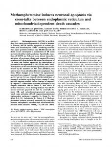
Cyclophilin
A
Figure 4. A) CsA but not FK506 blocks nucleosome-size DNA ladder formation in T5-treated epithelial cells. The primary epithelial cells were treated with or without 100 nM T5 and/ or CsA (600 ng/ml) or FK506 (10 ng/ml) for 1 day. The genomic DNA was isolated, analyzed on an agarose gel, stained with ethidium bromide, and visualized under ultraviolet light. B) Both CsA and FK506 inhibit the ladder formation in activated T cell hybridomas. Al .1 cells were treated under the conditions indicated and the genomic DNA was analyzed on a 1% agarose gel.
FKBP complex is incapable of inhibiting the calcineurin-dependent tyrosine phosphatase and thus has no effect on AICD (Fig. 2C; 27). Rap was also found to be incapable of inhibiting T3-induced epithelial cell death (Fig. 3A, B). These results indicate that AICD and the Ts-dependent epithelial apoptosis have distinct mechanisms but share a common, GsA-sensitive step.
A has a distinct
nuclease
activity
The molecular basis of these differential effects of GsA and FK506 on T3-induced apoptosis is unclear at present. One possible explanation for the different effects of the two immunosuppressants is that although both CsA and FK506 may inhibit AIGD in T cells through inhibition of calcineurin-dependent phosphatase (22), CsA can also exert its effect on one or more other steps leading to apoptosis. The lack of a detailed understanding of the molecular pathway leading to apoptosis makes it difficult to pinpoint the step (or steps) at which CsA acts during T3-induced apoptosis. On the other hand, a major difference between GsA and FK506 is their binding proteins, i.e., cyclophilin A and FKBP, respectively (30). It has been suggested that cyclophilin A has DNA degradation activity based on radioactive gel nuclease assay (31). We have investigated this possibility by examining the ability of cyclophilin A to digested supercoiled plasmid DNA. We found that purified cyclophilin A could digest supercoiled plasmid DNA in a dose-dependent manner to produce nicked circular and linearized DNA (Fig. 5). However, no further degradation of the linear DNA was observed under the incubation conditions, suggesting that the cyclophilin A has a distinct nuclease activity. The observed nuclease activity by cyclophilin A was relatively weak, requiring high concentrations of cyclophilin A. Nevertheless, we could not demonstrate any inhibition of this nuclease activity by GsA. It is possible that this intrinsic nuclease activity of cyclophilin A needs to be further activated during apoptosis and that CsA can effectively inhibit this activation process or the activated nuclease activity.
DISCUSSION T3 induces a typical nucleosomal DNA ladder can be blocked by CsA but not F.K506
that
A major characteristic of mammalian apoptosis is the generation of a ladder of multinucleosomal-sized genomic DNA fragments. To investigate whether the T3-induced intestinal epithelial apoptosis has such a property, we analyzed genomic DNA after 1 day of T3 treatment by agarose-gel electrophoresis. As depicted in Fig. 4A, the DNA from control epithelial cells appeared to be intact. However, a 1-day T3 treatment resulted in classical nucleosomal-sized DNA fragmentation. In agreement with results of the ELISA assay shown above, GsA also blocked the DNA ladder formation both in T3-induced tadpole epithelial cell death (Fig. 4A) and activation-induced T cell apoptosis, whereas FK506 inhibited only DNA fragmentation in T cells (Fig. 4B).
THYROID
HORMONE-INDUCED
EPITHELIAL
CELL DEATH
The causative role of thyroid hormone during phibian metamorphosis has been demonstrated
-
el
In
(1
If)
-
el
If)
-
Cydophilin -
amnot
A
(-) Circular Njcked Lmear
Closed Figure 5. Cleavage of supercoiled DNA by kb supercoiled plasmid (closed circular) indicated
amounts
of cyclophilin
A and
Circular
cyclophilin A. A 7.2 was incubated with then analyzed on a
0.8% agarose gel.
563
only by its ability to induce precocious metamorphosis when added to rearing water of premetamorphic tadpoles but also by its ability to cause organ-autonomous transformations in vitro (5, 14, 24). We have now extended these observations to show that isolated intestinal primary epithelial cells cultured in vitro can respond to T3 to reproduce the apoptotic changes that occur during metamorphosis, indicating that certain features of metamorphosis are cell autonomous. More important, our biochemical characterizations, especially the demonstration of the formation of a nucleosomal DNA ladder upon T3 treatment of the cells, indicate that epithelial cell death during intestinal remodeling shares considerable similarity to a variety of cell death in mammals, implying a conservation at the mechanistic level. We have shown here that the immunosuppressant GsA can inhibit intestinal epithelial cells death. Its exact mechanism of action is yet unclear. Two earlier studies of tadpole tail tissues suggest that adult-type non-T leukocytes might participate in the specific elimination of tadpole tail tissues (32, 33). This raises the possibility that the immunosuppressant GsA might suppress the function of these non-T leukocytes that may be present in the intestinal epithelial cell cultures, thus blocking epithelial cell death. However, such a mechanism seems to be very unlikely, as two other immnosuppressants (FK507 and rapamycm) failed to inhibit the epithelial cell death. Furthermore, the levels of such intestinal adult-type non-T leukocytes, if they exist, in the young tadpoles used in our study are likely to be to low to account for the massive apoptosis within the short treatment period in vitro. Thus, it is likely that thyroid hormone induces apoptosis directly within the epithelial cells and that GsA inhibits this process. Thyroid hormone is known to exert its effects by regulating gene transcription through its nuclear receptors, which are transcription factors (34-36). Indeed, many thyroid hormone-regulated genes have been isolated from different amphibian tissues (3, 9, 10, 12, 13). None of the genes that have been shown to be involved in cell death regulation are among these amphibian thyroid hormone response genes. Instead, many of the early response genes encode transcription factors and proteins that could potentially affect cell-cell and cell-extracellular matrix interactions (9, 15). Thus, it is likely that these early genes are involved in regulating the expression of downstream genes that are ultimately responsible for cell fate determination. The mechanisms by which these early genes affect downstream events and what the downstream genes are remain to be elucidated. Our studies with the immunosuppressants have provided some clues for this thyroid hormone induction pathway, i.e., the presence of a GsA-sensitive step (or steps) during larval epithelial cell death. On the other hand, both immunosuppressants GsA and
564
Vol. 11
June 1997
FK506 can inhibit AICD (22, 27). This is believed to be due to the inhibition of calcineurin-dependent tyrosine phosphatase by the complexes of GsA and FK506 with their binding proteins, cyclophilin and FKBP, respectively (22, 30). However, the data presented here clearly show the inability of FK506 to inhibit T3-induced epithelial cell death. These results may suggest that FKBP is absent in larval epithelial cells. This seems unlikely, as it has previously been demonstrated that FKBP is present even in bacteria (37). Furthermore, FKBP amino acid sequence is highly conserved from bacteria, yeast, to humans (38), indicating the likelihood of the existence of this protein in amphibians. So it is possible that the calcineurin-dependent tyrosine phosphatase may not be involved in thyroid hormone-dependent epithelial cell death. One possible explanation for our observation is that although both GsA and FK506 may inhibit AICD in T cells through inhibition of calcineurin-dependent phosphatase, GsA can also exert its effect on one or more other steps leading to apoptosis. We have shown here that cyclophilin A has a distinct nuclease activity toward a supercoiled plasmid. Little nuclease activity toward linear DNA was detected under our conditions. This seems to differ from the results of Montague et al. (31), who, using a radioactive gel nuclease assay, reported the degradation of linear DNA by cyclophilin A (31). However, much higher ratios of cyclophilin/DNA were used in their experiments compared to those in our experiments. In any case, the observed nuclease activity raises the possibility that cyclophilin A participates directly in DNA degradation during apoptosis. Our preliminary experiments using purified human cyclophilin A have so far failed to detect any nuclease activity toward isolated nuclei (unpublished observation). In addition, the observed nuclease activity is relatively low and is insensitive to GsA inhibition under the conditions tested. However, this activity might be further activated during apoptosis to cleave nuclear DNA, and GsA could effectively block this activation in vivo. Alternatively, the inhibition of this nuclease activity of cyclophilin A by CsA may require some other cellular factors. Another possibility is suggested by the recent report that GsA inhibits the DNA binding activity of the transcription factor Nur77 in T cells (39). Nur77 is a transcription factor belonging to the superfamily of the steroid/thyroid hormone receptors and is required for the activation-induced T cell apoptosis (40, 41). It is therefore suggested that the inhibition of Nur77 activity by GsA may block the ability of Nur77 to regulate its target genes, thus preventing AIGD. As both thyroid hormone receptors and Nur77 belong to the same receptor family, a possibility exists that GsA may directly block the function of thyroid hormone receptors. Alternatively, a gene encoding
The FASEB Journal
SU ET AL.
the frog Nur77 may be involved in the pathway leading to the thyroid hormone-induced apoptosis, and its inhibition by GsA may be responsible for the observed effects of GsA. Future experiments should be able to test these different possibilities directly.
21.
22.
23.
24.
REFERENCES 1. Schwartzman,
R. A., and Cidlowski, J. A. (1993 ) Apoptosis: The an molecular biology of programmed cell death. EndocrR,v. 14, 133-151 2. Wyllie, A. H., Kerr,J. F. R., and Curie, A. R. (1980) Cell death: the significance of apoptosis. mt. Rev. Cttol. 68, 251-306 3. Gilbert, L. 1., Tata,J. R., and Atkinson, B. G. (1996) Metamorphosis: Post-embryonic Reprogramming of Gene Expression in Amphibian and Insect Cells, Academic Press, New York 4. Ishizuya-Oka, A., and Shimozawa, A. (1992) Programmed cell death and heterolysis of larval epithelial cells by macrophagelike cells in the anuran small intestine in vivo and in vitro. J. Morphol. 213, 185-195 5. Tata,J. R., Kawahara, A., and Baker, B. S. (1991) Prolactin inhibits both thyroid hormone-induced morphogenesis and cell death in cultured amphibian larval tissues. Dev. Biol.146, 72-80 6. Dodd, M. H. I., and Dodd, J. M. (1976). The biology of metamorphosis. In Physiology oftheAmphibia (Lofts, B., ed) pp. 467599. Academic Press, New York 7. Gilbert, L. I., and Frieden, E. (1981).Metamorphosis:A Problem in Developmental Biology, 2nd Ed, Plenum Press, New York 8. Tata,J. R. (1993) Gene expression during metamorphosis: An ideal model for post-embryonic development. BioEssays15.239248 9. Brown, D. D., Wang, Z., Furlow, J. D., Kanamon, A., Schwartzman, R. A., Remo, B. F., and Pinder, A. (1996) The thyroid hormone-induced tail resorption program during Xenoptis laevis metamorphosis. Proc. Nat!. Acad. Sci, USA 93. 1924-1929 10. Buckbinder, L., and Brown, D. D. (1992) Thyroid hormone-induced gene expression changes in the developing frog limb. j Biol. C/tern. 267, 25786-25791 11. Shi, Y.-B. (1994) Molecular biology of amphibian metamorphosis: A new approach to an old problem. Trends Endocrinol. Metab. 5, 14-20 12. Shi, Y. B., and Brown, D. D. (1993) The earliest changes in gene expression in tadpole intestine induced by thyroid hormone.]. Biol.Chem. 268, 20312-20317 13. Wang, Z., and Brown, D. D. (1993) Thyroid hormone-induced gene expression program for amphibian tail resorption.]. BioL Chem. 268, 16270-16278 14. Dauca, M., and Hourdry,J. (1985) Transformations in the intestinal epithelium during anuran metamorphosis. In Metamorphosis (Balls, M., and Bownes, M., eds) pp. 36-58, The Clarendon Press, Oxford, U.K 15. Shi, Y.-B., and lshizuya-Oka, A. (1996) Biphasic intestinal development in amphibians: Embryogensis and remodeling during metamorphosis. Cur,. Topics Dev. BioL 32, 205-235 16. Yoshizato, K. (1989) Biochemistry and cell biology of amphibian metamorphosis with a special emphasis on the mechanism of removal of larval organs. mt. Rev. Cytol. 119, 97-149 17. Kerr, J. F. R., Harmon, B., and Searle, J. (1974) An electronmicroscope study of cell eletion in the anuran tadpole tail during spontaneous metamorphosis with special reference to apoptosis of striated muscle flbres.j Gell Sci.14, 571-585 18. Nieuwkoop. P. D.. and Faber,J. (1956) Normal table of Xenopus laevis, North Holland Publishing, Amsterdam 19. Ishizuya-Oka, A., and Shimozawa, A. (1992) Connective tissue is involved in adult epithelial development of the small intestine during anuran metamorphosis in vitro. Rowe’s Arch. Den. Biol. 201, 322-329 20. Marshall,J. A., and Dixon, K. E. (1978) Cell specialization in the epithelium of the small intestine of feeding Xenopus laevis. j Anal. 126, 133-144
25.
biochemistry
THYROID
HORMONE-INDUCED
EPITHELIAL
CELL DEATH
26.
27.
28. 29. 30.
31.
32.
33.
34.
35.
36.
37.
38.
39.
40.
41.
Ishizuya-Oka. A., and Shimozawa, A. (1987) Development of the connective tissue in the digestive tract of the larval and metamorphosing Xenopu.s laevis. Anat. Anz.]ena 164, 81-93 Shi, V., Sahai, B. M., and Green, D. R. (1989) Cyclosporin A inhibits activation-induced cell death in T-cell hybridomas and thymocytes. Nature (London) 339, 625-626 Mosmann, T. (1983) Rapid colorimetric assay for cellular growth and survival: Application to proliferation and cytotoxicity assays. j Immunol. Methods 65, 55-63 Ishizuya-Oka, A., and Shimozawa, A. (1991) Induction of metamorphosis b’ thyroid hormone in anuran small intestine cultured organotypically in vitro. In Vitro Cell. Dee. Biol.27A, 853857 Shi, V. B.. and Hayes, W. P. (1994) Thyroid hormone-dependent regulation of the intestinal fatty acid-binding protein gene during amphibian metamorphosis. Den. Biol. 161, 48-58 Telford, W. G., King, L. E., and Fraker, P.J. (1991) Evaluation of glucocorticoid-induced DNA fragmentation in mouse thymocytes by flow cytometrv. Cell Prolj 24, 447-459 Birer, B. E., Matilla, P. S., Standaert, R. F.. Hcrzenburg, I.. A., Burakoff, S. J., Crabtree, C., and Schreiher, S. L. (1990) Two distinct signal transmission pathways in T lymphocytes are inhibited by complexes formed between an immunophilin and either FK506 or rapamycin. Proc. Nat!.Acad. Sri. USA 87, 9231-9235 McKeon, F. (1991) When worlds collide: immunosuppressants meet protein phosphatases. Cell 66, 823-826 Schreiber, S. L. (1992) Immunophilin-sensitive protein phosphatase action in cell signaling pathways. (jell 70, 365-368 Liu,J., Farmer,J. D., Lane, W. S., Friedman,J., Weissman, I., and Schreiber, S. L. (1991) Calcineurin is a common target of cyclophilin-cyclosporin A and FKBP-FK506 complexes. Cell66, 807815 Montague, J. W., Gaido, M. L., Frye. C., and Cidlowski, J. A. (1994) A calcium-dependent nuclea.se from apoptotic rat thymocytes is homologous with cyclophilin. Recombinant cyclophilins A, B, and C have nuclease activity. J. Biol.C/tern. 269, 18877-18880 Izutsu, V., and Yoshizato, K. (1993) Metamorphosis-dependent recognition of larval skin as non-self by inbred adult frogs (Xenopus laevis).j Exp. Zoo!. 266, 163-167 Izutsu, V., Yoshizato, K., and Tochinai, S. (1996) Adult-type splenocytes of Xenopus induce apoptosis of histocompatible larval tail cells in vitro. D[ferentiation 60, 277-286 Mangelsdorf, D. J., Thummel, C., Beato, B., Herrlich, P., Schutz, C., Uniesono, K., Blumberg, B., Ka.stner, P., Mark, M., Chambon, P., and Evans, R. M. (1995) The nuclear receptor superfamily: The second decade. Cell 83, 835-839 Shi, Y.-B., Wong,J., and Puzianowska-Kuznicka, M. (1996) Thyroid hormone receptors: Mechanisms of transcriptional regulation and roles during frog development. j Biomed.Sci. 3. 307318 Tsai, M.-J., and O’Malley, B. W. (1994) Molecular mechanisms of action of steroid/thyroid receptor superfamily members. Annu. Rev. Biochem. 63, 45 1-486 PahI, A., and Keller, U. (1992) FK-506-hinding proteins from streptomycetes producing immunosuppressive macrolactones of the FK-506 type.]. Bacteriol. 174, 5888-5894 Home, S. M., and Young, K. D. (1995) Escherichia coliand other species of the Enterobacteriaceae encode a protein similar to the family of Mip-like-hinding proteins. Arch.Mierobiol. 163, 357-365 Yazdanbakhsh, K., Choi, J.-W.. Li, V., [.au, I.. F.. and Choi, Y. (1995) Cyclosporin A blocks apoptosis by inhibiting the DNA binding activity of the transcription factor Nur77. Proc. NatI. Acad. Sri. USA 92, 437-441 Liii, Z., Smith, S. W., McLaughlin, K A.. Schwartz, L. M., and Osborne, B. A. (1994) Apoptotic signals delivered through the T-cell receptor of a T-cclI hybrid require the immediate-early gene nur77. Nature (London) 367, 28 1-284 Woronicz,J., Calnan, B., Ngo, V., and Winoto, A (1994) Requirement for the orphan steroid receptor Nur77 in apoptosis ofTcell hybridomas. Nature (London) 367, 277-281 Received forpublication ]anuary Accepted for publication April
14, 1997. 18, 1997.
565