attracting neutrophils to the lung and the mediators of pathogenesis in BPD have not been ... and epithelial cell-derived neutrophil-activating peptide (ENA-.
INFECTION AND IMMUNITY, Dec. 1997, p. 5131–5136 0019-9567/97/$04.0010 Copyright © 1997, American Society for Microbiology
Vol. 65, No. 12
Induction of Neutrophil Chemoattractant Cytokines by Mycoplasma hominis in Alveolar Type II Cells THOMAS KRUGER
AND
JOHN BAIER*
Department of Pediatrics, Louisiana State University Medical Center, Shreveport, Louisiana 71130-3932 Received 5 June 1997/Returned for modification 16 July 1997/Accepted 23 September 1997
Bronchopulmonary dysplasia (BPD) is a chronic lung disease of premature infants who are mechanically ventilated due to respiratory distress. The disease consists of an initial inflammatory influx of neutrophils to the lungs, followed by long-term chronic fibrosis of the lung tissue. The antigenic repertoire that initiates the inflammatory component of BPD has not been defined. Furthermore, the repertoire of cytokines responsible for attracting neutrophils to the lung and the mediators of pathogenesis in BPD have not been characterized. Mycoplasmas such as Mycoplasma hominis and Ureaplasma urealyticum have been isolated from the lungs of infants that developed BPD and yet have not been widely recognized as potential initiators of the inflammatory component of BPD. In the studies described here, we examined the ability of both viable and heat-killed Mycoplasma hominis to elicit type II epithelial cell production of cytokines that are chemotactic for polymorphonuclear leukocytes (PMNs), particularly interleukin-8 (IL-8) and epithelial cell-derived neutrophil-activating peptide (ENA-78). The results of these studies demonstrate that M. hominis and M. hominis antigen are potent stimulators of type II epithelial cell-derived IL-8 and ENA-78. Thus, these data strongly suggest that the presence of M. hominis in the lungs of premature infants may initiate the inflammatory component of BPD by inducing epithelial cell production of cytokines chemotactic for PMNs. Furthermore, these data suggest that the onset of the inflammatory component of BPD likely precedes, and is independent of, the recruitment and activation of alveolar macrophages. ENA-78 in direct response to mycoplasmas, particularly Mycoplasma hominis.
Bronchopulmonary dysplasia (BPD) is a chronic lung disease that develops primarily in premature very-low-birthweight infants (,1,500 g) who require mechanical ventilation and oxygen therapy. The disease is typified by an initial inflammatory influx of polymorphonuclear leukocytes (PMNs) into the lungs and is followed by long-term chronic fibrosis lasting from weeks to years (10, 17). The inflammatory response that precedes BPD is poorly characterized. Neutrophil chemoattractant and activating cytokines such as interleukin-8 (IL-8) and epithelial cell-derived neutrophil-activating peptide (ENA78) presumably play a central role in the recruitment of PMNs into the lung (18). In support of this assumption is the finding that the level of one of these chemokines, IL-8, is elevated in the tracheal aspirates of infants that subsequently develop BPD (10, 11, 16, 17, 22, 25). The role of ENA-78 in BPD and the levels found in the neonatal tracheal aspirates of infants that develop BPD have not been ascertained. Recent studies demonstrate that respiratory epithelial cells are capable of producing many inflammatory cytokines (IL-8, ENA-78, and monocyte chemoattractant protein 1) in response to both macrophage-derived cytokines (e.g., IL-1 and tumor necrosis factor alpha) and respiratory pathogens (2, 3, 8, 12, 13, 15, 20, 21, 24, 26). However, the “inducing antigens” that trigger the inflammatory component of BPD are unknown. Further, the role (if any) in the development of BPD by activated alveolar macrophages and their proinflammatory cytokines remains undefined. Previous studies demonstrate that mycoplasmas are frequently isolated from the tracheal aspirates of infants with BPD (7, 27). Thus, the present studies were designed to determine if pulmonary type II epithelial cells produce IL-8 and
MATERIALS AND METHODS Cytokines and antibodies. Recombinant human IL-8, monoclonal rat (immunoglobulin G1k [IgG1k]) anti-human IL-8 antibodies, and polyclonal rabbit (IgG) anti-human IL-8 antibodies used in the enzyme-linked immunosorbent assay (ELISA) were purchased from Endogen (Cambridge, Mass.). Recombinant human ENA-78, monoclonal mouse (IgG1) anti-human ENA-78 antibodies, and polyclonal goat (IgG) anti-human ENA-78 antibodies were obtained from R&D Systems (Minneapolis, Minn.). Recombinant human IL-1b was obtained from Endogen. Peroxidase-conjugated polyclonal goat (IgG) anti-rabbit IgG detecting antibody was obtained from Bio-Rad Laboratories (Hercules, Calif.). Cell cultures. A549 type II epithelial cells (American Type Culture Collection, Rockville, Md.) were plated in 24-well plates at 3 3 105 cells/well/ml in F12K medium containing 10% fetal calf serum, and the plates were incubated overnight at 37°C in 5% CO2. The medium from confluent monolayers was removed, and quadruplicate wells were either stimulated for 48 h with a heat-killed M. hominis preparation equivalent to 108 bacteria/ml or infected with 3.8 3 106 M. hominis bacteria/well (100 ml) for 1 h at 37°C. After viable M. hominis was allowed to attach to the cell monolayers, fresh antibiotic-free F12K medium containing 1% fetal calf serum was added to the wells to a final volume of 1 ml and the cultures were incubated at 37°C in 5% CO2 for 48 h. Recombinant human IL-1b was added to quadruplicate A549 culture wells (without mycoplasma) as a positive control stimulator of IL-8 and ENA-78. Culture supernatants were then collected and frozen until the IL-8 and ENA-78 concentrations in the supernatants could be determined by ELISA as described below. Total RNA was isolated from quadruplicate A549 cultures, and the cytokine-specific mRNA was assessed by semiquantitative reverse transcription-PCR (RT-PCR) as described below. Bacterial culture. M. hominis, serotype 5, ATCC 23114 was grown in arginineenriched broth (AB) for 48 h at 37°C in 5% CO2 (14). M. hominis was harvested and concentrated by centrifugation for 15 min at 14,000 3 g. The resulting M. hominis pellet was reconstituted in fresh AB, and aliquots were prepared for cryopreservation in liquid nitrogen by the addition of 20% glycerol. The M. hominis stock used for all viable M. hominis studies contained approximately 7.6 3 107 M. hominis cells/ml after recovery from liquid nitrogen as determined by backtitration in AB culture media and was used in our studies at a multiplicity of infection of approximately 10:1. A heat-inactivated M. hominis stock, equivalent in titer to the viable M. hominis stock, was prepared by exposing viable M. hominis (108 M. hominis cells/ml) to 95°C for 10 min. This stock was used as a heat-inactivated M. hominis antigen preparation. Lethality to M. hominis was
* Corresponding author. Mailing address: Department of Pediatrics, Louisiana State University Medical Center, 1501 King’s Highway, Shreveport, LA 71130-3932. Phone: (318) 675-7808. Fax: (318) 6754790. 5131
5132
KRUGER AND BAIER
confirmed by the inability of this preparation to contain viable M. hominis growth activity after 7 days of culture in AB. ELISA. The ELISA for human IL-8 and ENA-78 was performed as described. Briefly, 96-well ELISA plates (Costar, Cambridge, Mass.) were coated overnight at 4°C with 100 ml of monoclonal anti-human IL-8 or ENA-78 antibodies per well at 1.3 mg/ml in carbonate coating buffer (pH 9.6). Plates were then washed three times for 5 min each with 200 ml of phosphate-buffered saline (PBS)-Tween 20 (pH 7.4) per well and then blocked for 2 h at ambient temperature (or overnight at 4°C) with 200 ml of PBS-Tween 20 containing 2% (wt/vol) bovine serum albumin (Sigma Chemical Co., St. Louis, Mo.). Plates were then washed three times with PBS-Tween as described above and incubated for 2 h at ambient temperature with 100 ml of blocking buffer per well containing either A549 culture supernatant or recombinant cytokine standards at the indicated concentrations. Plates were then washed three times in PBS-Tween and incubated overnight at 4°C with 100 ml of the appropriate polyclonal anti-human IL-8 or ENA-78 antibodies per well at 1.3 mg/ml in blocking buffer. Next, the plates were washed three times and incubated for 2 h at ambient temperature with 100 ml of blocking buffer containing polyclonal goat (IgG) anti-rabbit IgG peroxidaseconjugated antibody (Bio-Rad) at a 1:3,000 final dilution. Plates were then washed three times, developed with a 2,29-azinobis(3-ethylbenzthiazolinesulfonic acid) (ABTS) peroxidase substrate kit (Bio-Rad) according to the manufacturer’s specifications, and read at 405 nm with an ELISA Autoreader (Caymen Chemical, Ann Arbor, Mich.). All experimental IL-8 and ENA-78 levels were determined by comparing the mean absorbance at 405 to those obtained for a curve generated from recombinant human IL-8 and ENA-78 standards. Correlation coefficients of .0.96 are typical for this standard curve. Total RNA isolation. Isolation of total RNA was performed with RNeasy minikits from Qiagen (Chatsworth, Calif.) according to the manufacturer’s specifications. Briefly, after culture supernatants for ELISA analysis were removed, the remaining quadruplicate cell monolayers were lysed and applied to silica gel microcentrifuge spin columns. The columns were washed in appropriate buffers, and total RNA was eluted with 50 ml of RNase-free double-distilled water. The total RNA recovered from each sample was quantified and assessed for purity by spectrophotometric analysis at 260 and 280 nm. Equivalent amounts (0.5 mg) of the resulting total RNA were then subjected to RT-PCR analysis to assess the relative concentrations of cytokine-specific mRNA between treatments. Primers. The oligonucleotide primers used for RT-PCR were synthesized (20 mM each) commercially by Integrated DNA Technologies Inc. (Coralville, Iowa). The cytokine-specific PCR primer set for IL-8 consists of a 26-base sense primer (59-TAA ACA TGA CTT CCA AGC TGG CCG TG-39) and a 25-base antisense primer (59-CTT CTC CAC AAC CCT CTG CAC CCA G-39) that span genomic intron sequences and result in a 278-bp IL-8-specific cDNA PCR product (19). We developed a cytokine-specific PCR primer set for ENA-78 consisting of a 21-base sense primer (59-GCT GGT CCT GCC GCT GCT GTG-39) and antisense primer (59-GTT TTC CTT GTT TCC ACC GTC-39), using the published sequence for human ENA-78 (9, 23). These primers were derived from unique ENA-78-specific sequences that span genomic intron sequences to eliminate priming from DNA or heteronuclear RNA. The predicted PCR product with these primers would be 237 bp in length. The 21-base sense primer spans intron 1 (133 bases) and encompasses the base codons for the last three amino acids (IAS) in the leader sequence and the first four amino acids (AGPA) at the amino terminus of the mature peptide. The 21-base antisense primer is complementary to that which codes for seven amino acids (ILDGGNK) found in the carboxy terminus of the mature peptide. This primer spans intron 3, which consists of 355 bases. The specific PCR primer set for b-actin was a 21-base sense primer (59-CTG GCA CCC AGC ACA ATG AAG-39) and a 20-base antisense primer (59-ACC GAC TGC TGT CAC CTT CA-39), resulting in a 362-bp cDNA PCR product (6). Reverse transcription of total mRNA into cDNA was performed with a 20-base oligo(dT) primer. PCR. The mRNA from 0.5 mg of total RNA isolated from the different culture conditions was reverse transcribed (first strand synthesis) into single-stranded cDNA, and the cytokine-specific cDNA was amplified by PCR with an Access RT-PCR System kit (Promega). The concentration of each primer was approximately 0.5 mM in a 50-ml reaction mixture. Cycling parameters consisted of an initial oligo(dT)-primed reverse transcription step for 1 h at 37°C. The AMV reverse transcriptase was then inactivated for 2 min at 94°C. The cytokinespecific primary cDNA transcripts were then amplified for 40 cycles, each cycle consisting of an initial melting step at 94°C for 30 s, annealing at 58°C for 1 min, and extension at 68°C for 2 min. All PCR amplifications included a final extension step at 68°C for 10 min to ensure full-length transcripts. Initially, samples of the PCR products were taken at 15, 20, 25, and 35 cycles to determine the minimal number of cycles required to detect products and to optimize differences between treatment groups. As a control, we examined the b-actin mRNA in A549 cells in order to demonstrate a specific effect on IL-8 and/or ENA-78 gene transcription by M. hominis or M. hominis antigen. All RT-PCR products were visualized by 2% agarose gel electrophoresis as described below. Agarose gel electrophoresis. All RT-PCR products were visualized on ethidium bromide-stained 2% agarose gels. Briefly, 10 ml of each RT-PCR reaction mixture was diluted 1:2 in 10 ml of sample buffer (63) containing 40% (wt/vol) sucrose and 0.25% (wt/vol) bromophenol blue as a tracking dye and loaded onto a 15 by 15 cm 2% agarose gel. Electrophoresis was performed in Tris-acetate–EDTA (TAE) buffer at 100 V for 1.5 h. Gels were stained with 0.5
INFECT. IMMUN.
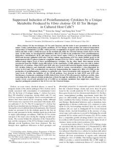
FIG. 1. M. hominis induction of IL-8 and ENA-78 in A549 cells as determined by ELISA. Quadruplicate wells of confluent A549 cells were untreated (Control) or cultured with either fresh AB, M. hominis (Mh) at 3.8 3 106 M. hominis cells/ml, or 5 U of IL-1b per ml for 48 h at 37°C in 5% CO2. Culture supernatants were harvested, and IL-8 (A) and ENA-78 (B) levels were determined by ELISA. Values are the mean cytokine concentrations in nanograms per milliliter 6 the SEMs from multiple experiments (IL-8, n 5 6; ENA-78, n 5 12). Asterisks indicate statistically significant differences compared to untreated controls.
mg of ethidium bromide per ml, and the cytokine-specific cDNA was visualized with a UV transilluminator. The molecular weights of the RT-PCR products were confirmed by comparison to a DNA ladder consisting of standard cDNA that ranges from 100 bp to 1,000 bp in 100-bp increments (Promega). Restriction analysis. Since the ENA-78 primers used in this study have not been described previously, we performed restriction analysis on the ENA-78specific RT-PCR product(s) to confirm that amplification of the appropriate region of the mRNA had occurred. A 10-ml RT-PCR sample from IL-1bstimulated A549 cells was treated with either 1 U of MspAlI or BclI overnight at 37°C in a 50-ml final volume containing the appropriate 13 restriction buffer. The total samples were then electrophoresed and visualized as described above. The molecular weights of the restriction fragments were determined to establish identity with molecular weights predicted from the ENA-78 gene sequence. The predicted number and molecular weights for the restriction fragments when an appropriate ENA-78 product is cut with MspAlI or BclI are two fragments each of 215 and 19 bp or 151 and 83 bp, respectively. Statistical analysis. ELISA data from pooled experiments are reported as the mean cytokine concentrations in nanograms per milliliter 6 the standard errors of the means (SEMs). Data were analyzed by analysis of variance, followed by the Student-Newman-Keuls test of multiple means. P value(s) of less than 0.05 were considered significant.
RESULTS Figure 1 illustrates the effect of M. hominis on the production of IL-8 and ENA-78 by A549 type II epithelial cells as determined by ELISA. Values are the mean cytokine concentrations in nanograms per milliliter 6 SEMs. As can be seen in this figure, M. hominis infection of type II epithelial cells with 3.8 3 106 M. hominis cells/ml/well (multiplicity of infection, 10:1) results in a significant increase in epithelial cell produc-
VOL. 65, 1997
tion of both IL-8 (n 5 6) (Fig. 1A) and ENA-78 (n 5 12) (Fig. 1B) compared to that of either untreated controls or cultures incubated with AB. Infection of cultures with M. hominis for 48 h caused IL-8 production to increase approximately 14-fold, from 0.8 6 0.2 ng/ml (control) to 11.3 6 4 ng/ml (M. hominis). The increase in IL-8 production by M. hominis was similar to that observed in cultures receiving 5 U (50 pg) of IL-1b. In fact, there was no statistically significant difference between IL-8 levels obtained in cultures receiving M. hominis alone and those receiving IL-1b at 5 U/ml. Additionally, there was no significant difference in IL-8 levels between the untreated control and those cultures receiving AB, a host cell-free medium used for culturing M. hominis. Similarly, we observed a significant (P 5 0.02) increase in ENA-78 production in M. hominisinfected A549 cells compared to that in untreated controls (Fig. 1B), even though A549 cells constitutively produce significant amounts of ENA-78 in culture. Infection of cultures with M. hominis for 48 h caused ENA-78 production to double from 37.3 6 6.9 ng/ml (control) to 71.9 6 12.3 ng/ml (M. hominis). Again, M. hominis stimulation of ENA-78 production in these cells was similar to that observed with 50 pg of IL-1b per ml. Taken together, these data suggest that M. hominis directly stimulates the production and secretion of IL-8 and ENA-78 by pulmonary type II epithelial cells. Alternatively, it is possible that M. hominis indirectly stimulates type II cell production of IL-8 and ENA-78 through the action of another factor (cytokine) not yet described. The kinetics of IL-8 and ENA-78 production following M. hominis infection of A549 cultures are illustrated in Fig. 2. Samples harvested at 24 h postinfection reveal little change in either IL-8 (Fig. 2A) or ENA-78 (Fig. 2B) production compared to that of controls. However, the levels of M. hominisinduced IL-8 and ENA-78 were greater after 48 h postinfection than that obtained from untreated 72-h controls (Fig. 2A). While the amount of M. hominis-induced IL-8 and ENA-78 appears to increase further by 72 h postinfection, the difference observed between M. hominis-infected cultures harvested at 48 h and those harvested at 72 h was not statistically different. Thus, these studies demonstrate that the production of IL-8 and ENA-78 increases dramatically from 24 to 48 h after infection with M. hominis. Since it was clear that M. hominis directly stimulates IL-8 and ENA-78 production in A549 type II epithelial cells, we wanted to determine if the enhancement effect required an active M. hominis infection or if heat-inactivated M. hominis could also induce cytokine production in these cells. To test this, we prepared a heat-inactivated M. hominis preparation (;108 M. hominis cells/ml) and assessed its ability to induce the production of IL-8 and ENA-78 in A549 cultures. A typical M. hominis antigen dose-response curve is shown in Fig. 3 which illustrates the amount of cytokine produced (in nanograms/millimeter) in response to heat-inactivated M. hominis at the indicated concentrations (no. of M. hominis cells 3 105/ml/well). As can be seen in this figure, the heat-killed preparation was a potent stimulator of IL-8 and ENA-78 (Fig. 3A and B, respectively) production in A549 cultures. Furthermore, the induction of these cytokines was dose dependent. The IL-8 levels produced in these cultures ranged from 0.6 ng/ml in response to 2 3 105 heat-killed organisms/ml to 5.3 ng/ml in cultures receiving 5 3 106 heat-killed organisms/ml (Fig. 3A). The concentration of IL-8 produced in cultures stimulated with 5 3 106 heat-killed organisms/ml represents an increase of approximately 17.6-fold above background levels (0.3 ng of IL-8/ml). Heat-killed M. hominis also stimulated increased production of ENA-78 in A549 cell cultures (Fig. 3B). The ENA-78 levels produced in these cultures ranged
MYCOPLASMA HOMINIS INDUCTION OF CYTOKINES
5133
FIG. 2. Kinetics of IL-8 and ENA-78 induction by M. hominis in A549 cells as determined by ELISA. Quadruplicate wells of confluent A549 cells were untreated for 72 h or cultured with M. hominis at 3.8 3 106 cells/ml as described. The 1-ml culture supernatants were harvested at 24, 48, and 72 h postinfection, and IL-8 (A) and ENA-78 (B) levels were determined by ELISA. Values are the mean cytokine concentrations in nanograms per milliliter 6 the SEMs from multiple experiments (n 5 3).
from 65 to 163 ng/ml in response to 2 3 105 and 5 3 106 heat-killed organisms per milliliter, respectively. The concentration of ENA-78 produced in cultures stimulated with 5 3 106 heat-killed organisms/ml represents an increase of approximately 3.2-fold above background levels (49.9 ng of ENA-78/ ml). Taken together, the results of these studies suggest that both viable and nonviable M. hominis (M. hominis antigen) are capable of stimulating the production and secretion of IL-8 and ENA-78 in A549 type II epithelial cells. To determine if M. hominis stimulates cytokine gene transcription, we performed RT-PCR to assess IL-8- and ENA-78specific mRNA in M. hominis-infected and untreated A549 cells. While the IL-8 primers have been described previously (19), we derived appropriate ENA-78-specific primers from the published amino acid sequence and gene sequence for human ENA-78 (GenBank accession no. L37036). The predicted size of the specific ENA-78 RT-PCR product is 237 bp. Total RNA was isolated from control and M. hominis-infected A549 cultures, and equal amounts (0.5 mg) were used as a template for cytokine-specific RT-PCR. Hence, the differences observed in the cytokine-specific RT-PCR products directly reflect differences in the relative frequency of cytokine-specific mRNAs induced by the different cell culture treatments. Figure 4 shows the IL-8- and ENA-78-specific mRNA-dependent RT-PCR products from control and M. hominis-infected A549 cells after 20 amplification cycles. Twenty amplification cycles
5134
KRUGER AND BAIER
INFECT. IMMUN.
FIG. 4. RT-PCR analysis of M. hominis-induced cytokine mRNA. Relative differences in cytokine-specific mRNA-dependent RT-PCR products following electrophoresis on 2% agarose gels and subsequent staining with ethidium bromide are shown. Template RNAs were isolated from untreated (Cont.), ABtreated, M. hominis-infected (Mh), IL-1b-stimulated, or heat-killed M. hominis antigen-stimulated (Mh Ag) A549 cultures, and equal amounts of total RNA were subjected to oligo(dT)-primed RT-PCR with amplification primers specific for IL-8 (top), ENA-78 (middle), or b actin (bottom). The sizes of the different cytokine-specific RT-PCR products are shown. All RT-PCR products were compared to a standard DNA ladder containing DNA fragments ranging from 100 to 1,000 bp in 100-bp increments (Fig. 5).
of b-actin mRNA in any of the different culture conditions. To confirm that the ENA-78 primers were resulting in the amplification of the appropriate RT-PCR product, we performed restriction analysis on the RT-PCR product, using two restriction enzymes, MspAlI and BclI. As can be seen in Fig. 5, the restriction analysis resulted in the appropriate fragments predicted by the published ENA-78 gene sequence of 215 and 19 FIG. 3. Dose-dependent induction of IL-8 and ENA-78 by heat-inactivated M. hominis as determined by ELISA. A typical dose-response curve is shown. Quadruplicate wells of confluent A549 cells were cultured with heat-inactivated M. hominis (Mh) at the indicated concentrations for 48 h at 37°C in 5% CO2. The 1-ml culture supernatants were harvested, and IL-8 (A) and ENA-78 (B) levels were determined by ELISA. Values represent a single typical dose-response curve and are expressed as the mean cytokine concentrations in nanograms per milliliter 6 standard deviations.
produced PCR products from the linear phase of the amplification process; this was the lowest number of cycles that would yield cytokine-specific products that could be visualized. It is evident in Fig. 4 that both viable M. hominis and M. hominis antigen increase the relative abundance of IL-8- and ENA-78specific mRNAs compared to controls, without influencing the transcription of other genes, such as b-actin. While the IL-8specific RT-PCR product from untreated cultures was barely visible, infection with viable M. hominis or stimulation with heat-inactivated M. hominis resulted in a dramatic increase in the amount of IL-8-specific 278-bp RT-PCR product. These results mirror those of the ELISA analysis, which demonstrated negligible constitutive IL-8 production in A549 cultures and significantly enhanced production of IL-8 in cultures receiving M. hominis and M. hominis antigen (Fig. 1A). The amount of 237-bp ENA-78-specific RT-PCR product was significantly increased in M. hominis- and M. hominis antigentreated cultures, reflecting an increase in the abundance of cytokine-specific mRNA compared to that of untreated controls. Again, these results reflect the constitutive ENA-78 production and the enhancement by M. hominis and M. hominis antigen observed in the ELISA analysis (Fig. 1B). No change was observed in the 362-bp b-actin-specific RT-PCR product, suggesting that there was no effect on the relative abundance
FIG. 5. Restriction analysis of the ENA-78 RT-PCR amplification product. Restriction analysis of IL-1b-induced ENA-78-specific RT-PCR amplification product following incubation overnight with restriction enzymes, electrophoresis on 2% agarose gels, and subsequent staining with ethidium bromide. Equal amounts of ENA-78-specific RT-PCR amplification product were incubated overnight at 37°C in the absence or presence of 1 U of either MspAlI or BclI. The sizes of the restriction fragments are indicated. All products were compared to a standard DNA ladder containing DNA fragments ranging from 100 to 1,000 bp in 100-bp increments (right lane).
MYCOPLASMA HOMINIS INDUCTION OF CYTOKINES
VOL. 65, 1997
bp or 151 and 83 bp when the RT-PCR product was digested with MspAlI and BclI, respectively. Thus, taken together, these data strongly suggest that M. hominis and M. hominis antigen potentiate IL-8 and ENA-78 production in A549 type II epithelial cells by increasing cytokine gene transcription. DISCUSSION The mechanisms that underlie the onset and development of BPD have not been adequately characterized. It is likely that the factors responsible for initiating the inflammatory component of BPD are complex and multifactorial. The microorganisms in the lungs of premature infants who require mechanical ventilation must be considered potential cofactors in the development of BPD, even when these microorganisms are of low virulence. This finding is particularly evident in light of the present studies demonstrating that lung epithelial cells are capable of responding to viable and heat-inactivated M. hominis by secreting inflammatory cytokines. Particularly, these studies were designed to determine if M. hominis could elicit the type II epithelial cell production of IL-8 and ENA-78, two inflammatory cytokines thought to play a central role in recruiting neutrophils to the lungs. These studies demonstrate that both viable and heat-inactivated M. hominis cells are potent stimulators of IL-8 and ENA-78 production by A549 pulmonary type II cells. When A549 cell cultures were infected with viable M. hominis, the amount of IL-8 production increased approximately 14-fold above background production levels. Similarly, the amount of ENA-78 produced by these cells in response to M. hominis was double the amount that was constitutively produced in these cultures. The increased production of both cytokines by A549 cells is likely due to the M. hominis-induced enhancement of cytokine gene transcription. While increased levels of ENA-78 have not been shown in the tracheal aspirates of infants with BPD, ENA-78 production is elevated in the lungs of children with cystic fibrosis, supporting a role for ENA-78 in chronic lung inflammation (5). Since ENA-78 is not constitutively expressed in the lung fluids of normal infants, the magnitude of M. hominis-induced ENA-78 production observed in the present studies may be much less than the potential increase in M. hominis-induced ENA-78 production in vivo (5). Taken together, these data suggest that an active pulmonary M. hominis infection in preterm infants would likely result in type II cell production of both IL-8 and ENA-78, two inflammatory cytokines capable of attracting the PMNs that mediate the inflammatory component of BPD. The induction of IL-8 and ENA-78 in type II cells does not require an active infection by M. hominis. When A549 cultures were stimulated with a heat-killed M. hominis antigen preparation, significant increases in IL-8 and ENA-78 production were observed compared to levels of production in untreated controls. As demonstrated previously, this result is likely due to an increase in transcription of the IL-8 and ENA-78 genes. Thus, these data suggest that the presence of M. hominis in the lung may result in cytokine (IL-8 and ENA-78) production by type II cells in the absence of an ongoing infection. Hence, an active lung infection by mycoplasmas may not be necessary to initiate the onset of BPD, since both viable and nonviable M. hominis cells are potent stimulators of cytokine production in type II cells. These data are important, since the presence of M. hominis in the lungs of infants who developed BPD has been considered incidental and of less significance than the presence of another mycoplasma, Ureaplasma urealyticum, which has also been isolated from the lungs of infants who subsequently developed BPD (7, 27). In fact, recent studies demonstrate that M. hominis infections frequently occur as
5135
coinfections with U. urealyticum (4, 27). Thus, it is possible that the role of M. hominis in the development of BPD has been underestimated due to a lack of routine culturing for M. hominis in tracheal aspirates from children at high risk for developing BPD. Finally, these data suggest that the activation of alveolar macrophages and the subsequent production of macrophagederived proinflammatory cytokines such as IL-1b and tumor necrosis factor alpha are not prerequisites for the initiation of the inflammatory component of BPD. In fact, studies demonstrate that except in children born with congenital pneumonia, alveolar macrophages are nonexistent at birth but begin colonizing the lungs by 48 h after birth (1). Thus, the type II cell may function as an immune cell by mediating inflammatory responses that occur in the first two days of life. The ability of different types of epithelial cells (including pulmonary epithelial cells) to serve as immune accessory cells by producing cytokines in response to antigen has been documented for a number of different antigens and/or pathogens (2, 3, 13, 14, 21). Recent studies suggest an essential role for gut epithelial cells in the development of intraepithelial T lymphocytes (28). In light of the data presented in this report, it is interesting to speculate that perhaps pulmonary type II cells provide the first line of defense against infections in both premature and fullterm infants prior to the recruitment of alveolar macrophages to the lungs. REFERENCES 1. Alenghat, E., and J. R. Esterly. 1984. Alveolar macrophages in perinatal infants. Pediatrics 74:221–223. 2. Amin, R., R. Wilmott, Y. Schwarz, B. Trapnell, and J. Stark. 1995. Replication-deficient adenovirus induces expression of interleukin-8 by airway epithelial cells in vitro. Hum. Gene Therapy 6:145–153. 3. Arnold, R., B. Humbert, H. Werchau, H. Gallati, and W. Konig. 1994. Interleukin-8, interleukin-6 and soluble tumour necrosis factor receptor type I release from a human pulmonary epithelial cell line (A549) exposed to respiratory syncytial virus. Immunology 82:126–133. 4. Baier, J., J. Bocchini, and E. Brown. 1994. Are genital mycoplasmas risk factors for intraventricular hemorrhage and hydrocephalus in very low birth weight infants? Pediatr. Res. 35:A293. 5. Bozic, C. R., N. P. Gerard, and C. R. Gerard. 1996. Receptor binding specificity and pulmonary gene expression of the neutrophil-activating peptide ENA-78. Am. J. Respir. Cell Mol. Biol. 14:302–308. 6. Buckley, M. G., C. M. Williams, J. Thompson, P. Pryor, K. Ray, J. H. Butterfield, and J. W. Coleman. 1995. IL-4 enhances IL-3 and IL-8 gene expression in a human leukemic mast cell line. Immunology 84:410–415. 7. Cassell, G. H., K. B. Waites, D. T. Crouse, P. T. Rudd, K. C. Canupp, S. Stagno, and G. R. Cutter. 1988. Association of Ureaplasma urealyticum infection of the lower respiratory tract with chronic lung disease and deaths in very-low-birth-weight infants. Lancet ii:240–244. 8. Choi, A. M., and D. B. Jacoby. 1992. Influenza virus A infection induces interleukin-8 gene expression in human airway epithelial cells. FEBS Lett. 309:327–329. 9. Corbett, M. S., I. Schmitt, O. Riess, and A. Walz. 1994. Characterization of the gene for human neutrophil-activating peptide-78. Biochem. Biophys. Res. Commun. 205:612–617. 10. Groneck, P., B. Go¨tze-Speer, M. Oppermann, H. Eiffert, and C. P. Speer. 1994. Association of pulmonary inflammation and increased microvascular permeability during the development of bronchopulmonary dysplasia: a sequential analysis of inflammatory mediators in respiratory fluids of high-risk neonates. Pediatrics 93:712–718. 11. Groneck, P., and C. P. Speer. 1993. Interleukin-8 in pulmonary effluent fluid of preterm infants. J. Pediatr. 123:839–840. 12. Humbert, R. A., H. Werchau, H. Gallati, and W. Ko¨nig. 1994. Interleukin-8, interleukin-6 and soluble tumour necrosis factor receptor type I release from a human pulmonary epithelial cell line (A549) exposed to respiratory syncytial virus. Immunology 82:126–133. 13. Inoue, H., P. P. Massion, I. F. Ueki, K. M. Grattan, M. Hara, A. F. Dohrman, B. Chan, J. A. Lausier, J. A. Golden, and J. A. Nade. 1994. Pseudomonas stimulates interleukin-8 mRNA expression selectively in airway epithelium, in gland ducts, and in recruited neutrophils. Am. J. Respir. Cell Mol. Biol. 11:651–663. 14. Isenberg, H. D. (ed.). 1994. Clinical microbiology procedures handbook. American Society for Microbiology, Washington, D.C. 15. Kwon, O. J., B. T. Au, P. D. Collins, I. M. Adcock, J. C. Mak, R. R. Robbins, K. F. Chung, and P. J. Barnes. 1994. Tumor necrosis factor-induced inter-
5136
16. 17. 18. 19. 20. 21. 22.
KRUGER AND BAIER
leukin-8 expression in cultured human airway cells. Am. J. Physiol. 267: L398–L405. McColm, J. R., and N. McIntosh. 1994. Interleukin-8 in bronchoalveolar lavage fluid samples as predictor of chronic lung disease in premature infants. Lancet 343:729. Merritt, T. A., J. M. Puccia, and I. D. Stuard. 1981. Cytologic evaluation of pulmonary effluent in neonates with respiratory distress syndrome and bronchopulmonary dysplasia. J. Pediatr. 98:949–955. Miller, M. D., and M. S. Krangel. 1992. Biology and biochemistry of the chemokines: a family of chemotactic and inflammatory cytokines. Crit. Rev. Immunol. 12:17–46. Napoli, J., G. A. Bishop, and G. W. McCaughan. 1994. Increased intrahepatic messenger RNA expression of interleukins 2, 6, and 8 in human cirrhosis. Gastroenterology 107:789–798. Paine, R., III, M. W. Rolfe, T. J. Standiford, M. D. Burdick, B. J. Rollins, and R. M. Strieter. 1993. MCP-1 expression by rat type II alveolar epithelial cells in primary culture. J. Immunol. 150:4561–4570. Palfreyman, R. W., M. L. Watson, C. Eden, and A. W. Smith. 1997. Induction of biologically active interleukin-8 from lung epithelial cells by Burkholderia (Pseudomonas) cepacia products. Infect. Immun. 65:617–622. Papoff, P., P. Fiorucci, F. Ficuccilli, F. Midulla, I. Capodici, L. Giannini, G. Bucci, and F. Laurenti. 1994. Interleukin-8 levels in bronchoalveolar lavage
Editor: R. E. McCallum
INFECT. IMMUN. fluid of infants with RDS. Pediatr. Res. 36:33A. 23. Power, C. A., R. B. Furness, C. Brawand, and T. N. C. Wells. 1994. Cloning of a full-length cDNA encoding the neutrophil-activating peptide ENA-78 from human platelets. Gene 151:333–334. 24. Standiford, T. J., S. L. Kunkel, M. A. Basha, S. W. Chensue, J. P. Lynch III, G. B. Toes, J. Westwick, R. M. Strieter, and J. P. Lynch. 1990. Interleukin-8 gene expression by a pulmonary epithelial cell line: a model for cytokine networks in the lung. J. Clin. Invest. 86:1945–1953. 25. Tullus, K., G. W. Noack, L. G. Burman, R. Nilsson, B. Wretland, and A. Brauner. 1996. Elevated cytokine levels in tracheobronchial aspirate fluids from ventilator treated neonates with bronchopulmonary dysplasia. Eur. J. Pediatr. 155:112–116. 26. Walz, A., R. Burgener, B. Car, M. Baggiolini, S. L. Kunkel, and R. M. Strieter. 1991. Structure and neutrophil-activating properties of a novel inflammatory peptide (ENA-78) with homology to interleukin 8. J. Exp. Med. 174:1355–1362. 27. Wang, E. E. L., H. Frayha, J. Watts, O. Hammerberg, M. A. Chernesky, J. B. Mahony, and G. H. Cassell. 1988. Role of Ureaplasma urealyticum and other pathogens in the development of chronic lung disease of prematurity. Pediatr. Infect. Dis. J. 7:547–552. 28. Wang, J., M. Whetsell, and J. R. Klein. 1997. Local hormone networks and intestinal T cell homeostasis. Science 275:1937–1939.