INFECTION AND IMMUNITY, Dec. 2008, p. 5843–5852 0019-9567/08/$08.00⫹0 doi:10.1128/IAI.01176-08 Copyright © 2008, American Society for Microbiology. All Rights Reserved.
Vol. 76, No. 12
Infected-Host-Cell Repertoire and Cellular Response in the Lung following Inhalation of Francisella tularensis Schu S4, LVS, or U112䌤 Joshua D. Hall, Matthew D. Woolard, Bronwyn M. Gunn, Robin R. Craven, Sharon Taft-Benz, Jeffrey A. Frelinger, and Thomas H. Kawula* Department of Microbiology and Immunology, School of Medicine, University of North Carolina at Chapel Hill, Chapel Hill, North Carolina 27599 Received 22 September 2008/Accepted 26 September 2008
Francisella tularensis causes systemic disease in humans and other mammals, with high morbidity and mortality associated with inhalation-acquired infection. F. tularensis is a facultative intracellular pathogen, but the scope and significance of cell types infected during disease is unknown. Using flow cytometry, we identified and quantified infected-cell types and assessed the impact of infection on cell populations following inhalation of F. tularensis strains U112, LVS, and Schu S4. Initially, alveolar macrophages comprised over 70% of Schu S4- and LVS-infected cells, whereas approximately 51% and 27% of U112-infected cells were alveolar macrophages and neutrophils, respectively. After 3 days, roughly half of Schu S4- and LVS- and nearly 80% of U112-infected cells were neutrophils. All strains infected CD11bhigh macrophages, dendritic cells, monocytes, and alveolar type II cells throughout infection. Macrophage, monocyte, and dendritic-cell populations were reduced during U112 infection but not Schu S4 or LVS infection. These results demonstrate directly that F. tularensis is a promiscuous intracellular pathogen in the lung that invades and replicates within cell types ranging from migratory immune cells to structural tissue cells. However, the proportions of cell types infected and the cellular immune response evoked by the human pathogenic strain Schu S4 differ from those of the human avirulent U112. been determined. In order to understand the mechanisms pivotal to the virulence of this intracellular organism, it is important to identify which cell types interact with the bacterium during different stages of disease progression. Therefore, we identified and quantified the repertoire of infected lung cells and characterized the cellular immune response in mice following inhalation of three different F. tularensis subspecies.
Since 2001, there has been increased interest in understanding pathogens with virulence characteristics that make them dangerous for purposeful release. One such organism is the gram-negative bacterium Francisella tularensis, the etiological agent of tularemia. Virulent strains of Francisella can cause incapacitating or lethal disease in humans, mice, and other mammals (24). Tularemia can be acquired via insect bites (13), by handling infected animal carcasses (30), from contaminated water (11), or by inhalation (15). Inhalation exposure results in the most-acute, rapidly progressing manifestation of disease (6). When inhaled, as few as 10 organisms can cause a debilitating, and potentially fatal, infection in humans (22). Given the seriousness of inhalation-acquired tularemia, surprisingly little is known about Francisella biology in the host lung. Within 1 hour after inhalation, Francisella is found in airway macrophages and dendritic cells (DCs) (3, 4). However, the infection of these cells does not trigger production of tumor necrosis factor alpha or interleukin-6 (3, 4). Instead, Francisella infection induces immunosuppressive mediators, such as transforming growth factor  (TGF-) (3) and prostaglandin E2 (31), through yet-unknown mechanisms. F. tularensis also infects other cell types important for host defense against lung infections, such as monocytes (23), neutrophils (16), and alveolar type II (ATII) epithelial cells (10). Growth within host cells is recognized as an important aspect of Francisella pathogenesis; however, the range and scope of cells infected throughout disease in an animal host have not
MATERIALS AND METHODS Bacterial culture. Francisella strains were maintained on chocolate agar supplemented with Isovitalex (BD Biosciences). U112 was a gift from Colin Manoil, LVS was obtained from the CDC in Atlanta, GA, and Schu S4 was obtained from BEI Resources. Green fluorescent protein (GFP)-expressing strains contained a modified pKK214gfp plasmid (a kind gift from Mats Forsman). Mouse infections. Female 7- to 10-week-old C57BL/6 mice were inoculated with Francisella strains diluted in sterile phosphate-buffered saline (PBS) and enumerated by Klett reading or the optical density at 600 nm. Actual doses were calculated from the average CFU of inoculums plated in triplicate. Mice were anesthetized with avertin until unresponsive to toe pinch, and 50 l of bacterial suspension was dispensed onto the nares of each mouse. The results of previous studies demonstrated that roughly 20% of the inoculum CFU is recovered from the lung 2 h postinoculation (8a) and that the procedure is effective for establishing a pulmonary Francisella infection (10). All animal experiments were conducted in accordance with animal care and use guidelines, and animal protocols were approved by the IACUC at UNC-Chapel Hill. Bacterial burden. The lungs were aseptically removed, and tissues were homogenized, diluted, and plated on chocolate agar to determine the bacterial burden. Lung cell isolation. The mice were anesthetized with avertin and heparin (1,000 U/ml) and perfused with 4 to 7 ml of PBS and heparin (200 U/ml). The tracheas were cannulated using a 16-gauge blunt-tipped needle, and the lungs were inflated with approximately 1 ml of dispase (BD Biosciences). The tracheas were tied off with surgical sutures, and the lungs were removed and incubated in 3.0 ml dispase at room temperature for 45 min. The tracheas were removed, the lungs were transferred to a petri plate along with 7 ml of PBS-DNase I (250 g/ml), and the tissue was teased apart using forceps. The cells were gently
* Corresponding author. Mailing address: Department of Microbiology and Immunology, CB# 7290, 804 MEJB, University of North Carolina at Chapel Hill, Chapel Hill, NC 27599-7290. Phone: (919) 966-9699. Fax: (919) 962-8103. E-mail:
[email protected]. 䌤 Published ahead of print on 13 October 2008. 5843
5844
HALL ET AL.
INFECT. IMMUN. TABLE 1. Comparison of Francisella tularensis strains Result for F. tularensis strain
Parameter U112
Subspecies Genome (19) Size (bp) G⫹C content (%) No. of predicted open reading frames No. of pseudogenes Sequence similarities (%) of common genes Intracellular replication occurs in: Macrophages (2, 27; our unpublished data) ATII cells (10; our unpublished data) Virulence in mice (inhalation) (8, 17) Virulence in humansa (inhalation) (22, 26) Dissemination to distal organs postinhalation (5, 8, 18; our unpublished data) Lethal inhalation dose in mice (7, 8; our unpublished data) a
LVS
Schu S4
novicida
holarctica
tularensis
1,910,031 32.47 1731
1,895,998 32.15 1380
1,892,819 32.26 1445
14 97.8 to LVS, 98.1 to Schu S4
303 97.8 to U112, 99.2 to Schu S4
254 98.1 to U112, 99.2 to LVS
Yes
Yes
Yes
Yes
Yes
Yes
High None reported
Moderate Low
High High
Yes
Yes
Yes
10–100
500–5,000
⬍10
Data are for immunocompetent adults.
swirled for 1 to 2 min, and the suspension was filtered through 40-m mesh. The filtered suspensions were pelleted by centrifugation at 300 ⫻ g for 5 min at 4°C and resuspended in 1 ml red blood cell lysis solution for 2 min at room temperature before 9 ml PBS was added to neutralize osmolarity. The cells were pelleted, resuspended in PBS, and enumerated. Cell sorting and fluorescence microscopy. Cell sorting was performed using a MoFlo (Dako) cell sorter, and collected cells were dispensed onto poly-L-lysinecoated glass coverslips and allowed to adhere for 1 h at 4°C. Cells were stained with 4⬘,6⬘-diamidino-2-phenylindole (DAPI) mounting media, observed by using a Zeiss Axioplan 2 epifluorescence microscope, and analyzed using SlideBook digital deconvolution software (Intelligent Imaging Innovation). Staining of lung cells for flow cytometry. Cells were kept on ice, and all incubations were done at 4°C. To block Fc receptors, lung cells were incubated in clone 2.4G2 culture supernatant for 20 min. Cells (106) were incubated with the following fluorescently labeled antibodies to cell surface components: F4/80 phycoerythrin (clone BM8; eBioscience), GR-1 peridinin chlorophyll protein (clone RB6-8C5; BD Biosciences), CD11b phycoerythrin-Cy7 (clone M1/70; eBioscience), and CD11c Alexa 647 (clone N418; eBioscience) in flow buffer (1% bovine serum albumin and 0.09% sodium azide in PBS) for 30 min. Cells were washed with PBS and fixed with 4% paraformaldehyde in PBS for 30 min. Cells were washed and resuspended in PBS and stored at 4°C until analysis by flow cytometry. Intracellular staining for ATII cells. 3C9 antibody (Abcam), which is specific for LBM180 expressed in ATII cells (32), was labeled using an Alexa 647 Zenon mouse immunoglobulin G2a antibody labeling kit (Invitrogen) according to the manufacturer’s instructions. Lung cells were incubated in clone 2.4G2 culture supernatant for 20 min to block Fc receptors, fixed, permeabilized with Cytofix/ Cytoperm buffer (BD Biosciences) for 30 min, and washed with Cytoperm buffer (BD Biosciences). Permeabilized cells were incubated with labeled 3C9 antibody in Cytoperm buffer for 30 min and washed with Cytoperm buffer before being resuspended in PBS and stored at 4°C until analysis by flow cytometry. Flow cytometry of lung cells and data analysis. Cells were analyzed using a CyAn ADP LX 9 color flow cytometer (Dako). The data were analyzed using Summit version 4.3 (Dako). Compensation was performed using lung cells stained with each labeled antibody individually, and gates were drawn based on n-1 controls. Data bars in figures represent the means of the results for 3 to 6 mice, and error bars represent the standard deviations of the means. Significance was determined by using an unpaired two-tailed t test assuming unequal variance. P values of ⬍ 0.05 were characterized as significant.
Identifying lung cell types. Lung homogenates were treated with cell-typespecific fluorescently labeled antibodies for the identification of cell populations. The cell types we have defined as alveolar macrophages and DCs have the phenotypic characteristics of these cell types (28). ATII cells were defined as having high levels of expression of LBM180. No LBM180-expressing cells were observed in splenocytes (data not shown), and ⬎96% of cells staining positive for LBM180 were GFP-positive (GFP⫹) in an enhanced GFP/surfactant protein C (SP-C) transgenic mouse (data not shown) in which GFP expression is driven by the SP-C promoter, which is only active in ATII cells (20).
RESULTS Identifying Francisella-infected lung cells. U112, live vaccine strain (LVS), and Schu S4 are commonly studied, closely related F. tularensis strains that exhibit differences in host range, virulence, and host response (Table 1). To directly compare differences in pulmonary disease progression among Francisella strains, we sought to inoculate mice with a similar number of CFU for each strain. Mice were intranasally inoculated with an average of 531 CFU of U112, 436 CFU of LVS, or 457 CFU of Schu S4. Twenty-four hours after inoculation, U112infected mice had the highest bacterial burden in the lungs (6.74 ⫻ 105 ⫾ 3.89 ⫻ 105 CFU [mean ⫾ standard deviation]), followed by Schu S4 (7.58 ⫻ 104 ⫾ 3.57 ⫻ 104 CFU) and LVS (1.16 ⫻ 104 ⫾ 1.75 ⫻ 103 CFU) (Fig. 1A). By day 3, the lung bacterial burden was greatest in Schu S4-infected (1.59 ⫻ 108 ⫾ 1.11 ⫻ 108 CFU) mice, followed by U112-infected (6.97 ⫻ 107 ⫾ 1.47 ⫻ 107 CFU) and LVS-infected (2.81 ⫻ 106 ⫾ 1.03 ⫻ 106 CFU) mice. These data indicate that U112 may have a faster initial growth rate in the lung, but Schu S4 achieves the highest bacterial burden by day 3. To identify the host-cell niche occupied by these organisms, single-cell suspensions from whole-lung digests of mice inocu-
FIG. 1. Identifying Francisella-infected lung cells. Lung cells infected with GFP⫹ Francisella are readily detectable by flow cytometry. Mice were intranasally inoculated with GFP⫹ Francisella strains, and on days 1 and 3 postinoculation, lungs were harvested and digested to produce single-cell suspensions which could be further analyzed. (A) Lung bacterial burden was determined for each strain at day 1 and day 3. (B) GFP⫹ lung cells were detected on day 1 following intranasal inoculation with GFP⫹ LVS (LVSgfp). SSC, side scatter. (C) High-GFP, low-GFP, and GFP-negative populations were collected and plated on coverslips for analysis by fluorescence microscopy. (D) The absolute numbers of GFP⫹ lung cells from mice infected with U112, LVS, or Schu S4 were determined on day 1 and day 3 postinoculation. Error bars represent standard deviations of the means (n ⫽ 3 to 6 mice). Statistical significance of difference between results on day 1 and day 3 was determined by unpaired two-tailed t test assuming unequal variance (*, P ⬍ 0.05). 5845
5846
HALL ET AL.
INFECT. IMMUN.
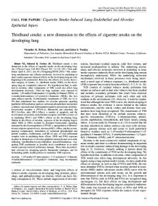
FIG. 2. Identifying cell types in the lung by flow cytometry. Cell types were identified based on differential expression of F4/80, CD11b, CD11c, and GR-1. Mouse lungs were digested with dispase, and cells were stained with fluorescently labeled cell-type-specific antibodies. (A, B) F4/80⫹ cells (A) that were CD11blow (B) were classified as alveolar macrophages, while F4/80high CD11bhigh cells were classified as CD11bhigh macrophages. (C) F4/80low CD11chigh cells were classified as DCs and subdivided into CD11blow/mid DCs and CD11bhigh DCs. F4/80low CD11clow CD11bmid cells were classified as monocytes. (D) F4/80low CD11clow CD11bhigh GR-1high cells were classified as neutrophils.
lated intranasally with Francisella strains expressing GFP were analyzed by flow cytometry and fluorescence microscopy. GFP⫹ lung cells, indicating association with Francisella, were readily detectable by flow cytometry (Fig. 1B). Microscopic examination of cells sorted from the high-GFP⫹ population presented significant punctate GFP⫹ fluorescence, indicative of highly infected cells (Fig. 1C), whereas low-GFP⫹ cells had as few as 1 GFP⫹ bacterium, indicating that cells containing a single bacterium could be detected by flow cytometry. No GFP⫹ bacteria were observed in the negative population. Projection images compiled from multiple planes throughout the z axis of membrane-stained cells revealed that the bacteria were intracellular (data not shown). For each strain, there were more infected cells 24 h postinoculation than were in the initial inoculum, indicating that the bacteria had replicated within this time frame (Fig. 1D). From day 1 to day 3 postinhalation, there was a significant increase in the number of infected lung cells for each strain (Fig. 1D). On day 3, there were similar numbers of infected cells in mice infected with U112 and Schu S4. Fewer GFP⫹ cells were observed in LVS-infected mice at this time point.
Composition of the Francisella host cell niche. To assess the suite of cells infected by Francisella following inhalation, we identified GFP⫹ cells with cell-type-specific staining patterns (Fig. 2 and Table 2). Twenty-four hours postinoculation, the predominant infected-cell type in mice infected with U112 (51.6%), LVS (70.3%), and Schu S4 (78.9%) was alveolar macrophages (Fig. 3). Other cell types infected at day 1 were CD11bhigh macrophages (11.0% for U112, 9.0% for LVS, and 6.2% for Schu S4), CD11blow/mid DCs (1.2% for U112, 10.6% for LVS, and 2.0% for Schu S4), and ATII cells (2.4% for U112, 5.1% for LVS, and 6.0% for Schu S4). Only 0.4% of LVS-associated cells were neutrophils and no Schu S4-associated neutrophils were observed on day 1. In contrast, 27.3% of U112-infected cells at day 1 were neutrophils. By day 3, neutrophils were the most-common infected-cell type for each strain (79.3% for U112, 45.0% for LVS, and 56.2% for Schu S4), although they were more prevalent in U112-infected mice (Fig. 3). The percentage of infected cells that were alveolar macrophages decreased to 2.3% for U112, 23.5% for LVS, and 14.3% for Schu S4. We observed a higher fraction of CD11blow/mid DCs among LVS-infected cells than
FRANCISELLA INTERACTIONS WITH HOST LUNG CELLS
VOL. 76, 2008 TABLE 2. Identification of specific cell types from lung homogenates Cell type
Propertiesa
Alveolar macrophage ..........F4/80high CD11blow CD11chigh GR-1low FShigh SShigh CD11bhigh Macrophage ......F4/80high CD11bhigh CD11cvar GR-1low FSvar SSvar CD11blow/mid DC .................F4/80low CD11blow/mid CD11chigh GR-1low FSmid SSmid CD11bhigh DC ......................F4/80low CD11bhigh CD11chigh GR-1low FSlow/mid SSlow/mid Monocyte ..............................F4/80low CD11bmid CD11clow/mid GR-1low/mid FSlow SSlow Neutrophil ............................F4/80low CD11bhigh CD11clow GR-1high FSvar SSvar ATII epithelial .....................LBP18high FSmid SShigh (low for other markers) a FS, forward light scatter; SS, side light scatter; mid, medium level; var, variable level.
for either of the other strains. Also, a greater proportion of Schu S4-infected cells (8.8%) than of LVS- (1.6%) or U112infected cells (1.4%) at day 3 were monocytes. Numbers of infected cells. Examining the proportions of Francisella-infected-cell types in the lung gave us insight into the Francisella host cell suite over time, but another important consideration for understanding inhalation tularemia is how the abundance of infected cells changes in the lung during the course of disease. Therefore, we quantified the number of each cell type infected at days 1 and 3 following inhalation. There were significant increases in the numbers of infected CD11bhigh macrophages, CD11bhigh DCs, monocytes, neutrophils, and ATII cells in mice infected with each strain between
5847
day 1 and day 3 (Fig. 4B, D, E, F, and G). In contrast, the number of infected alveolar macrophages and CD11blow/mid DCs increased from day 1 to day 3 in mice infected with LVS or Schu S4, whereas the number of these cells infected with U112 was unchanged (Fig. 4A and C). Most strikingly, on day 1 following inoculation, there were 2,670 ⫾ 1,287 U112-infected neutrophils compared to 19 ⫾ 17 for LVS and none detected for Schu S4. (Fig. 4F). These data demonstrate that increasing numbers of alveolar macrophages and DCs continue to become infected from day 1 to day 3 following inhalation of LVS or Schu S4 but not during infection with U112. Schu S4-infected lungs had more infected monocytes than U112- or LVS-infected lungs, indicating a potential propensity by this strain to infect these cells. Effects on cell populations during disease progression. In addition to analyzing the numbers and proportions of infectedcell types, we determined how Francisella infections impacted total cell populations in the lung as disease progressed. Strain U112 caused a 2.4-fold decrease in the number of alveolar macrophages from day 1 to day 3 (Fig. 5A). The alveolar macrophage numbers in LVS- or Schu S4-infected mice did not change significantly from day 1 to day 3. Similar to alveolar macrophages, DC populations decreased significantly from day 1 to day 3 in U112-infected mice (Fig. 5C and D). CD11blow/mid DCs decreased 2.3-fold and CD11bhigh DCs decreased 1.8-fold from day 1 to day 3 in U112-infected mice. There was no significant change in the number of DCs in LVS- or Schu S4-infected mice from day 1 to day 3. Also, no difference was observed between the numbers of DCs from infected mice and PBS control mice at day 1. There was no significant difference in the number of monocytes on day 1 following infection with each strain and the
FIG. 3. Lung cell types infected by Francisella following inhalation. Proportions of lung cell types infected with Francisella strain U112, LVS, or Schu S4 on day 1 and day 3 postinoculation.
5848
HALL ET AL.
INFECT. IMMUN.
FIG. 4. Numbers of Francisella-infected cells following inhalation. Absolute numbers of infected alveolar macrophages (A), CD11bhigh macrophages (B), CD11blow/mid DCs (C), CD11bhigh DCs (D), monocytes (E), neutrophils (F), and ATII cells (G) from mouse lungs on day 1 and day 3 following intranasal inoculation with U112, LVS, or Schu S4. Error bars represent standard deviations of the means (n ⫽ 3 to 6 mice). Statistical significance of difference between results on day 1 and day 3 was determined by unpaired two-tailed t test assuming unequal variance.
number in PBS control mice (Fig. 5E). The number of monocytes in LVS- and Schu S4-infected mice increased significantly from day 1 to day 3, whereas U112-infected mice had fewer monocytes in the lung over time. Lungs from LVS- and Schu S4-infected mice had more CD11bhigh macrophages at day 3 than day 1, while U112-
infected mice displayed no significant change in CD11bhigh macrophage numbers (Fig. 5B). Considering that alveolar macrophage, DC, and monocyte populations decreased in response to U112 infection by day 3, it is plausible that there is a balance between macrophage recruitment to the lung and macrophage killing which would result in no net change from
VOL. 76, 2008
FRANCISELLA INTERACTIONS WITH HOST LUNG CELLS
5849
FIG. 5. Effects of Francisella infection on cell numbers within lung populations. Absolute numbers of alveolar macrophages (A), CD11bhigh macrophages (B), CD11blow/mid DCs (C), CD11bhigh DCs (D), monocytes (E), neutrophils (F), and ATII cells (G) from mouse lungs on day 1 and day 3 following intranasal inoculation with U112, LVS, or Schu S4. Error bars represent standard deviations of the means (n ⫽ 3 to 6 mice). Statistical significance of difference between day 1 and day 3 was determined by unpaired two-tailed t test assuming unequal variance.
day 1 to day 3, though more experimental work is needed to determine if this is the case. There was no difference between the number of CD11bhigh macrophages in infected lungs and lungs from PBS control mice at day 1. In response to Francisella infection, there was a significant increase in the number of neutrophils in the lungs of mice infected by each strain from day 1 to day 3 (Fig. 5F), indicating that infection resulted in neutrophil recruitment, but not until at least 24 h following exposure. One day postinhalation, there
were no increases in the numbers of neutrophils in the lungs of infected mice compared to the number in PBS-inoculated control mice. We consistently observed fewer neutrophils at day 1 in Schu S4-infected mice than in PBS control mice (P ⬍ 0.05), suggesting that Schu S4 may initially suppress neutrophil recruitment more efficiently than U112 or LVS. Unlike other cell types analyzed, ATII cells are stationary structural cells. As expected, there was no significant change in the number of ATII cells in response to Francisella infection by
5850
HALL ET AL.
INFECT. IMMUN.
TABLE 3. Comparison of infected and total cell populations from mice infected with 400 CFU versus 40 CFU of Schu S4 Cell type
Alveolar macrophage CD11bhigh macrophage CD11blow/mid DC CD11bhigh DC Monocyte Neutrophil ATII epithelial a
Ratio for mice infected with 400 CFU/ mice infected with 40 CFUa Infected cells
Total cells
3.66 ⫾ 2.25 7.90 ⫾ 6.97 3.72 ⫾ 1.29 10.80 ⫾ 4.42 16.03 ⫾ 6.78 34.81 ⫾ 13.44 13.11 ⫾ 3.46
1.24 ⫾ 0.41 0.82 ⫾ 0.16 0.52 ⫾ 0.02 2.01 ⫾ 0.12 1.20 ⫾ 0.37 2.08 ⫾ 0.89 0.85 ⫾ 0.24
Numbers represent means ⫾ standard deviations.
U112, LVS, or Schu S4 from day 1 to day 3 (Fig. 5G), but these studies do not address any effects that infection may have on ATII cell physiology or function. Taken together, these data demonstrate that, with regard to cell recruitment, the innate immune response to LVS and Schu S4 in the lung is similar despite differences in the lethal dose of each strain. Upon inhalation, Francisella infects a variety of host cells, though alveolar macrophages are the predominant infected cell early during infection while neutrophils are recruited and become the main host cell in the later stages of pulmonary tularemia. U112 differed from both LVS and Schu S4 in that it was taken up by neutrophils more rapidly following inhalation, and this apparently occurs without significant recruitment of new neutrophils to the lung. U112 infection also affected macrophage, DC, and monocyte populations differently during the course of disease than LVS or Schu S4. Dose effects on host cell response and infected-cell populations. In order to reproducibly detect infected-cell populations in the early stages of infection, it was necessary to inoculate animals with a dose of at least 400 CFU. Since this dose represents at least a 10-fold increase over the 100% lethal dose of Schu S4, we wanted to determine if the magnitude of the inoculum impacted host cell infiltration or the composition of infected-cell types over time. Mice were inoculated intranasally with 42 CFU of GFP⫹ Schu S4. Infected- and uninfected-hostcell populations were quantified 3 days postinoculation, and the results compared to those for 3-day-infected animals that received the 10-fold-higher dose. Not surprisingly, there were more infected cells of each type in the high-dose-inoculated mice, but the proportions of infected-cell types resulting from the different doses were essentially the same (Table 3). There were differences in the total numbers of some cell types present in the lungs of the high- and low-dose-inoculated animals. Specifically, there were 2.08-fold ⫾ 0.89-fold more neutrophils and 2.01-fold ⫾ 0.12-fold more CD11bhigh DCs in mice infected with the higher dose of Schu S4. Interestingly, there were almost twofold more CD11bhigh macrophages in mice infected with the lower Schu S4 dose. These data indicate that the magnitude of the dose primarily affected the absolute numbers of infected cells but not the proportions of infectedcell types or the magnitude of the cellular response to Francisella infection in the lung.
DISCUSSION A goal of this study was to determine which cells in the lung become infected by F. tularensis following inhalation and identify how these populations are altered during pulmonary tularemia. At the same time, we sought to compare three commonly studied Francisella strains with regard to cell infection and impact on cell population dynamics in the lung. Understanding the similarities and differences among strains will enable more-accurate interpretations of the results of other studies using these strains, as well as facilitate more-specific inquiries into Francisella disease mechanisms. To directly compare lung infections by Francisella strains, we initiated infection with similar numbers of each strain. Our dose range allowed the detection of infected-cell types from whole-lung digests 24 h postinoculation. Infected cells from mice inoculated with less than 100 bacteria were not consistently distinguished from the background at day 1. However, mice infected in a similar way with a low dose of Schu S4 displayed a suite of infected cells and recruitment of monocytes and neutrophils at day 3 similar to those in mice inoculated with 400 CFU, indicating that the host response in the lung at a higher dose was not appreciably different from the response to a very low dose, with the exception of the total number of infected cells. During the early stages of infection, LVS- and Schu S4mediated disease appeared similar, while U112-mediated disease displayed fundamental differences. The most-common infected-cell type 24 h after inhalation of each strain was alveolar macrophages, although CD11bhigh macrophages, CD11blow/mid DCs, and ATII cells were also infected. In contrast, U112 was found in neutrophils at 24 h but LVS and Schu S4 were not. While there was no apparent influx of neutrophils overall at this stage of infection, there were over 1,000-fold more neutrophils associated with U112 than with LVS or Schu S4, indicating that neutrophils responded to and phagocytosed U112 to a significantly greater extent than they did LVS or Schu S4. Also, U112-infected mice lost alveolar macrophages, DCs, and monocytes during the course of disease, whereas depletion of these cell types was not observed in LVS- or Schu S4infected mice. Potentially, phagocytes that are infected in the early stages of disease begin to undergo apoptosis or necrosis by day 3. More studies are needed to determine if U112 infection results in more-rapid killing of infected cells than infection by LVS or Schu S4. Other studies have noted differences in U112 compared to LVS or Schu S4. For example, “F. tularensis subspecies novicida” (U112) exhibits different immunostimulatory properties in a mammalian host than F. tularensis subspecies holarctica (LVS) or tularensis (Schu S4). U112 lipopolysaccharide (LPS) stimulates macrophages to produce proinflammatory cytokines, such as tumor necrosis factor alpha, which are important in signaling the recruitment of neutrophils and monocytes, whereas LPS of LVS and Schu S4 does not (21). We therefore might have expected that U112 would provoke a more-rapid or -robust neutrophil influx than LVS or Schu S4, but this was not the case. Though significantly more U112 organisms than LVS or Schu S4 organisms were observed in neutrophils early during infection, there was no difference between the abundance of neutrophils in U112-infected lungs and their abundance in
VOL. 76, 2008
FRANCISELLA INTERACTIONS WITH HOST LUNG CELLS
lungs infected with the other strains or in lungs of PBS control mice. A different study reported that inhalation of U112 did not lead to neutrophil recruitment in the lung after 4 h (9), which is similar to our observations at 24 h. In addition to neutrophil recruitment, we observed an increase in the number of monocytes in LVS- and Schu S4-infected mouse lungs; however, the number of these cells in U112-infected mice decreased. Different studies are needed to determine whether monocyte recruitment is being blocked by U112 or whether these cells are being killed at a more-rapid rate than they are recruited. Francisella’s high capacity to cause disease in mammals is not entirely explained by lack of host recognition. Francisellainfected macrophages are unable to respond to proinflammatory stimuli, such as E. coli LPS (25), indicating an active suppression of this response. Though the complete mechanism for inflammatory cytokine suppression is unclear, there is evidence that LVS and Schu S4 stimulate production of the immunosuppressive cytokine TGF- within infected pulmonary macrophages and DCs (3, 4). TGF- leads to upregulation of Fc receptor on macrophages, which increases the phagocytic capability of these cells (29) and at the same time limits the production of gamma interferon and other proinflammatory molecules (12). Normally, increased phagocytosis by macrophages would expedite the clearance of bacterial infection. However, given Francisella’s ability to survive and replicate within these cells, this may exacerbate the replication of the organism within the host. While the production of TGF- and other immunosuppressive mediators may delay the influx of inflammatory cells, the results of this study demonstrate that eventual neutrophil and monocyte recruitment did occur in response to Francisella infection in the lung; however, these cells contribute to the progression of the disease by becoming host cells for Francisella replication. Normally, neutrophils are key to controlling bacterial infections with a significant intramacrophage-growth component (14). Despite the evolution of neutrophils as efficient microbial killers, the data presented here demonstrate that interactions with neutrophils are a key component of Francisella pathogenesis within the lung. The results of recent in vitro studies demonstrated that LVS is rapidly taken up by human neutrophils, but the respiratory burst is prevented due to disruption of NADPH oxidase assembly within the phagosome. This process is apparently due to an undefined active bacterial process, as Francisella infection also prevented neutrophils from responding to potent heterologous stimuli (1, 16). Another observation from this study was that infected ATII cells, which are nonmigratory, were as abundant as infected DCs or interstitial macrophages during the early stages of disease. Francisella interactions with ATII cells could play a unique role in pulmonary tularemia. Cell-cell junctions of the epithelium and endothelium are affected by infection-induced cytokines and chemokines which facilitate chemotaxis of neutrophils and monocytes from the bloodstream to infected tissue. The alveolar epithelium is closely associated with blood vessels to provide efficient oxygen exchange, and in addition to facilitating inflammatory-cell recruitment into the lung, increasing numbers of Francisella within ATII cells during disease progression could provide a method of bacterial entry into the bloodstream. In this way, ATII cells could provide a “gate-
5851
way” to the bloodstream where bacteria could then disseminate to distal organs, such as the liver and spleen. Further studies are ongoing in our lab to decipher the unique contribution of ATII cells during inhalation-acquired tularemia. ACKNOWLEDGMENTS We thank Jo Rae Wright for helpful advice and for providing proSP-C/enhanced GFP mice. We also thank James Fuller and Todd Kijek for helpful assistance in preparing the manuscript. This work was supported by a Southeast Regional Center of Excellence in Biodefense and Emerging Infections grant (NIH/NIAID U54AI057157) and by the National Institutes of Health (1R56-AI069339). REFERENCES 1. Allen, L. A., and R. L. McCaffrey. 2007. To activate or not to activate: distinct strategies used by Helicobacter pylori and Francisella tularensis to modulate the NADPH oxidase and survive in human neutrophils. Immunol. Rev. 219:103–117. 2. Anthony, L. D., R. D. Burke, and F. E. Nano. 1991. Growth of Francisella spp. in rodent macrophages. Infect. Immun. 59:3291–3296. 3. Bosio, C. M., H. Bielefeldt-Ohmann, and J. T. Belisle. 2007. Active suppression of the pulmonary immune response by Francisella tularensis Schu4. J. Immunol. 178:4538–4547. 4. Bosio, C. M., and S. W. Dow. 2005. Francisella tularensis induces aberrant activation of pulmonary dendritic cells. J. Immunol. 175:6792–6801. 5. Conlan, J. W., X. Zhao, G. Harris, H. Shen, M. Bolanowski, C. Rietz, A. Sjostedt, and W. Chen. 2008. Molecular immunology of experimental primary tularemia in mice infected by respiratory or intradermal routes with type A Francisella tularensis. Mol. Immunol. 45:2962–2969. 6. Ellis, J., P. C. Oyston, M. Green, and R. W. Titball. 2002. Tularemia. Clin. Microbiol. Rev. 15:631–646. 7. Eyles, J. E., M. G. Hartley, T. R. Laws, P. C. Oyston, K. F. Griffin, and R. W. Titball. 2008. Protection afforded against aerosol challenge by systemic immunisation with inactivated Francisella tularensis live vaccine strain (LVS). Microb. Pathog. 44:164–168. 8. Fortier, A. H., M. V. Slayter, R. Ziemba, M. S. Meltzer, and C. A. Nacy. 1991. Live vaccine strain of Francisella tularensis: infection and immunity in mice. Infect. Immun. 59:2922–2928. 8a.Fuller, J. R., R. R. Craven, J. D. Hall, T. M. Kijek, S. Taft-Benz, and T. H. Kawula. 2008. RipA, a cytoplasmic membrane protein conserved among Francisella species, is required for intracellular survival. Infect. Immun. 76: 4934–4943. 9. Hajjar, A. M., M. D. Harvey, S. A. Shaffer, D. R. Goodlett, A. Sjostedt, H. Edebro, M. Forsman, M. Bystrom, M. Pelletier, C. B. Wilson, S. I. Miller, S. J. Skerrett, and R. K. Ernst. 2006. Lack of in vitro and in vivo recognition of Francisella tularensis subspecies lipopolysaccharide by Toll-like receptors. Infect. Immun. 74:6730–6738. 10. Hall, J. D., R. R. Craven, J. R. Fuller, R. J. Pickles, and T. H. Kawula. 2007. Francisella tularensis replicates within alveolar type II epithelial cells in vitro and in vivo following inhalation. Infect. Immun. 75:1034–1039. 11. Leblebicioglu, H., S. Esen, D. Turan, Y. Tanyeri, A. Karadenizli, F. Ziyagil, and G. Goral. 2008. Outbreak of tularemia: a case-control study and environmental investigation in Turkey. Int. J. Infect. Dis. 12:265–269. 12. Letterio, J. J., and A. B. Roberts. 1998. Regulation of immune responses by TGF-beta. Annu. Rev. Immunol. 16:137–161. 13. Lopez, C. E., A. N. Kornblatt, R. K. Sikes, and O. E. Hanes. 1982. Tularemia: review of eight cases of tick-borne infection and the epidemiology of the disease in Georgia. South. Med. J. 75:405–407. 14. Mandic-Mulec, I., J. Weiss, and A. Zychlinsky. 1997. Shigella flexneri is trapped in polymorphonuclear leukocyte vacuoles and efficiently killed. Infect. Immun. 65:110–115. 15. Matyas, B. T., H. S. Nieder, and S. R. Telford III. 2007. Pneumonic tularemia on Martha’s Vineyard: clinical, epidemiologic, and ecological characteristics. Ann. N. Y. Acad. Sci. 1105:351–377. 16. McCaffrey, R. L., and L. A. Allen. 2006. Francisella tularensis LVS evades killing by human neutrophils via inhibition of the respiratory burst and phagosome escape. J. Leukoc. Biol. 80:1224–1230. 17. Owen, C. R., E. O. Buker, W. L. Jellison, D. B. Lackman, and J. F. Bell. 1964. Comparative studies of Francisella tularensis and Francisella novicida. J. Bacteriol. 87:676–683. 18. Pammit, M. A., V. N. Budhavarapu, E. K. Raulie, K. E. Klose, J. M. Teale, and B. P. Arulanandam. 2004. Intranasal interleukin-12 treatment promotes antimicrobial clearance and survival in pulmonary Francisella tularensis subsp. novicida infection. Antimicrob. Agents Chemother. 48:4513–4519. 19. Rohmer, L., C. Fong, S. Abmayr, M. Wasnick, T. J. Larson Freeman, M. Radey, T. Guina, K. Svensson, H. S. Hayden, M. Jacobs, L. A. Gallagher, C. Manoil, R. K. Ernst, B. Drees, D. Buckley, E. Haugen, D. Bovee, Y. Zhou, J. Chang, R. Levy, R. Lim, W. Gillett, D. Guenthener, A. Kang, S. A. Shaffer,
5852
20.
21. 22. 23. 24. 25.
HALL ET AL.
G. Taylor, J. Chen, B. Gallis, D. A. D’Argenio, M. Forsman, M. V. Olson, D. R. Goodlett, R. Kaul, S. I. Miller, and M. J. Brittnacher. 2007. Comparison of Francisella tularensis genomes reveals evolutionary events associated with the emergence of human pathogenic strains. Genome Biol. 8:R102. Roper, J. M., R. J. Staversky, J. N. Finkelstein, P. C. Keng, and M. A. O’Reilly. 2003. Identification and isolation of mouse type II cells on the basis of intrinsic expression of enhanced green fluorescent protein. Am. J. Physiol. Lung Cell Mol. Physiol. 285:L691–L700. Sandstrom, G., A. Sjostedt, T. Johansson, K. Kuoppa, and J. C. Williams. 1992. Immunogenicity and toxicity of lipopolysaccharide from Francisella tularensis LVS. FEMS Microbiol. Immunol. 5:201–210. Saslaw, S., H. T. Eigelsbach, J. A. Prior, H. E. Wilson, and S. Carhart. 1961. Tularemia vaccine study. II. Respiratory challenge. Arch. Intern. Med. 107: 702–714. Schulert, G. S., and L. A. Allen. 2006. Differential infection of mononuclear phagocytes by Francisella tularensis: role of the macrophage mannose receptor. J. Leukoc. Biol. 80:563–571. Sjostedt, A. 2007. Tularemia: history, epidemiology, pathogen physiology, and clinical manifestations. Ann. N. Y. Acad. Sci. 1105:1–29. Telepnev, M., I. Golovliov, T. Grundstrom, A. Tarnvik, and A. Sjostedt. 2003. Francisella tularensis inhibits Toll-like receptor-mediated activation of intracellular signalling and secretion of TNF-alpha and IL-1 from murine macrophages. Cell. Microbiol. 5:41–51.
Editor: W. A. Petri, Jr.
INFECT. IMMUN. 26. Tigertt, W. D. 1962. Soviet viable Pasteurella tularensis vaccines. A review of selected articles. Bacteriol. Rev. 26:354–373. 27. Twine, S., M. Bystrom, W. Chen, M. Forsman, I. Golovliov, A. Johansson, J. Kelly, H. Lindgren, K. Svensson, C. Zingmark, W. Conlan, and A. Sjostedt. 2005. A mutant of Francisella tularensis strain SCHU S4 lacking the ability to express a 58-kilodalton protein is attenuated for virulence and is an effective live vaccine. Infect. Immun. 73:8345–8352. 28. Vermaelen, K., and R. Pauwels. 2004. Accurate and simple discrimination of mouse pulmonary dendritic cell and macrophage populations by flow cytometry: methodology and new insights. Cytometry A 61:170–177. 29. Welch, G. R., H. L. Wong, and S. M. Wahl. 1990. Selective induction of Fc gamma RIII on human monocytes by transforming growth factor-beta. J. Immunol. 144:3444–3448. 30. Wherry, W. B., and B. H. Lamb. 2004. Infection of man with Bacterium tularense. 1914. J. Infect. Dis. 189:1321–1329. 31. Woolard, M. D., J. E. Wilson, L. L. Hensley, L. A. Jania, T. H. Kawula, J. R. Drake, and J. A. Frelinger. 2007. Francisella tularensis-infected macrophages release prostaglandin E2 that blocks T cell proliferation and promotes a Th2-like response. J. Immunol. 178:2065–2074. 32. Zen, K., K. Notarfrancesco, V. Oorschot, J. W. Slot, A. B. Fisher, and H. Shuman. 1998. Generation and characterization of monoclonal antibodies to alveolar type II cell lamellar body membrane. Am. J. Physiol. 275:L172– L183.