JOURNAL OF VIROLOGY, June 2007, p. 5841–5849 0022-538X/07/$08.00⫹0 doi:10.1128/JVI.00096-07 Copyright © 2007, American Society for Microbiology. All Rights Reserved.
Vol. 81, No. 11
Infectivity Determinants of the Hepatitis B Virus Pre-S Domain Are Confined to the N-Terminal 75 Amino Acid Residues䌤 Matthieu Blanchet1 and Camille Sureau1,2* Laboratoire de Virologie Mole´culaire, INTS, Paris, France,1 and Department of Virology and Immunology, Southwest Foundation for Biomedical Research, San Antonio, Texas 782282 Received 15 January 2007/Accepted 9 March 2007
The N-terminal pre-S domain of the large hepatitis B virus (HBV) envelope protein plays a pivotal role at the initial step of the viral entry pathway. In the present study, the entire pre-S domain was mapped for infectivity determinants, following a reverse-genetics approach and using in vitro infection assays with hepatitis delta virus (HDV) or HBV particles. The results demonstrate that lesions created within the N-terminal 75 amino acids of the pre-S region abrogate infectivity, whereas mutations between amino acids 76 and 113, overlapping the matrix domain, had no effect. In contrast to the results of a recent study (L. Stoeckl, A. Funk, A. Kopitzki, B. Brandenburg, S. Oess, H. Will, H. Sirma, and E. Hildt, Proc. Natl. Acad. Sci. 103:6730–6734, 2006), the deletion of a cell membrane translocation motif (TLM) located between amino acids 148 and 161 at the C terminus of pre-S2 did not interfere with the infectivity of the resulting HDV or HBV mutants. Furthermore, a series of large deletions overlapping the pre-S2 domain were compatible with infectivity, although the efficiency of infection was reduced when the deletions extended to the pre-S1 domain. Overall, the results demonstrate that the activity of the pre-S domain at viral entry solely depends on the integrity of its first 75 amino acids and thus excludes any function of the matrix domain or TLM. determinant is likely to map to the antigenic loop (AGL) in the S domain of the HBV envelope proteins (24). It is still unclear whether the AGL function at viral entry resides in binding to a coreceptor. Within pre-S1, the integrity of the first 77 residues is essential for HBV infectivity (29). As suggested by a series of experiments, it appears that the N-terminal moiety of pre-S1 mediates attachment to the hepatocyte membrane. This attachment is inhibited by pre-S1-specific monoclonal antibodies that also exert neutralizing activity (23, 33, 34, 36, 37). The pre-S1 binding function is also responsible for host range specificity (1, 12), and more interestingly, pre-S1 receptor binding can be competed with synthetic peptides specific for the Nterminal 48 amino acids of pre-S1 (2, 17, 18). In this study, we sought to map the entire pre-S domain of L-HBsAg for the presence of infectivity determinants, because the region had not been fully explored in previous studies. It was clearly demonstrated by Le Seyec et al. (29) that internal deletions in the first 77 amino acids of pre-S1 had an adverse effect on infectivity, whereas mutations in the 78-to-87 sequence were tolerated. However, the matrix domain (amino acids 92 to 113) had not been examined because lesions in this domain would block the formation of HBV virions. Hence, the reverse-genetics analysis of the matrix domain activity at viral entry can be carried out only with the HDV model, since assembly of HDV virions is independent of an L-HBsAg matrix function (26, 47). We also focused our attention on the C-terminal domain of pre-S2 because of conflicting information between two independent studies: the first study, conducted by Le Seyec et al. (30), failed to identify any infectivity determinant in L-HBsAg pre-S2, whereas a second study by Stoeckl et al. (42) identified a pre-S2-specific cell membrane translocation motif (TLM) that the authors presented as crucial for HBV entry. Our results demonstrate that the pre-S functions in infectivity are borne essentially by the first 75
Hepatitis B virus (HBV) is characterized by a narrow host range that reflects the specificity of interaction between envelope proteins and human hepatocyte receptors at viral entry (41). The HBV envelope proteins designated large (L-HBsAg), middle (M-HBsAg), and small (S-HBsAg) are transmembrane glycoproteins that differ from each other in the sizes of their N-terminal ectodomains (22). L-HBsAg contains an N-terminal pre-S1 domain, a central pre-S2 region, and a C-terminal S domain. M-HBsAg is composed of the pre-S2 and S domains (Fig. 1), and S-HBsAg consists of the S domain only. Envelope protein synthesis occurs at the endoplasmic reticulum membrane, and particles, mostly empty subviral particles (SVPs), assemble from aggregates at a pre-Golgi membrane. Envelopment of the HBV nucleocapsid requires the presence of LHBsAg (but not that of M-HBsAg) in addition to S-HBsAg (6). L-HBsAg displays a dual-membrane topology: N-terminal pre-S (pre-S1 plus pre-S2) is either external (Le-HBsAg), at the surface of the particles, or internal (Li-HBsAg), facing the inner side of the particle (9, 35, 38). Le-HBsAg is thought to assume a receptor-binding function at viral entry, and LiHBsAg is thought to serve as a matrix protein for HBV nucleocapsid envelopment (10, 29). In addition to their propensity to form SVPs, the HBV envelope proteins can package the hepatitis delta virus (HDV) ribonucleoprotein (RNP) in the case of HBV-HDV coinfection (4, 47). This interaction leads to the formation of HDV particles, which are infectious when L-HBsAg is included in the envelope (44). L-HBsAg contains a major infectivity determinant in its pre-S1 domain, including a myristoyl anchor linked to glycine 2 (8, 20), whereas a second * Corresponding author. Mailing address: Laboratoire de Virologie Mole´culaire, Institut National de la Transfusion Sanguine, 6 Rue Alexandre-Cabanel, 75739 Paris, France. Phone: (33) 1 44 49 30 56. Fax: (33) 1 44 49 30 59. E-mail:
[email protected]. 䌤 Published ahead of print on 21 March 2007. 5841
5842
BLANCHET AND SUREAU
J. VIROL.
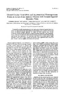
FIG. 1. Schematic representation of wt and mutant L-HBsAg. (A) The L-HBsAg polypeptide is depicted by a horizontal thin line. The pre-S1, pre-S2, and S domains are indicated. The sequence of the wt preS-1 domain is indicated, and each deletion mutant is designated by the positions of the first and last deleted amino acids. The letters KL correspond to an insertion of a Lys-Leu dipeptide at the site of the deletion. (B) The sequences of the wt and mutant polypeptides overlapping part of the pre-S1 domain and the pre-S2 region are indicated. Deletion mutants are designated by the positions of the first and last deleted amino acids. ⌬TLM corresponds to the deletion of amino acids 149 to 160 of L-HBsAg.
amino acids, clearly excluding a TLM function in the entry pathways of HDV and HBV. MATERIALS AND METHODS Plasmids. The HDV recombinant plasmid pSVLD3 was used for replication of HDV RNA and production of HDV RNP (27). The HBV recombinant plasmid pCIHBenv(⫺) was used for the production of HBV nucleocapsid (3). It was engineered to drive the transcription of HBV pregenomic RNA (D genotype, ayw3 subtype) in which the pre-S1, pre-S2, and S start codons were mutated to ACG to prevent translation of the envelope proteins. The HBV expression vectors p123 and p124 were used for the production of the wild-type (wt) S- and L-HBsAg proteins, respectively (43). Vectors for the expression of L-HBsAg mutants were derived from p124 using the PCR technique with two complementary mutagenic oligonucleotides as described previously (25). Mutations in preS1, referred to as KL insertion/deletion, consisted of a five-residue deletion and the insertion of a Lys-Leu dipeptide. Larger deletions in pre-S2 or pre-S1/pre-S2 were designated by the positions of the first and last deleted residues. Viral-particle production in Huh-7 cells. Huh-7 cells were maintained in Williams’ medium E supplemented with 10% fetal bovine serum. The cells (106/well) were transfected with 1 g of p123 and 1 g of p124 (or derivatives) and either 1 g of pSVLD3 for the production of HDV particles or 1 g of pCIHBenv(⫺) for the production of HBV virions. Particles were thus devoid of M-HBsAg. Transfections were conducted using the FuGENE 6 reagent (Roche) as described previously (43). For HDV virion production, culture medium was collected at days 5, 7, 9, and 12 posttransfection. For HBV virion production, culture medium was harvested at days 3, 6, 9, and 11 posttransfection. Analysis of secreted HBV envelope proteins. Immunoblot analysis of HBV envelope proteins was conducted as described previously (3). Briefly, culture medium from transfected cells was clarified and subjected to ultracentrifugation on a 20% sucrose cushion in phosphate-buffered saline (PBS). Viral-particle pellets were resuspended in Laemmli buffer containing -mercaptoethanol and subjected to sodium dodecyl sulfate-polyacrylamide gel electrophoresis (SDSPAGE) before transfer to a polyvinylidene difluoride (PVDF) membrane and immunodetection with anti-S and anti-pre-S1 or anti-pre-S2 antibodies. The immunoblot was developed using the enhanced chemiluminescence reagents, followed by exposure to Kodak film for signal detection. For PNGase F treatment, particles sedimented from 2 ml of culture supernatant were resuspended in 40 l of Laemmli buffer supplemented with 4 l of G7 buffer (New England Biochemical), 4 l of 10% Nonidet P-40, and 500 units of PNGase F (New England Biochemical). After incubation overnight at 37°C, the digestion products were analyzed as described above. Detection of HDV RNA in the supernatant of transfected cells. RNA from 140 l of culture supernatant was extracted using the QIAamp Viral RNA Mini Kit (QIAGEN), precipitated in the presence of isopropanol, and resuspended in RNase-free water (3). The purified RNA was subjected to electrophoresis through a 2.2 M formaldehyde, 1.2% agarose gel and transferred to a nylon
membrane. The membrane-bound RNA was hybridized to a 32P-labeled RNA probe specific for genomic HDV RNA. Quantification of radioactive signals was achieved using a phosphorimager (BAS-1800 II; Fuji). Detection of HBV DNA in the supernatant of transfected cells. HBV virions from clarified supernatants were immunoprecipitated with anti-pre-S1 antibodies by overnight incubation at 4°C (3), followed by incubation on ice for 2 h in the presence of 100 l of a Pansorbin suspension in PBS prepared as recommended by the manufacturer (Calbiochem). The Pansorbin was subjected to sedimentation by centrifugation at 13,000 rpm for 1 min, and the pellet was washed three times in cold PBS. Bound particles were disrupted in lysis buffer containing SDS and proteinase K (QIAamp DNA Mini Kit; QIAGEN). After incubation at 56°C for 10 min, followed by centrifugation for 1 min at 13,000 rpm, the supernatant was collected, and DNA was extracted using the QIAamp DNA Mini Kit (QIAGEN). After DNA precipitation in the presence of ethanol and centrifugation, the pellet was resuspended in 10 mM Tris-HCl (pH 8.0), 1 mM EDTA and subjected to agarose gel electrophoresis, transfer to a positively charged nylon membrane (Roche), and hybridization to a 32P-labeled RNA probe specific for negative-strand HBV DNA. Quantification of radioactive signals was achieved using a phosphorimager. In vitro infection assays of HepaRG cells. HepaRG cells were maintained and differentiated as described previously (21). For infection with HDV virions, clarified Huh-7 supernatants containing wt or mutant virions were used as inocula after normalization of their HDV RNA titers. HepaRG cells (3.3 ⫻ 105 cells/20-mm-diameter well) were exposed to 108 genome equivalents (ge) of HDV virions for 16 h in the absence or presence of 5% polyethylene glycol (PEG) 8000 (Sigma). Cells were harvested at day 8 postinoculation in RLT buffer (QIAGEN). Total cellular RNA was purified using the Rneasy Mini Kit and tested for the presence of genomic or antigenomic HDV RNA as a marker of infection (3). For infection with HBV virions, clarified supernatants containing wt or mutant virions at approximately 1.5 ⫻ 107 ge/ml were concentrated by precipitation with PEG as described previously (21). HepaRG cells (3.3 ⫻ 105 cells/20-mm-diameter well) were exposed to HBV virions at a multiplicity of infection of 50 ge per cell for 16 h in the presence of 5% PEG. The cells were harvested at day 12 postinoculation in RLT buffer (QIAGEN), and total cellular RNA was purified using the Rneasy and Oligotex mRNA Mini Kits (QIAGEN). The purified mRNA was subjected to electrophoresis, transferred to a nylon membrane, and hybridized to a 32P-labeled RNA probe specific for HBV mRNA. Quantification of radioactive signals was achieved using a phosphorimager. In vitro infection assays of human primary hepatocyte cultures. Primary cultures of human hepatocytes were purchased from Biopredic Inc. (Rennes, France). The cells were obtained after isolation from residual human liver tissue that was not usable for liver transplantation (the donor patients were free of HBV). Three days postseeding, hepatocytes (2 ⫻ 106 cells/12.5-cm2 flask) were exposed to normalized amounts of wt or mutant HDV virions at a multiplicity of infection of 100 ge per cell for 16 h in the absence or presence of 5% PEG. Cells were harvested at day 9 postinoculation and disrupted in RLT buffer
VOL. 81, 2007
LARGE HBV ENVELOPE PRE-S DOMAIN INFECTIVITY DETERMINANTS
5843
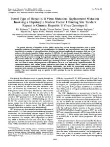
FIG. 2. Characterization of HDV particles carrying preS-1 mutations. Production of wt and mutant HDV particles was achieved by transfection of 106 Huh-7 cells with 1 g each of pSVLD3, p123, and p124 (or mutant derivatives) as described in Materials and Methods. (A) Particles from the culture fluids of transfected cells were concentrated and assayed for the presence of HBV envelope (Env.) proteins after SDS-PAGE, transfer to a PVDF membrane, and immunodetection with a mixture of rabbit anti-S and anti-preS2 antibodies (1:500 dilution each). Signals are from 1 ml of culture medium. The S-HBsAg (S) and L-HBsAg (L) HBV envelope proteins are indicated. The glycosylated (gp) and nonglycosylated (p) forms of S- and L-HBsAg are indicated. (B) RNA extracted from supernatants was subjected to electrophoresis through a 2.2 M formaldehyde, 1.2% agarose gel and analyzed by Northern blotting using a 32P-labeled RNA probe specific for detection of genomic HDV RNA. Signals are from 70 l of culture medium. The size of genomic HDV RNA is indicated. Wt SL, HDV particles coated with wt S- and L-HBsAg. Wt S, HDV particles coated with the S-HBsAg only. Mutant HDV particles bearing KL insertion/deletions in L-HBsAg are designated by the positions of the first and last deleted amino acids.
(QIAGEN). Total cellular RNA was purified using the Rneasy Mini Kit and assayed for the presence of genomic or antigenomic HDV RNA as a marker of infection.
RESULTS To study the functions of the HBV envelope protein at viral entry, we used an in vitro assay in which the envelope proteins were expressed at the surfaces of HDV virions and tested for their functions at viral entry on the HepaRG cell cultures. Production of infectious HDV virions was achieved in Huh-7 cells by cotransfection with a plasmid providing the HDV RNP and plasmids providing the S- and L-HBsAg envelope proteins; M-HBsAg was omitted because it is dispensable for morphogenesis and infectivity (7, 16, 45). The infectivity of HDV particles was assayed by inoculation into susceptible HepaRG cells and measurement of the intracellular accumulation of HDV RNA at days 6 to 9 postinoculation. Since infections were nonproductive in the absence of the helper HBV, the level of intracellular viral RNA that accumulated in an infected cell was proportional to the HDV titer in the inoculum (3). The HDV-HepaRG system was utilized to examine the in vitro infectivities of HDV mutants that presented lesions in the pre-S domain of L-HBsAg. One infection assay was conducted on primary cultures of human hepatocytes, and for one particular L-HBsAg mutant bearing a deletion of the TLM (⌬TLM), the effects of the mutation were also analyzed at the surfaces of HBV virions. The 76-to-109 amino acid sequence of pre-S1 is free of infectivity determinants. We first sought to precisely map the pre-S1 domain, including the matrix domain (residues 92 to 113), for determinants of infectivity (5, 30). Because of its crucial role in HBV morphogenesis, the matrix domain had not been studied for implication in viral entry by a reverse-genetics analysis. Therefore, we took advantage of the HDV model
because it lacked the requirement for the HBV matrix domain to assemble HDV virions. In addition, this analysis was expected to reveal whether pre-S1 determinants for HDV infectivity map exactly to those previously identified for HBV infectivity. For the production of wt and mutant particles coated with S-HBsAg and L-HBsAg (SL HDV particles), Huh-7 cells were cotransfected with pSVLD3, p123, and p124 or derivatives carrying sequential KL insertions/deletions between residues 10 and 111 as described in Materials and Methods. Because the very N terminus of L-HBsAg contains a myristoylation signal that is essential to infectivity (20), deletions within the first 10 residues were not considered. Following transfection, culture supernatants were collected and tested for the presence of envelope proteins and HDV RNA. All insertions/deletions were permissive for the production of HDV particles in amounts sufficient for infectivity assays (Fig. 2). The variations in envelope protein secretion between mutants paralleled those recorded for viral RNA, indicating that the ratio of SVPs to HDV virions was not affected by the mutations. To assess the effects of mutations on infectivity, preparations of mutant HDV particles were normalized to approximately 108 ge/ml prior to inoculation into HepaRG cells in the presence of 5% PEG. Cells were harvested 9 days postinoculation and assayed for the presence of genomic and antigenomic HDV RNA. As shown in Fig. 3B, insertions/deletions in the 10-to-75 sequence of the pre-S1 domain were deleterious to HDV infectivity, whereas deletions in the 76-to-110 sequence were tolerated. These results clearly demonstrate the absence of an infectivity determinant in the pre-S1 domain that overlaps with the 92-to-113 sequence previously identified as the HBV matrix domain (5, 30). They are also in agreement with a prior analysis that mapped a determinant of HBV infectivity in the first 77 residues of pre-S1 (29).
5844
BLANCHET AND SUREAU
J. VIROL.
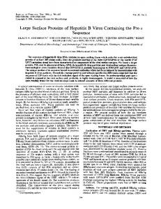
FIG. 3. Infectivity of pre-S1 HDV mutants in HepaRG cell cultures. HepaRG cells (3.3 ⫻ 105 cells/20-mm-diameter well) were inoculated with 108 ge of wt or mutant HDV particles in the presence of 5% PEG. (A) (Left) Amount of HDV RNA in 70 l of undiluted or serially diluted (twofold) wt HDV particles. (Right) HDV RNA signal from 70 l of supernatants containing wt or mutant HDV particles after normalization to approximately 108 ge/ml. (B) At day 8 postinoculation, HepaRG cells were analyzed by Northern blot hybridization with 32P-labeled RNA probes specific for detection of antigenomic (AG) or genomic (G) HDV RNA. Signals are from 6.6 ⫻ 104 cells. Signals of nonspecific hybridization of the antigenomic-specific HDV RNA probe to rRNA served as a loading control. Quantification of HDV RNA signals by phosphorimager is indicated as percentages of the wt value. The size of HDV RNA is indicated. Wt SL, HDV particles coated with wt S- and L-HBsAg. Wt S, HDV particles coated with the S-HBsAg only. Mutant HDV particles bearing KL insertion/deletions in L-HBsAg are designated by the positions of the first and last deleted amino acids.
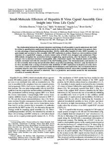
TLM is not essential for HDV infectivity in HepaRG cells. Recent studies proposed that a discrete domain referred to as TLM, located at the C terminus of pre-S2, would be essential for infectivity of duck HBV (DHBV) and HBV (42). However, as mentioned above, this hypothesis is in conflict with the conclusion of a prior study demonstrating that HBV infectivity was not affected by internal deletions in the pre-S2 region of L-HBsAg. To specifically evaluate the role of TLM in HDV infectivity, we produced HDV particles bearing S-HBsAg and ⌬TLM L-HBsAg (devoid of M-HBsAg). Supernatants from virus-producing cells were examined for the presence of HDV particles by SDS-PAGE and immunoblotting for the detection of envelope proteins and by agarose gel electrophoresis and Northern blotting for detection of HDV RNA. As shown in Fig. 4A, wt and mutant HDV particles displayed identical characteristics in terms of HBV envelope proteins and HDV RNA content. Each preparation was adjusted to 108 ge/ml, and 1 ml was inoculated into HepaRG cells in the presence of 5% PEG. The HepaRG cells were harvested at day 8 postinoculation and analyzed for the presence of genomic and antigenomic HDV RNAs. As shown in Fig. 4B, ⌬TLM HDV infected the HepaRG cells with an efficiency estimated at 75% of that of the wt HDV. We concluded that TLM is not essential for infectivity of HDV in HepaRG cells under the conditions utilized in this experiment, i.e., in the presence of PEG at the time of inoculation. TLM is not essential for HDV infectivity in HepaRG cells in the absence of PEG. Since PEG has been shown to drastically increase the efficiency of in vitro infection by a still-unknown mechanism (1, 19), we questioned whether the addition of PEG during the inoculation period could have compensated
for the absence of a TLM function in the mutant HDV. The infectivity of ⌬TLM HDV was therefore tested in the absence of PEG during inoculation. To ensure an efficient infection in the absence of PEG, we used inocula containing a high concentration of viral particles (approximately 16 ⫻ 108 ge/ml). For a precise evaluation of infectivity, preparations of mutant and wt virions were serially diluted (twofold), and each dilution was inoculated into HepaRG cells. As shown in Fig. 5, wt and ⌬TLM HDV particles were similarly infectious, as evidenced by the detection of HDV RNA in equivalent amounts in HepaRG cells exposed to wt or mutant particles. Measurement of the intracellular antigenomic HDV RNA at day 8 postinfection was achieved by quantification of the radioactive signals with the phosphorimager. Overall, we calculated the infectivity of ⌬TLM HDV in the absence of PEG at 75% of that of the wt, a value identical to the one estimated in the presence of PEG. TLM is not essential for HDV infectivity in primary cultures of human hepatocytes. We sought to assay the infectivity of ⌬TLM HDV particles in primary cultures of human hepatocytes to ascertain that the results obtained in HepaRG cells were not specific to the particular cell line. Primary cultures of human hepatocytes (2 ⫻ 106 cells/flask) were inoculated with 2 ml of wt and ⌬TLM HDV particles containing approximately 2 ⫻ 108 ge, either in the absence or in the presence of 5% PEG. At day 9 postinoculation, cells were harvested and analyzed for the presence of HDV RNA. Figure 6 shows evidence of an efficient infection with recombinant ⌬TLM HDV. In the presence or absence of PEG at inoculation time, the infectivity of ⌬TLM particles was estimated at 70% of that of the wt. This value was not significantly different from the one obtained with the HepaRG cells. Whether to consider the average difference between ⌬TLM
VOL. 81, 2007
LARGE HBV ENVELOPE PRE-S DOMAIN INFECTIVITY DETERMINANTS
FIG. 4. Infectivity of ⌬TLM HDV particles in HepaRG cell cultures. Production of wt or mutant HDV particles was achieved by transfection of 106 Huh-7 cells with 1 g each of pSVLD3, p123, and p124 (or a ⌬TLM derivative) plasmids. (A) Culture fluids from transfected cells were harvested, and viral particles were concentrated and assayed for the presence of HBV envelope proteins as described in the legend to Fig. 2. Signals are from 1 ml of culture medium. RNA was extracted from the clarified supernatant, subjected to electrophoresis, and analyzed by Northern blotting using a 32P-labeled RNA probe specific for genomic HDV RNA. Signals are from 70 l of supernatant. (B) HepaRG cells (3.3 ⫻ 105 cells/20 mm-diameter well) were exposed to 1 ml of inoculum containing approximately 108 ge in the presence of 5% PEG. At day 8 postinoculation, cells were harvested and cellular RNA was analyzed by Northern blot hybridization with 32P-labeled RNA probes specific for detection of antigenomic (AG) or genomic (G) HDV RNA. Signals are from 6.6 ⫻ 104 cells. Quantification of HDV RNA signals by phosphorimager is indicated as percentages of the wt value. The size of HDV RNA is indicated. The glycosylated (gp) and nonglycosylated (p) forms of S-HBsAg (S) and L-HBsAg (L) proteins are indicated. Wt SL, HDV particles coated with wt S- and L-HBsAg. Wt S, HDV particles coated with the S-HBsAg only. ⌬TLM, HDV particles coated with wt S-HBsAg and mutant L-HBsAg carrying a deletion of the TLM domain (residues 149 to 160).
and wt HDV (30%) as significant is questionable, since we observed that ⌬TLM L-HBsAg was expressed at a level slightly lower than that of its wt counterpart (Fig. 4A). This would translate into a preparation of ⌬TLM HDV particles presenting a reduced proportion of L-HBsAg in the envelope (i.e., reduced infectivity). Overall, our data clearly demonstrate that the 149-to-160 sequence of pre-S2, previously described as a TLM crucial for HBV infectivity, is dispensable for the HDV entry process. TLM is not essential for HBV infectivity in HepaRG cells. To directly assay the TLM activity at the surface of HBV particles, we produced recombinant HBV virions (devoid of M-HBsAg) bearing ⌬TLM L-HBsAg. For the production of wt and mutant SL HBV particles, Huh-7 cells were transfected with pCIHBenv(⫺), p123, and p124 (or a ⌬TLM derivative).
5845
As a noninfectious HBV control, we prepared particles in which wt L-HBsAg was replaced with a ⌬26-30 L-HBsAg, a mutant carrying a deletion in the pre-S1 receptor binding site and hence deficient in viral entry (Fig. 3). Supernatants from virus-producing Huh-7 cells were adjusted to approximately 1.5 ⫻ 107 ge/ml. This quantification was achieved by precipitating mature HBV virions from cell culture fluids with an anti-pre-S1 antibody and testing the precipitate for viral DNA by Southern blot analysis as described previously (3). Note that the anti-pre-S1 antibody was raised in rabbits against a synthetic peptide specific for the 83-to-106 amino acid sequence (D genotype, ayw3 subtype) and therefore capable of reacting with ⌬TLM and ⌬26-30 L-HBsAg mutants. Inocula normalized for their HBV DNA titers were assayed for viral envelope proteins by SDS-PAGE and immunoblotting and for HBV DNA as described above. Figure 7A shows that wt and mutant L-HBsAg had equivalent efficiencies for HBV virion production. HepaRG cells (3.3 ⫻ 105/well) were inoculated with 1.5 ⫻ 7 10 ge of HBV virions, and cells were harvested at day 9 postinoculation for detection of de novo-synthesized HBV mRNA as a marker of infection. As shown in Fig. 7B, viral mRNA was detected in cells exposed to ⌬TLM HBV particles at levels similar to those observed for wt HBV. As expected, ⌬26-30 HBV was noninfectious. The efficiency of ⌬TLM HBV infectivity was evaluated at 136% of that of the wt, based on measurement of mRNA-specific signals by phosphorimager and normalization to GAPDH (glyceraldehyde-3-phosphate dehydrogenase) mRNA. We considered a 30% difference between wt and ⌬TLM HBV nonsignificant, since the HBV DNA content of ⌬TLM inoculum was still 24% above that of the wt after normalization (Fig. 7A). Large deletions encompassing the pre-S1 and pre-S2 domains are tolerated for HDV infectivity. To further investigate the role of pre-S2 at viral entry, we constructed a set of four L-HBsAg mutants carrying large deletions (80 to 153, 97 to 153, 107 to 153, and 115 to 153) extending upstream to the pre-S1 domain. Production of HDV virions bearing S-HBsAg and mutant L-HBsAg was achieved as described above. Supernatants of transfected cells were analyzed for the presence of both envelope proteins and HDV RNA. As shown in Fig. 8A, envelope proteins and viral RNA were detected at similar levels in wt and mutant HDV particles. Note that the distance of the L-HBsAg mutants’ migration directly correlated with the size of the deletion, except for ⌬80-153, which displayed a migration pattern slower than expected. When envelope protein extracts were treated with PNGase F prior to SDS-PAGE and subsequent immunoblotting, the migration of the unglycosylated ⌬80-153 L-HBsAg mutant was as expected, indicating that the protein had undergone an additional glycosylation at Asn-4 in pre-S1, as previously reported (31). In addition, when Asn-4 of ⌬80-153 was mutated to Gln, the resulting N4Q ⌬80-153 proteins presented the expected migration profile in SDS-PAGE (i.e., the unglycosylated and monoglycosylated forms) (data not shown). The fact that the levels of ⌬83-153 and ⌬97-153 proteins appeared lower than that of the wt is likely due to an incomplete transfer to the PVDF membrane in the immunoblotting procedure, because when the samples were treated with PNGase F prior to SDS-PAGE, all mutant and wt proteins were detected at similar levels (Fig. 8A). Gly-
5846
BLANCHET AND SUREAU
J. VIROL.
FIG. 5. Infectivity of ⌬TLM HDV viral particles in HepaRG cell cultures in the absence of PEG. HepaRG cells (3.3 ⫻ 105 cells/20 mm-diameter well) were exposed for 16 h to 1 ml of inoculum containing approximately 16, 8, 4, 2, 1, and 0.5 ⫻ 108 ge, as indicated, in the absence of PEG. (A) Signals corresponding to viral RNA purified from 70 l of inoculum after Northern blot hybridization using a 32P-labeled RNA probe specific for detection of genomic (G) HDV RNA. (B) At day 8 postinoculation, HepaRG cells were assayed for the presence of antigenomic (AG) or genomic (G) HDV RNA. Signals are from 6.6 ⫻ 104 cells. The size of HDV RNA is indicated. Quantification of HDV RNA signals by phosphorimager is indicated below each panel as percentages of the wt value. Wt SL, HDV particles coated with wt S- and L-HBsAg. Wt S, HDV particles coated with the S-HBsAg only. ⌬TLM, HDV particles coated with wt S-HBsAg and mutant L-HBsAg bearing a TLM deletion.
cosylation at Asn-4 does not occur on the wt polypeptide, because the pre-S domain is not directly translocated in the endoplasmic reticulum lumen upon synthesis, and therefore, Asn-4 glycosylation may reflect the occurrence of a rapid translocation of the ⌬80-153 pre-S domain. Prior to infection assays, supernatants from transfected cells
were adjusted to approximately 108 ge/ml, and 1 ml was added to 3.3 ⫻ 105 HepaRG cells in a 20-mm-diameter well. Cells were harvested at day 8 postinoculation before being assayed for the presence of intracellular HDV RNA (Fig. 8B). HDV mutants carrying the deletion ⌬115-153 demonstrated a near-wt level of infectivity, whereas a significant decrease in HDV RNA was observed in cells inoculated with virions carrying larger L-HBsAg deletions. The failure to detect any evidence of infection for the ⌬80-153 HDV mutant could be explained either by the requirement for a minimal distance between the pre-S1 receptor-binding domain and the first transmembrane domain (TMD1) of L-HBsAg, by the presence of an N-linked glycan on Asn-4 of pre-S1, or by a lack of myristoylation at glycine 2 (see Discussion). The progressive loss of infectivity that is observed when the pre-S2 deletion is extended upstream of residue 107 in the pre-S1 region (compare mutants ⌬115-153, ⌬107-153, and ⌬97-153) is likely to result from the size of the deletion rather than from the removal of a functional motif, since as shown in Fig. 3, there are no specific amino acid sequence requirements for infectivity between residues 75 and 111 of L-HBsAg. DISCUSSION
FIG. 6. Infectivity of ⌬TLM HDV particles in primary cultures of human hepatocytes. Two ml of each inoculum (2 ⫻ 108 ge) described in Fig. 4 was added to primary cultures of human hepatocytes (2 ⫻ 106 cells/12.5-cm2 flask) at day 3 postseeding and incubated with the cells for 16 h in the absence or the presence of 5% PEG. At day 8 postinoculation, cells were harvested and assayed for the presence of antigenomic (AG) or genomic (G) HDV RNA. Signals are from 2 ⫻ 105 cells. Quantification of HDV RNA signals by phosphorimager is indicated as percentages of the wt value. The size of HDV RNA is indicated. Wt SL, HDV particles coated with wt L- and S-HBsAg. Wt S, HDV particles coated with the S-HBsAg only. ⌬TLM, HDV particles coated with wt S-HBsAg and ⌬TLM L-HBsAg mutant.
In the present study, we demonstrate that pre-S1 infectivity determinants are confined to the N-terminal 75 amino acid residues. Previous observations assigned to this region a receptor-binding activity mediated by the N-terminal 48 amino acids in which the myristoyl anchor linked to glycine 2 participates (2, 18). The function of amino acids 49 to 75 is unclear; they may pertain to the stabilization of the interaction with the receptor or to an intramolecular masking of the receptorbinding site at a prebinding stage (18, 46). Our results ruled out the presence of infectivity determi-
VOL. 81, 2007
LARGE HBV ENVELOPE PRE-S DOMAIN INFECTIVITY DETERMINANTS
FIG. 7. Infectivity of ⌬TLM HBV particles in HepaRG cell cultures. Production of wt and mutant HBV particles was achieved by transfection of Huh-7 cells (106 cells) with a mixture of 1 g each of pCIHBenv(⫺), p123, and p124 (or a ⌬TLM mutant derivative) plasmids. (A) Culture fluids of transfected cells were assayed for the presence of HBV envelope proteins by SDS-PAGE and immunoblot analysis with a mixture of rabbit anti-S and anti-preS2 antibodies (1:500 dilution each). Signals are from 1 ml of supernatant. HBV virions were immunoprecipitated from the culture fluids of transfected cells using anti-pre-S1 antibodies, and viral DNA was extracted from the precipitate using the QIAamp DNA Mini Kit. Purified DNA was subjected to electrophoresis through a 1% agarose gel, transfer to a positively charged nylon membrane, and hybridization to a 32P-labeled RNA specific for detection of negative-strand HBV DNA. Signals are from 1 ml of supernatant. (B) HepaRG cells (3.3 ⫻ 105 cells/20-mmdiameter well) were inoculated with 1 ml of inoculum containing approximately 1.5 ⫻ 107 ge. Cells were harvested at day 12 postinoculation, and mRNAs were purified and assayed by Northern blot analysis using a 32P-labeled RNA specific for detection of HBV mRNA and a probe specific for detection of GAPDH mRNA (as a loading control). Signals are from 1.1 ⫻ 105 cells. Quantification of HBV RNA signals by phosphorimager is indicated below each panel as percentages of the wt value. RC, relax circular; L, linear; SS, single strand. The size of HBV mRNAs is indicated. The glycosylated (gp) and nonglycosylated (p) forms of S-HBsAg (S) and L-HBsAg (L) proteins are indicated. Wt SL, HBV particles coated with wt S- and L-HBsAg. ⌬26-30, HBV particles coated with wt S-HBsAg and L-HBsAg carrying a 26-to-30 deletion in the pre-S1 domain. ⌬TLM, HBV particles coated with wt S-HBsAg and ⌬TLM L-HBsAg mutant.
nants in the cytoplasmic anchorage domain between amino acids 70 and 94 (31) and in the matrix domain (amino acids 92 to 113). However, our analysis of the pre-S1 deletion mutants was conducted on HDV particles, the morphogenesis of which does not require an interaction of the L-HBsAg-borne matrix domain with the HDV RNP. It remains possible that in the case of HBV, the matrix domain, which is assumed to bind the nucleocapsid within Dane particles, might be instrumental at viral entry by controlling the intracellular release of the nucleocapsid at a postattachment step. The failure of the ⌬80-153 mutant to provide infectivity was surprising, considering that the deletion overlaps a domain devoid of any amino acid sequence requirements for function at viral entry (this study). This defect could result from insuf-
5847
FIG. 8. Effects of large deletions in the pre-S domain on HDV infectivity in HepaRG cell cultures. Production of wt and mutant HDV particles was achieved by transfection of 106 Huh-7 cells with 1 g each of pSVLD3, p123, and p124 (or mutant derivatives) plasmids. (A) Culture fluids from transfected cells were harvested, and particles were concentrated and assayed for the presence of HBV envelope proteins before and after incubation with PNGase F as indicated. After SDSPAGE and transfer to a PVDF membrane, the proteins were probed with a mixture of rabbit anti-S and anti-pre-S1 antibodies (1:500 dilution each). Signals are from 1 ml of culture medium. RNA was extracted from the clarified supernatant, subjected to electrophoresis, and analyzed by Northern blotting using a 32P-labeled RNA probe specific for detection of genomic HDV RNA. Signals are from 70 l of supernatant. (B) Prior to infection assays, supernatants were normalized to approximately 108 ge/ml, and 1 ml of each preparation was inoculated into 3.3 ⫻ 105 HepaRG cells in the presence of 5% PEG. At day 8 postinoculation, HepaRG cells were harvested and cellular RNA was analyzed by Northern blot hybridization with 32P-labeled RNA probes specific for antigenomic (AG) or genomic (G) HDV RNA. Signals are from 6.6 ⫻ 104 cells. Quantification of HDV RNA signals by phosphorimager is indicated below each panel as percentages of the wt value. The size of HDV RNA is indicated. The glycosylated (gp) and nonglycosylated (p) forms of S-HBsAg (S) and LHBsAg (L) proteins are indicated. Wt SL, HDV particles coated with wt L- and S-HBsAg. Wt S, HDV particles coated with the S-HBsAg only. Pre-S deletions mutants are designated by the positions of the first and last deleted amino acids.
ficient spacing between the N-terminal receptor binding domain and TMD1 of the S domain. In support of this interpretation is the fact that increasing the size of the deletion in pre-S1/preS-2 from ⌬115-153 to ⌬107-153 to ⌬97-153 correlated with a progressive loss of infectivity (Fig. 8). With regard
5848
BLANCHET AND SUREAU
to ⌬80-153, we showed evidence of its glycosylation at Asn-4, which might exert an adverse effect at viral entry by interfering with receptor attachment. Glycosylation at Asn-4 also indicates that the pre-S1 domain of ⌬80-153 undergoes a rapid posttranslational translocation that would be incompatible with its acylation at glycine 2 by cytosolic N-myristoyl transferase. In fact, the lack of an N-terminal myristoyl anchor would be sufficient to abrogate infectivity. In an attempt to better understand the phenotype of this mutant, we substituted a glutamine for Asn-4 in the ⌬80-153 mutant to prevent glycosylation while leaving the myristoylation signal unaffected, but we observed no restoration of infectivity as a consequence of the mutation. Unfortunately, a role of the N-linked carbohydrates could not be ruled out, because in a control experiment with a full-length L-HBsAg mutant carrying a glutamine in place of Asn-4, infectivity was lost (data not shown), indicating that the Asn-4 side chain is important. With regard to the pre-S2 domain, we first questioned the relevance of a TLM function for viral entry because of conflicting data published in the literature. A study conducted by Le Seyec et al. (30) demonstrated that HBV virions presenting internal deletions in pre-S2 of L-HBsAg were fully infectious. In contrast, a more recent study by Stoeckl et al. (42) showed that lesions in the TLM of the DHBV envelope protein interfered with in vitro infectivity, leading the authors to ascribe to the TLM a crucial function in the entry pathway of all hepadnaviruses. We also questioned the function of the TLM because its sequence, PLSSIFSRIGDP, (D genotype, ayw3 subtype) is not conserved in the large envelope protein of the closely related woolly monkey HBV (28). In fact, the woolly monkey HBV TLM sequence, TTSSSFSTTGVP, is not even predicted to adopt the amphipathic alpha-helical profile that is supposedly responsible for the membrane translocation activity engaged in at viral entry (42). In theory, the conflicting reports mentioned above could be reconciled if one considers that Le Seyec’s study was performed with HBV particles coated with wt M-HBsAg in addition to TLM-deficient LHBsAg, because in that case, there was a remote possibility that a TLM function could have been provided by the wt M-HBsAg. In the present study, infections were conducted using particles lacking M-HBsAg, and the results clearly indicate that TLM is of no relevance to infectivity. This is sustained by several complementary experiments in the HBV and HDV models; however, it may not apply to DHBV, the pre-S domain of which includes two separate TLMs and shares limited amino acid sequence homology with its HBV counterpart. Together with previous reports, our analysis helps to refine the infectivity determinants of the HBV envelope proteins. (i) Infectivity requires only the S- and L-HBsAg proteins in the viral envelope. (ii) Pre-S1 is central to the entry process, and the N-terminal 75 amino acid residues bear all the amino acids sequence requirements for activity, including the receptorbinding function. (iii) The S domain of S-HBsAg and/or LHBsAg contains a determinant located in the AGL, which is likely to govern the disassembly of the viral envelope at a postbinding step (24). No function could be assigned to cytosolic loops I and II or to the carbohydrates linked to Asn-146 in the AGL (3, 43). (iv) Based on the present analysis, there seems to be a requirement for a minimal distance between the pre-S1 binding site and the downstream TMD1. (v) There is
J. VIROL.
evidence for a potential fusion peptide within TMD1 of the DHBV large envelope protein (11) and, to a lesser extent, within TMD1 of L-HBsAg, but for the latter, a fusion activity at viral entry remains to be demonstrated (32, 39, 40). Considering that additional determinants may lie within the C terminus of the S domain, for which no investigation has been carried out, the HBV entry pathway appears already complex and possibly involves several receptors, as in the case of hepatitis C virus (13). Obviously the identification of cellular receptors for HBV would help to sort determinants that mediate receptor binding from those responsible for membrane fusion (or breaching) or dismantling of the viral envelope. The present findings, the recent developments in the identification of cellular receptor candidates (14), and the demonstration of the potent inhibition activity of pre-S1-specific lipopeptides hold considerable promise for our general understanding of the HBV entry pathway and for the development of a new class of antiviral molecules (2, 15, 17). ACKNOWLEDGMENTS We are grateful to C. Tre´po and O. Hantz for providing the HepaRG cell line. We thank G. Abou Jaoude´ for assistance with the HepaRG cell culture. This work was supported by a grant from the ANRS. C.S. is a CNRS investigator. M.B. was supported by a predoctoral fellowship from ANRS. REFERENCES 1. Barrera, A., B. Guerra, H. Lee, and R. E. Lanford. 2004. Analysis of host range phenotypes of primate hepadnaviruses by in vitro infections of hepatitis D virus pseudotypes. J. Virol. 78:5233–5243. 2. Barrera, A., B. Guerra, L. Notvall, and R. E. Lanford. 2005. Mapping of the hepatitis B virus pre-S1 domain involved in receptor recognition. J. Virol. 79:9786–9798. 3. Blanchet, M., and C. Sureau. 2006. Analysis of the cytosolic domains of the hepatitis B virus envelope proteins for their function in viral particle assembly and infectivity. J. Virol. 80:11935–11945. 4. Bonino, F., K. H. Heermann, M. Rizzetto, and W. H. Gerlich. 1986. Hepatitis delta virus: protein composition of delta antigen and its hepatitis B virusderived envelope. J. Virol. 58:945–950. 5. Bruss, V. 1997. A short linear sequence in the pre-S domain of the large hepatitis B virus envelope protein required for virion formation. J. Virol. 71:9350–9357. 6. Bruss, V., and D. Ganem. 1991. Mutational analysis of hepatitis B surface antigen particle assembly and secretion. J. Virol. 65:3813–3820. 7. Bruss, V., and D. Ganem. 1991. The role of envelope proteins in hepatitis B virus assembly. Proc. Natl. Acad. Sci. USA 88:1059–1063. 8. Bruss, V., J. Hagelstein, E. Gerhardt, and P. R. Galle. 1996. Myristylation of the large surface protein is required for hepatitis B virus in vitro infectivity. Virology 218:396–399. 9. Bruss, V., X. Lu, R. Thomssen, and W. H. Gerlich. 1994. Post-translational alterations in transmembrane topology of the hepatitis B virus large envelope protein. EMBO J. 13:2273–2279. 10. Bruss, V., and K. Vieluf. 1995. Functions of the internal pre-S domain of the large surface protein in hepatitis B virus particle morphogenesis. J. Virol. 69:6652–6657. 11. Chojnacki, J., D. A. Anderson, and E. V. Grgacic. 2005. A hydrophobic domain in the large envelope protein is essential for fusion of duck hepatitis B virus at the late endosome. J. Virol. 79:14945–14955. 12. Chouteau, P., J. Le Seyec, I. Cannie, M. Nassal, C. Guguen-Guillouzo, and P. Gripon. 2001. A short N-proximal region in the large envelope protein harbors a determinant that contributes to the species specificity of human hepatitis B virus. J. Virol. 75:11565–11572. 13. Cocquerel, L., C. Voisset, and J. Dubuisson. 2006. Hepatitis C virus entry: potential receptors and their biological functions. J. Gen. Virol. 87:1075– 1084. 14. Deng, Q., J. W. Zhai, M. L. Michel, J. Zhang, J. Qin, Y. Y. Kong, X. X. Zhang, A. Budkowska, P. Tiollais, Y. Wang, and Y. H. Xie. 27 December 2006. Identification and characterization of peptides that interact with hepatitis B virus via the putative receptor binding site. J. Virol. doi:10.1128/ JVI.01270-06. 15. Engelke, M., K. Mills, S. Seitz, P. Simon, P. Gripon, M. Schnolzer, and S. Urban. 2006. Characterization of a hepatitis B and hepatitis delta virus receptor binding site. Hepatology 43:750–760.
VOL. 81, 2007
LARGE HBV ENVELOPE PRE-S DOMAIN INFECTIVITY DETERMINANTS
16. Fernholz, D., P. R. Galle, M. Stemler, M. Brunetto, F. Bonino, and H. Will. 1993. Infectious hepatitis B virus variant defective in pre-S2 protein expression in a chronic carrier. Virology 194:137–148. 17. Glebe, D., S. Urban, E. V. Knoop, N. Cag, P. Krass, S. Grun, A. Bulavaite, K. Sasnauskas, and W. H. Gerlich. 2005. Mapping of the hepatitis B virus attachment site by use of infection-inhibiting preS1 lipopeptides and tupaia hepatocytes. Gastroenterology 129:234–245. 18. Gripon, P., I. Cannie, and S. Urban. 2005. Efficient inhibition of hepatitis B virus infection by acylated peptides derived from the large viral surface protein. J. Virol. 79:1613–1622. 19. Gripon, P., C. Diot, and C. Guguen-Guillouzo. 1993. Reproducible high level infection of cultured adult human hepatocytes by hepatitis B virus: effect of polyethylene glycol on adsorption and penetration. Virology 192:534–540. 20. Gripon, P., J. Le Seyec, S. Rumin, and C. Guguen-Guillouzo. 1995. Myristylation of the hepatitis B virus large surface protein is essential for viral infectivity. Virology 213:292–299. 21. Gripon, P., S. Rumin, S. Urban, J. Le Seyec, D. Glaise, I. Cannie, C. Guyomard, J. Lucas, C. Trepo, and C. Guguen-Guillouzo. 2002. Infection of a human hepatoma cell line by hepatitis B virus. Proc. Natl. Acad. Sci. USA 99:15655–15660. 22. Heermann, K., and W. Gerlich. 1992. Surface proteins of hepatitis B viruses, p. 109–143. In A. Maclachlan (ed.), Molecular biology of HBV. CRC Press, Boca Raton, FL. 23. Hong, H. J., C. J. Ryu, H. Hur, S. Kim, H. K. Oh, M. S. Oh, and S. Y. Park. 2004. In vivo neutralization of hepatitis B virus infection by an anti-preS1 humanized antibody in chimpanzees. Virology 318:134–141. 24. Jaoude, G. A., and C. Sureau. 2005. Role of the antigenic loop of the hepatitis B virus envelope proteins in infectivity of hepatitis delta virus. J. Virol. 79:10460–10466. 25. Jenna, S., and C. Sureau. 1999. Mutations in the carboxyl-terminal domain of the small hepatitis B virus envelope protein impair the assembly of hepatitis delta virus particles. J. Virol. 73:3351–3358. 26. Komla-Soukha, I., and C. Sureau. 2006. A tryptophan-rich motif in the carboxyl terminus of the small envelope protein of hepatitis B virus is central to the assembly of hepatitis delta virus particles. J. Virol. 80:4648–4655. 27. Kuo, M. Y., M. Chao, and J. Taylor. 1989. Initiation of replication of the human hepatitis delta virus genome from cloned DNA: role of delta antigen. J. Virol. 63:1945–1950. 28. Lanford, R. E., D. Chavez, A. Barrera, and K. M. Brasky. 2003. An infectious clone of woolly monkey hepatitis B virus. J. Virol. 77:7814–7819. 29. Le Seyec, J., P. Chouteau, I. Cannie, C. Guguen-Guillouzo, and P. Gripon. 1999. Infection process of the hepatitis B virus depends on the presence of a defined sequence in the pre-S1 domain. J. Virol. 73:2052–2057. 30. Le Seyec, J., P. Chouteau, I. Cannie, C. Guguen-Guillouzo, and P. Gripon. 1998. Role of the pre-S2 domain of the large envelope protein in hepatitis B virus assembly and infectivity. J. Virol. 72:5573–5578. 31. Loffler-Mary, H., M. Werr, and R. Prange. 1997. Sequence-specific repression of cotranslational translocation of the hepatitis B virus envelope pro-
32. 33. 34. 35. 36. 37. 38. 39. 40.
41. 42. 43. 44. 45. 46. 47.
5849
teins coincides with binding of heat shock protein Hsc70. Virology 235:144– 152. Lu, X., T. M. Block, and W. H. Gerlich. 1996. Protease-induced infectivity of hepatitis B virus for a human hepatoblastoma cell line. J. Virol. 70:2277– 2285. Maeng, C. Y., C. J. Ryu, P. Gripon, C. Guguen-Guillouzo, and H. J. Hong. 2000. Fine mapping of virus-neutralizing epitopes on hepatitis B virus PreS1. Virology 270:9–16. Neurath, A. R., B. Seto, and N. Strick. 1989. Antibodies to synthetic peptides from the preS1 region of the hepatitis B virus (HBV) envelope (env) protein are virus-neutralizing and protective. Vaccine 7:234–236. Ostapchuk, P., P. Hearing, and D. Ganem. 1994. A dramatic shift in the transmembrane topology of a viral envelope glycoprotein accompanies hepatitis B viral morphogenesis. EMBO J. 13:1048–1057. Petit, M. A., S. Dubanchet, F. Capel, P. Voet, C. Dauguet, and P. Hauser. 1991. HepG2 cell binding activities of different hepatitis B virus isolates: inhibitory effect of anti-HBs and anti-preS1(21-47). Virology 180:483–491. Pontisso, P., M. G. Ruvoletto, W. H. Gerlich, K. H. Heermann, R. Bardini, and A. Alberti. 1989. Identification of an attachment site for human liver plasma membranes on hepatitis B virus particles. Virology 173:522–530. Prange, R., and R. E. Streeck. 1995. Novel transmembrane topology of the hepatitis B virus envelope proteins. EMBO J. 14:247–256. Rodriguez-Crespo, I., J. Gomez-Gutierrez, M. Nieto, D. L. Peterson, and F. Gavilanes. 1994. Prediction of a putative fusion peptide in the S protein of hepatitis B virus. J. Gen. Virol. 75:637–639. Rodriguez-Crespo, I., E. Nunez, J. Gomez-Gutierrez, B. Yelamos, J. P. Albar, D. L. Peterson, and F. Gavilanes. 1995. Phospholipid interactions of the putative fusion peptide of hepatitis B virus surface antigen S protein. J. Gen. Virol. 76:301–308. Seeger, C., and W. S. Mason. 2000. Hepatitis B virus biology. Microbiol. Mol. Biol. Rev. 64:51–68. Stoeckl, L., A. Funk, A. Kopitzki, B. Brandenburg, S. Oess, H. Will, H. Sirma, and E. Hildt. 2006. Identification of a structural motif crucial for infectivity of hepatitis B viruses. Proc. Natl. Acad. Sci. USA 103:6730–6734. Sureau, C., C. Fournier-Wirth, and P. Maurel. 2003. Role of N glycosylation of hepatitis B virus envelope proteins in morphogenesis and infectivity of hepatitis delta virus. J. Virol. 77:5519–5523. Sureau, C., B. Guerra, and R. E. Lanford. 1993. Role of the large hepatitis B virus envelope protein in infectivity of the hepatitis delta virion. J. Virol. 67:366–372. Sureau, C., B. Guerra, and H. Lee. 1994. The middle hepatitis B virus envelope protein is not necessary for infectivity of hepatitis delta virus. J. Virol. 68:4063–4066. Urban, S., C. Schwarz, U. C. Marx, H. Zentgraf, H. Schaller, and G. Multhaup. 2000. Receptor recognition by a hepatitis B virus reveals a novel mode of high affinity virus-receptor interaction. EMBO J. 19:1217–1227. Wang, C. J., P. J. Chen, J. C. Wu, D. Patel, and D. S. Chen. 1991. Small-form hepatitis B surface antigen is sufficient to help in the assembly of hepatitis delta virus-like particles. J. Virol. 65:6630–6636.