M. Simo˜es, S. Cleto, M.O. Pereira and M.J. Vieira Centro de Engenharia Biolo´gica – CEB, Universidade do Minho, 4710-057 Braga, Portugal (E-mail:
[email protected];
[email protected];
[email protected];
[email protected]) Abstract Bacillus cereus and Pseudomonas fluorescens were used to develop monoculture biofilms in a bioreactor rotating system using a stainless steel cylinder for biofilm formation. The biofilms were allowed to grow for 7 days, exposed continuously to a Reynolds number of agitation (ReA) of 2,400. Afterwards, the biofilms were characterised in terms of respiratory activity, amount of biomass, cellular density, cellular size and total and extracellular proteins and polysaccharides. The biofilm mechanical stability was assessed by sequential submission of the biofilms to increasing ReA, respectively, 4,000, 8,100, 12,100 and 16,100. The results showed that P. fluorescens biofilms were five times more active, had a higher amount of biomass, cellular density, a reduced cellular size and a four-fold higher amount of extracellular proteins and polysaccharides than B. cereus biofilms. The application of shear stress forces higher than the one under which the biofilm was formed (ReA ¼ 2,400) caused biomass removal. The high percentage of removal occurred with the implementation of a ReA of 8,100 for both B. cereus and P. fluorescens biofilms. The total series of ReA did not give rise to total biofilm removal, as only about 76% of P. fluorescens biofilm mass and 53% of B. cereus biofilm mass were detached from the cylinders. This latter result evidences that B. cereus had a higher mechanical stability than P. fluorescens biofilms. The overall results demonstrate that P. fluorescens and B. cereus formed physiologically distinct biofilms, B. cereus biofilms mainly being constituted by cells and P. fluorescens biofilms largely constituted by extracellular proteins and polysaccharides. B. cereus biofilms had a substantially higher mechanical stability than P. fluorescens biofilms. Keywords Biofilm behaviour; biofilm control; detachment; mechanical stability; mechanical stress; phenotypic characterisation
Water Science & Technology Vol 55 No 8–9 pp 473–480 Q IWA Publishing 2007
Influence of biofilm composition on the resistance to detachment
Introduction
A better understanding of biofilm behaviour is particularly important because of the many serious problems associated with their presence (Simo˜es et al., 2003a, b). Once developed, biofilms are harder to remove completely (Simo˜es et al., 2003a, b, 2005a, 2006). Mechanical forces are a parameter often involved simultaneously in the sanitation and removal of biofilms, since the application of sole chemical agents tends to leave the biofilm intact when no mechanical treatment is implemented in the control process (Simo˜es et al., 2005a). Furthermore, physical forces acting on the biofilm influence the biofilm structure and behaviour (Vieira et al., 1993; van Benthum et al., 1996; Stoodley et al., 1999; Pereira et al., 2002). An important biofilm feature involved in the recalcitrance to current biofilm control procedures is the mechanical stability. This biofilm parameter, i.e. the behaviour the biofilms face to external stress mechanical conditions, is an important factor in determining the structure and function of biofilm systems, playing a key role in the removal and/or control of biofilms in engineered systems (Poppele and Hozalski, 2003; Simo˜es et al., 2005a). To date, very limited studies have been reported regarding the mechanical stability of biofilms (Ohashi and Harada, 1994, 1996; Ohashi et al., 1999; Stoodley et al., 1999; Ko¨rstgens et al., 2001; Poppele and Hozalski, 2003; Simo˜es et al., 2005a). doi: 10.2166/wst.2007.293
473
M. Simo˜es et al.
According to Ko¨rstgens et al. (2001), the EPS matrix provides the biofilm with mechanical stability by filling and forming the space between the bacterial cells, keeping them together. The biofilm matrix develops an inherent internal tension, which is in equilibrium with the shear stress under which the biofilm is formed, and the removal of a well-established biofilm requires overcoming the forces which maintain the integrity of the biofilm (Stoodley et al., 1999; Ko¨rstgens et al., 2001). Several authors have also reported that EPS production is required for biofilm formation (initial attachment) and development (Danese et al., 2000; Watnick and Kolter, 1999). Furthermore, the biofilm matrix structure, function and composition vary greatly depending on the microbial cells and the prevailing environmental conditions (Ra¨tto¨ et al., 2005). In this study, biofilms formed by Bacillus cereus, a Gram-positive bacterium, and Pseudomonas fluorescens, a Gram-negative bacterium, were developed in a bioreactor rotating system that allows the formation and subsequent exposure of biofilms to different mechanical stresses. With this system, it was possible to characterise the phenotype of the developed biofilms and to assess the intrinsic biofilm mechanical stability. Materials and methods Microorganism and culture conditions
P. fluorescens (ATCC 13525) was obtained from the American Type Culture Collection. B. cereus was isolated from a disinfection solution and identified by 16 S ribosomal DNA sequence analysis. Bacterial growth was performed in a 0.5 L chemostat, aerated, agitated and continuously fed (10 mL/h) with growth media consisting of 5 g/L glucose, 2.5 g/L peptone and 1.25 g/L yeast extract, in phosphate buffer at pH 7 (Simo˜es et al., 2005a). Biofilm reactor system and operation
Biofilms were grown on ASI 316 stainless steel cylinders, with a surface area of 34.6 cm2 (diameter ¼ 2.2 cm; length ¼ 5 cm), inserted in a 3.5 L reactor and rotating at a constant Reynolds number of agitation (ReA) of 2,400. Three stainless steel cylinders were used in every experiment. This reactor was continuously fed (1.7 L/h) with sterile diluted medium, containing 50 mg/L glucose, 25 mg/L peptone, 12.5 mg/L yeast extract in phosphate buffer (pH 7, 0.02 M), and bacteria in the exponential phase of growth supplied by the above mentioned 0.5 L chemostat at a flow rate of 10 mL/h. The biofilms were allowed to grow for 7 days in order to obtain steady-state biofilms (Pereira et al., 2001; Simo˜es et al., 2005a). Biofilm scraping
The biofilm that covered the metal slides was entirely removed from the slides, using a stainless steel scraper, and then resuspended into 10 mL of extraction buffer (2 mM Na3PO4, 2 mM NaH2PO4, 9 mM NaCl and 1 mM KCl, pH 7) and homogenised in a vortex (Heidolph, model Reax top) for 30 sec with 100% power input, according to the methodology described by Simo˜es et al. (2005a). Homogenised biofilm suspensions were then used to assess, sequentially, respiratory activity, cellular density, cellular size, proteins’ and polysaccharides’ content and mass. The experiments were repeated on three different occasions by performing three independent biofilm formation experiments. Biofilm matrix extraction
474
Extraction of the extracellular components of the biofilm was carried out using Dowex resin (50X 8, NAþ form, 20 –50 mesh, Aldrich-Fluka 44445) according to the procedure
described by Frølund et al. (1996). The biofilms were resuspended in 20 mL of extraction buffer and 50 g of Dowex resin per g of volatile solids were added to the biofilm suspension and the extraction took place at 400 min21 for 4 hours at 4 8C. The extracellular components were separated from the cells through centrifugation (3,777 g, 5 min). Respiratory activity assessment
M. Simo˜es et al.
The respiratory activity of the biofilm was evaluated by measuring oxygen uptake rates due to glucose consumption in a biological oxygen monitor (BOM) in short-term assays. The assays were performed in a Yellow Springs Instruments BOM (Model 53) and the procedure used was described elsewhere (Simo˜es et al., 2005b). The respiratory activity results were expressed as mg O2/gbiofilm.min. Direct cell counts
After the extraction procedure the cells separated from the extracellular products were diluted to an adequate concentration, thereafter being stained with 40 ,6-diamidino-2phenylindole (DAPI) (Sigma Cat. No. D-9542) as described by Saby et al. (1997) and inspected by epifluorescence microscopy. A programme path (Sigma Scan Pro 5, Sigma) involving object measurement and data output was used to assess the cellular size and to quantify the number of cells. Proteins and polysaccharides
The proteins were determined by the Lowry modified method using bovine serum albumin as standard (Sigma, Portugal, Cat. No. P5656) and the polysaccharides by the phenol-sulphuric acid method of Dubois et al. (1956) using glucose as standard. Biofilm mass quantification
The wet biofilm mass was assessed by the difference between the cylinder plus biofilm before the treatment and the clean cylinder. The dry biofilm mass was assessed by the determination of the total volatile solids (TVS) of the homogenised biofilm suspensions, according to the Standard Methods (1989), method number 2490 A-D. The biofilm mass accumulated was expressed in g of TVS per cm2 of surface area of the stainless steel cylinder. Mechanical stability of the biofilm
This parameter was investigated by biomass loss after submitting the biofilms covering the stainless steel cylinders to increasing ReA, according to the methodology described by Simo˜es et al. (2005a). After 7 days of operation, the cylinders plus biofilm were removed from the reactor. After that, the cylinders were weighed, introduced into reactors (170 mL) with phosphate buffer, and consecutively subjected to serial ReA, i.e. 4,000, 8,100, 12,100 and 16,100, for a period of 30 sec each. The wet weight of the cylinders plus attached biofilm was ascertained before and after each ReA. The wet mass of the biofilm removed from the cylinder surface, after each ReA, was expressed in the percentage of biofilm removal, and the biofilm that remained adhered to the cylinders after the serial ReA was expressed as the percentage of biofilm remaining, according to the following equations: Biofilm remaining ð%Þ ¼ ðX 16100 2 X c Þ=ðX biofilm 2 X c Þ £ 100
ð1Þ
Biofilm removal i ð%Þ ¼ ðX biofilm 2 X i Þ=ðX biofilm 2 X c Þ £ 100
ð2Þ
475
where Xbiofilm is wet mass of the biofilm plus cylinder before submission to the series of ReA; Xc is wet mass of the cylinder; i is ReA, i.e. 4,000, 8,100, 12,100 and 16,100; and Xi is wet mass of the biofilm plus cylinder after submission to a ReA of 4,000, 8,100, 12,100 and 16,100. Statistical analysis
M. Simo˜es et al.
The mean and standard deviation within samples were calculated for all cases. The statistical software package SPSS 14.0 was used for statistical analysis. The results were statistically tested by using a parametric test (one-way analysis) when data were normally distributed as assessed by the Levene’s test of homogeneity. Non-parametric statistical tests (Mann–Whitney or Kruskal –Wallis) were used for data that were not normally distributed. Statistical calculations were based on confidence level equal to or higher than 95%. Results and discussion Characterisation of the B. cereus and P. fluorescens biofilms formed on the bioreactor rotating system
Some characteristics of the B. cereus and P. fluorescens biofilms formed on the cylinders of the bioreactor rotating device, namely the biofilm activity, cellular density, cellular size, mass, protein and polysaccharide content, are presented in Table 1. The bioreactor rotating system allowed the formation of a high amount of biofilm by B. cereus and P. fluorescens that covered the surface of the stainless steel cylinders (results not shown). From Table 1 it can be verified that B. cereus and P. fluorescens biofilms were metabolically active, since they demonstrated the ability to oxidise glucose (Simo˜es et al., 2005b), and contained approximately 96 –97% water, which is in accordance with other authors (Vieira et al., 1993; Pereira et al., 2001). P. fluorescens biofilms were five times more active, had a higher amount of biomass (two times), cellular density (about 1 log), a reduced cellular size (when compared with the planktonic situation – results not shown) and a four-fold higher amount of extracellular proteins and polysaccharides than B. cereus biofilms. The amount of extracellular proteins was approximately 9 and 29% of the total biofilm proteins and the amount of extracellular polysaccharides was nearly 11 and 61.5% of the total biofilm polysaccharides, respectively, for biofilms formed by B. cereus and P. fluorescens. This result reveals that the matrix of P. fluorescens biofilms was highly composed of proteins and polysaccharides. The amount of EPS plays a significant role in the recalcitrance of biofilms, as the EPS strengthens the cohesive forces within the Table 1 Characteristics of the B. cereus and P. fluorescens biofilms formed on the surface of the stainless steel cylinders after 7 days of growth
476
Biofilm activity (mg O2/gbiofilm.min) Cellular density (cells/cm2) Cellular size (mm) Biofilm mass (mg/cm2) Dry Wet Proteins (mg/gbiofilm) Total Extracellular Polysaccharides (mg/gbiofilm) Total Extracellular
B. cereus
P. fluorescens
0.0332 ^ 0.0098 1.03 £ 1013 ^ 2.1 £ 1012 1.58 ^ 0.09
0.150 ^ 0.022 9.88 £ 1013 ^ 3.3 £ 1012 0.583 ^ 0.07
0.413 ^ 0.11 13.2 ^ 0.10
0.907 ^ 0.093 21.5 ^ 6.1
205 ^ 27 15.8 ^ 5.3
210 ^ 19 59.9 ^ 15
307 ^ 33 30.3 ^ 4.2
200 ^ 4.6 121 ^ 56
M. Simo˜es et al.
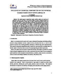
biofilm, thereby contributing to an enhanced inherent biofilm mechanical stability (Azeredo and Oliveira, 2000; Simo˜es, 2005). The characteristics of the P. fluorescens biofilms formed on the stainless steel cylinders (Table 1), namely the respiratory activity, biofilm mass and total content of proteins and polysaccharides, are similar to the ones observed in biofilms formed in a flow cell system under turbulent flow (Simo˜es et al., 2003b, 2005c), specifically the significant content of extracellular proteins and polysaccharides found in the composition of the biofilm matrix. Regarding B. cereus biofilms, no previous studies are available in terms of phenotypic characterisation. In fact, according to Kolari et al. (2001), B. cereus is an inefficient biofilm former. However, this study reveals the biofilm formation ability of B. cereus when using bioreactors that mimic real industrial situations. Furthermore, it is known that no strain can effectively represent its species (Fux et al., 2005). Monoculture B. cereus and P. fluorescens biofilms formed on stainless steel slides in a chemostat, according to the procedure described by Simo˜es et al. (2003a), were inspected by scanning electron microscopy (SEM) and the results are depicted in Figure 1. The existence of a more complex matrix of P. fluorescens biofilms when compared with the one existent in B. cereus biofilms is evident when visualising the biofilms by SEM (Figure 1). This microscopic evidence corroborates the extracellular proteins and polysaccharides results found in Table 1. The biofilm matrix has acquired great importance in biofilm architecture, and thus in biofilm mechanical stability, as EPS are responsible for keeping biofilm together and binding the biofilm to the support, forming a temporary network of fluctuating junction points (Ko¨rstgens et al., 2001). Biofilm removal due to mechanical stress
Figure 2 shows the biofilm removal obtained due to the increase in the Reynolds number of agitation (ReA) for B. cereus and P. fluorescens biofilms developing on the stainless steel cylinder. In this study the mechanical stability of the biofilm was assessed by submitting biofilms to different shear stress forces, correspondent to increasing ReA. The existence of shear stress forces higher than the one under which the biofilm was formed (ReA ¼ 2,400) caused biofilm removal. This mechanical stability experiment (Figure 2) shows that biofilms subjected to mechanical treatment were hardly removed with low shear stress (ReA # 4,000) since only approximately 13% (B. cereus) and 14% (P. fluorescens) of biofilm removal was achieved. However, when the ReA was raised from 4,000 to 8,100 a noticeable biofilm detachment was observed, but a layer remained on the surface even when the highest ReA was applied. In a biofilm control process, this biofilm remaining can act as an additional source of nutrients and/or as a suitable surface
a
b
Figure 1 SEM photomicrographs of a 6 old B. cereus (a) and P. fluorescens (b) and biofilms formed on stainless steel slides. Magnification 6,330 £ , bar ¼ 5 mm
477
M. Simo˜es et al.
Biofilm removal (%)
50
B. cereus
P. fluorescens
40 30 20 10 0 4,000
8,100
12,100
16,100
ReA Figure 2 Biofilm removal for the control assay due to change in the ReA
for further growth of cells (Simo˜es et al., 2005c). The high percentage of removal occurred with the implementation of a ReA of 8,100 for both biofilms (Figure 2), being biofilm removal similar to the other Reynolds numbers tested ( p . 0.05). Thus, it can be said that the biofilm removal is dependent on the hydrodynamic conditions ( p , 0.05). Azeredo and Oliveira (2000) reported that biofilm detachment is processed in layers, where the increase in the shear stress may progressively thin the biofilm, the ultimate effect expected being mechanical failure and total detachment. Figure 2 also shows that the total series of ReA numbers did not give rise to total biofilm removal. The total percentage of biofilm that was not removed by submission to the total series of ReA, considered as the biofilm remaining, is presented in Table 2. From Table 2 it is possible to ascertain that 47% of the total B. cereus biofilms and 24% of the P. fluorescens biofilms remained adhered to the stainless steel surface, after submission to the total series of ReA. This result demonstrates that B. cereus biofilms had a higher mechanical stability than biofilms formed by P. fluorescens. The slow growing biofilm phenotype, revealed by the decreased respiration rate (Table 1), and the increased motility (results not shown) of B. cereus when compared with P. fluorescens could account for the mechanical stability differences. According to several authors (Kwok et al., 1998; Villasen˜or et al., 2000), slower growing biofilms make more compact, with a smaller amount of EPS, but more stable biofilms, corroborating our results concerning the distinct amounts of B. cereus and P. fluorescens biofilm EPS (Table 1 and Figure 1). Also, motility is known to be essential for biofilm formation, probably to overcome the electrostatic repulsion of cells and surfaces (Pratt and Kolter, 1998; Heydorn et al., 2002; Harshey, 2003; Walker et al., 2004), being, apparently, important in the adhesion strength of the biofilm inner layers, as suggested by our results (Figure 2). This preliminary study suggests that further analyses are required in order to ascertain the basis of the increased mechanical stability of B. cereus biofilms. The comparison between biofilm composition (Table 1) and mechanical stability results (Figure 2 and Table 2) further suggests that other molecular mechanisms besides the EPS amount and bacterial motility, such as quorum sensing phenomenon and EPS composition, can account for the distinct behaviour that B. cereus and P. fluorescens biofilms face to mechanical stress. For example, Table 2 Total percentage of B. cereus and P. fluorescens biofilm remaining on the stainless steel surface after the submission to the total series of ReA
478
Biofilm remaining (%)
B. cereus
P. fluorescens
47.0 ^ 7.5
24.2 ^ 5.3
the existence of quorum sensing events is known to be expressed differently by Gramnegative and Gram-positive bacteria (El-Sayed et al., 2001; Dong et al., 2002), playing an important role in biofilm formation and behaviour (Davies et al., 1998). Also, microbial binding strength to surfaces depends on the chemical nature of the EPS (Perry et al., 2005).
The system presented in this work provides a reliable approach to investigate the mechanical stability of biofilms, leading to a better understanding of biofilms in different environments and the development of biofilm control strategies. The characterisation of the biofilms showed that the system tested allowed the formation of a great quantity of B. cereus and P. fluorescens biofilms which covered the surface of the stainless steel cylinders and had an inherent mechanical stability. B. cereus and P. fluorescens formed physiologically distinct biofilms, B. cereus biofilms mainly being constituted by cells while P. fluorescens biofilms were mostly constituted by EPS. The evidence of the more complex P. fluorescens biofilm matrix did not play a major role in the maintenance of the biofilm mechanical stability as proposed in previous studies (Stoodley et al., 1999; Ko¨rstgens et al., 2001; Poppele and Hozalski, 2003). In fact, B. cereus biofilms with a smaller content of EPS had a higher mechanical stability than P. fluorescens biofilms.
M. Simo˜es et al.
Conclusions
Acknowledgements
The authors acknowledge the financial support provided by IBQF, and the Portuguese Foundation for Science and Technology (Project CHEMBIO - POCI/BIO/61872/2004 and Post-Doc Grant - Manuel Simo˜es).
References Azeredo, J. and Oliveira, R. (2000). The role of exopolymers produced by Sphingomonas paucimobilis in biofilm formation and composition. Biofouling, 16, 17– 27. Danese, P.N., Pratt, L.A. and Kolter, R. (2000). Exopolysaccharide production is required for development of Escherichia coli K-12 biofilm architecture. J. Bacteriol., 182, 3593– 3596. Davies, D.G., Parsek, M.R., Pearson, J.P., Iglewski, B.H., Costerton, J.W. and Greenberg, E.P. (1998). The involvement of cell-to-cell signals in the development of a bacterial biofilm. Science, 280, 295 – 298. Dong, Y.-H., Gusti, A.R., Zhang, Q., Xu, J.-L. and Zhang, L.-H. (2002). Identification of quorum-quenching N-acyl homoserine lactonases from Bacillus species. Appl. Environ. Microbiol., 68, 1754– 1759. Dubois, M., Gilles, K.A., Hamilton, J.K., Rebers, A. and Smith, F. (1956). Colorimetric method for determination of sugars and related substances. Anal. Chem., 28, 350 – 356. El-Sayed, A.K., Hothersall, J. and Thomas, C.M. (2001). Quorum-sensing-dependent regulation of biosynthesis of the polyketide antibiotic mupirocin in Pseudomonas fluorescens NCIMB 10586. Microbiology, 147, 2127– 2139. Frølund, B., Palmgren, R., Keiding, A. and Nielsen, P.H. (1996). Extraction of extracellular polymers from activated sludge using a cation exchange resin. Wat. Res., 30, 1749– 1758. Fux, C.A., Shirtliff, M., Stoodley, P. and Costerton, J.W. (2005). Can laboratory reference strains mirror “real-world” pathogenesis. Trends Microbiol., 13, 58 – 63. Harshey, R.M. (2003). Bacterial motility on a surface: many ways to a common goal. Annu. Rev. Microbiol., 57, 249 – 273. Kolari, M., Nuutinen, J. and Salkinoja-Salonen, M.S. (2001). Mechanisms of biofilm formation in paper machine by Bacillus species: the role of Deinococcus geothermalis. J. Ind. Microbiol. Biotechnol., 27, 343 – 351. Ko¨rstgens, V., Flemming, H.-C., Wingender, J. and Borchard, W. (2001). Uniaxial compression measurement device for investigation of the mechanical stability of biofilms. J. Microbiol. Meth., 46, 9 – 17.
479
M. Simo˜es et al. 480
Kwok, W.K., Picioreanu, C., Ong, S.L., van Loosdrecht, M.C.M., Ng, W.J. and Heijnen, J.J. (1998). Influence of biomass production and detachment forces on biofilm structures in a biofilm airlift suspension reactor. Biotech. Bioeng., 58, 400 – 407. Ohashi, A. and Harada, H. (1994). Adhesion strength of biofilm developed in an attached-growth reactor. Wat. Sci. Tech., 29(10– 11), 281 – 288. Ohashi, A. and Harada, H. (1996). A novel concept foe evaluation of biofilm adhesion strength by applying tensile force and shear force. Wat. Sci. Tech., 34(5 –6), 201 – 211. Ohashi, A., Koyama, T., Syutsubo, S. and Harada, H. (1999). A novel method for evaluation of biofilm tensile strength resisting erosion. Wat. Sci. Tech., 39(7), 261 –268. Pereira, M.O., Morin, P., Vieira, M.J. and Melo, L.F. (2001). A versatile reactor for continuous monitoring of biofilm properties in laboratory and industrial conditions. Lett. Appl. Microbiol., 34, 22 – 26. Pereira, M.O., Kuehn, M., Wuertz, S., Neu, T. and Melo, L. (2002). Effect of flow regime on the architecture of a Pseudomonas fluorescens biofilm. Biotech. Bioeng., 78, 164 – 171. Perry IV, T.D., Klepac-Cera, J.V., Zhang, X.V., McNamara, C.J., Polz, M.F., Martin, S.T., Berke, N. and Mitchell, R. (2005). Binding of harvested bacterial exopolymers to the surface of calcite. Environ. Sci. Technol., 39, 8770– 8775. Poppele, E.H. and Hozalski, R.M. (2003). Micro-cantilever method for measuring the tensile strength of biofilms and microbial flocs. J. Microbiol. Meth., 55, 607 –615. Pratt, L.A. and Kolter, R. (1998). Genetic analysis of Escherichia coli biofilm formation: roles of flagella, motility, chemotaxis and type I pili. Mol. Microbiol., 30, 285 – 293. Ra¨tto¨, M., Suihko, M.-L. and Siika-Aho, M. (2005). Polysaccharide-producing bacteria isolated from paper machine slime deposits. J. Ind. Microbiol. Biotechnol., 32, 109 – 114. Saby, S., Sibille, L., Mathiew, J., Paquin, L. and Block, J.C. (1997). Influence of water chlorination on the counting of bacteria with DAPI (4,6-diamino-2-phenylindole). Appl. Environ. Microbiol., 63, 1564– 1569. Simo˜es, M. (2005). Use of biocides and surfactants to control Pseudomonas fluorescens biofilms – role of hydrodynamic conditions. PhD Thesis, University of Minho. Simo˜es, M., Carvalho, H., Pereira, M.O. and Vieira, M.J. (2003a). Studies on the behaviour of Pseudomonas fluorescens biofilms after ortho-phthalaldehyde treatment. Biofouling, 19, 151 – 157. Simo˜es, M., Pereira, M.O. and Vieira, M.J. (2003b). Monitoring the effects of biocide treatment of Pseudomonas fluorescens biofilms formed under different flow regimes. Wat. Sci. Technol., 47(5), 217 – 223. Simo˜es, M., Pereira, M.O. and Vieira, M.J. (2005a). Effect of mechanical stress on biofilms challenged by different chemicals. Wat. Res., 39, 5142– 5152. Simo˜es, M., Pereira, M.O. and Vieira, M.J. (2005b). Validation of respirometry as a short-term method to assess the efficacy of biocides. Biofouling, 21, 9 –17. Simo˜es, M., Pereira, M.O. and Vieira, M.J. (2005c). Action of a cationic surfactant on the activity and removal of bacterial biofilms formed under different flow regimes. Wat. Res., 39, 478 – 486. Simo˜es, M., Simo˜es, L.C., Machado, I., Pereira, M.O. and Vieira, M.J. (2006). Control of flow-generated biofilms using surfactants – evidence of resistance and recovery. Food Bioprod. Process., 84, 1 – 8. Standard Methods for the Examination of Water and Wastewater (1989). 17th edn, American Public Health Association/American Water Works Association/American Environment Federation, Washington DC, USA. Stoodley, P., Boyle, J.D., DeBeer, D. and Lappin-Scott, H.M. (1999). Evolving perspectives of biofilm structure. Biofouling, 14, 75 –90. van Benthum, W.A.J., Garrido-Ferna´ndez, J.M., Tijhuis, L., van Loosdrecht, M.C.M. and Heijnen, J.J. (1996). Formation and detachment of biofilms and granules in a nitrifying biofilm airlift suspension reactor. Biotechnol. Prog., 12, 764 – 772. Vieira, M.J., Melo, L. and Pinheiro, M.M. (1993). Biofilm formation: hydrodynamic effects on internal diffusion and structure. Biofouling, 7, 67 – 80. Villasen˜or, J.C., van Loosdrecht, M.C.M., Picioreanu, C. and Heijnen, J.J. (2000). Influence of different substrates on the formation of biofilms in a biofilm airlift suspension reactor. Wat. Sci. Technol., 41(4– 5), 323 – 330. Walker, S.L., Redman, J.A. and Elimelech, M. (2004). Role of cell surface lipopolysaccharides in Escherichia coli K12 adhesion and transport. Langmuir, 20, 7736– 7746. Watnick, P.I. and Kolter, R. (1999). Steps in the development of a Vibrio cholerae El Tor biofilm. Mol. Microbiol., 34, 586 – 595.