Insect Immunity. SEPTIC INJURY OF DROSOPHILA INDUCES THE SYNTHESIS OF A POTENT ANTIFUNGAL PEPTIDE WITH. SEQUENCE HOMOLOGY TO ...
Vol. 269, No. 52, Issue of December 30, pp. 33159-33163, 1994 Printed in U.S.A.
THEJOURNAL OF BIOLOGICAL CHEMISTRY 0 1994 by The American Society for Biochemistry and Molecular Biology, Inc.
Insect Immunity SEPTIC INJURY OFDROSOPHILA INDUCES THE SYNTHESIS OFA POTENT ANTIFUNGAL PEPTIDE WITH SEQUENCE HOMOLOGY TO PLANT ANTIFUNGAL PEPTIDES* (Received for publication, September 2, 1994)
Pascale FehlbaumS, Philippe BuletS, Lydia MichautS, Marie LagueuxS, Willem F. BroekaertO, Charles HetruS, andJules A. Hoffmannh From the vnstitut deBiologie Moliculaire et Cellulaire, UPR 9022 Centre National dela Recherche Scientifique “Riponse Immunitaire et De‘veloppement chez les Znsectes,” 15, rue Rene‘ Descartes, 67084 Strasbourg Cedex, France and the§F: A. Janssens Laboratory of Genetics, Catholic University of Leuuen, Willem De Croylaan 42, B-3001Heuerlee, Belgium
In response to a septic injury (pricking with a bacte-coccus luteus and Escherichiacoli 1106. The insects were kepta t 25 “C ria-soaked needle) larvae and adults of Drosophila pro- for 24 h and frozen in liquid nitrogen until extraction. duce considerable amounts of a 44-residue peptide con-Extraction and Purification-Abdomens and thoraces were ground to continuous presenceof liquid nitrogen. taining 8 cysteines engaged in intramolecular disulfide a finepowder in a mortar in the The powder was taken up in 10 volumes (w/v) of 25 mM ammonium bridges. The peptide is synthesized in the fat body, a acetate buffer, pH 5.2, containing 10pg/ml aprotinin, for 20 min in an functional homologue of the mammalian liver, and se- ice-cold water bath. After centrifugation(3,000x g, 30 min, 4 “C), the creted into the blood of the insect. It exhibits potent supernatant was loaded onto two linked Sep-PakC,, cartridges previantifungal activity butis inactive against bacteria. Thisously washed with methanol and equilibrated with25 mM ammonium novelinduciblepeptide,whichweproposetoname acetate bufferat pH 5.2. Elution wasperformed stepwise with various solutions of acetonitrile in the same buffer (10, 30, and 80% acetonidrosomycin, showsa significant homology with a family trile). Fractions were concentrated under vacuum, reconstituted Milin of5-kDa cysteine-richplantantifungalpeptidesrecently isolatedfrom seeds of Brassicaceae. This finding liQ water, and applied on an Aquapore OD300 C,, column (250 x 4.6 mm, BrownleeTM) equilibrated with the samebuffer. Elutions were perunderlines that plants and insects can rely on similar formed with a linear gradientof 7-57% acetonitrile in the samebuffer molecules in their innate host defense. over 90 min at a flow rate of 1 mumin. The fraction containing the strongly induced peptide (see below) was desalted by reversed-phase HPLC’ on an Aquapore RP300 C, column (30 x 2.1 mm, BrownleeTM) Drosophila, like o t h e r i n s e c t s , r e s p o n d s t o s e p t i c injury by with a linear gradientof 10-90% acetonitrile inacidified water (0.05% the r a p i d and transient synthesis of a battery of p o t e n t anti- trifluoroacetic acid) at a flow rate of 1mumin over 20 min. All HPLC purificationswereperformedwithaBeckman Gold HPLC system bacterial peptides (reviewed in Refs. 1and 2). The principal site equipped with a photodiode array detector Beckman 168. The column of synthesis of these peptides is the fat body, a functional ho- effluent was monitored by absorbance at 225 nm. mologue ofthe mammalian liver. The i n s e c t h o s tdefense lacks Microorganisms-Filamentous fungi were grown on a five-cereal mespecificity and memory, and it has been argued that it is ho- dium. Spores were harvestedas described previously (7). Thefollowing fungal strains were used: Alternaria brassicola(MUCL 20297), Altermologous to the mammalian a c u t e phase response (1,3). To date, a limited number of immune-induced peptideshave naria longipes (CBS 620-831, Ascochytu pisi (MUCL 301641, Botrytis cinerea (MUCL 30158), Fusarium culmorum (IMI 1804201, Fusarium been i s o l a t e d f r o m D r o s o p h i l aand their s t r u c t u r e s c h a r a c t e r - oxysporum (MUCL 9091, Nectria haematococca (Collection Van Etten ized (4,5). The isolation studies were based so f a r o nthe use of 160-2-2), Neurosporacrassa(CBS 327-541, Dichodermahamatum growth inhibition assays to detect antibacterial activities. We (MUCL 297361, and Dichoderma uiride (MUCL 19724). have taken here a n o v e l a p p r o a c h based on differential proBacteria were precultured in appropriate conditions (4). The followfrom colt e i n o g r a m s between e x t r a c t s f r o m untreated and immune-chal- ing are the bacterial strains used with their source (gifts lenged insects. Under o u r w o r k i n g c o n d i t i o n s , w h iused c h mild leagues): Bacillus megaterium from J. Millet and A. Klier (Pasteur acidic extraction techniques, we observed the massive appear- Institute, Paris); M. luteus A270 from the Pasteur Institut Collection, Paris; Staphylococcus aureus, Streptococcus sanguis, Streptococcus a n c e a f t e r c h a l l e n g e oaf small sized, slightly cationic peptide. agalatae, and Pseudomonas cepaciafrom H. Monteil (Institute of BacWe r e p o r t the a m i n o a c i d s e q u e n c e of this peptide and the teriologie, University of Strasbourg); Enterobacter cloacae p12 and E. cloning of the corresponding cDNA. This peptide exhibits pocoli D31 from H. G. Boman (Department of Microbiology, University tent a n t i f u n g a l activity and s h o w s sequence homology tosmall of Stockholm), and E. coli D22 from P. L. Boquet (Centre #Etudes Nuclbaires, Saclay). sized cationic cysteine-rich antifungal moleculesrecently Antifungal, Antimicrobial Assays-Fungal spores (final concentrai s o l a t e df r o m seeds ofBrassicaceae,includingArabidopsis tion: lo4 sporedml) were suspended in a growth medium containing thaliana (6). Potato Dextrose Broth (DIFCO, in half-strength, supplemented with tetracycline (10 pg/mV and cefotaxim (100 pg/ml)), dispensed by aliMATERIALANDMETHODS quots of 80 pl into wells of a microplate containing20 pl of either water or of the fractions to be analyzed, and incubatedfor 48 ha t 25 “C in the Immune Challenge of Insects-1-day-old adult males of Drosophila Oregon R were individually pricked, under CO,, with a needle dipped dark. Growth of fungi was evaluated by measuring the culture absorbinto a combined bacterial pellet of 37 “C overnight cultures of Micro- ance a t 595 nm using a microplate reader. Antibacterial activity was measured by a liquid growth inhibition as described previously (4). * The costsof publication of this article were defrayed in part by theassay payment of page charges. This article must therefore be hereby markedHemolytic Activity Assay-Bovine red blood cells were washed sev“advertisement” in accordance with 18 U.S.C. Section 1734 solely to eral times with phosphate-buffered saline. A 0.5% suspension of red blood cells was made in phosphate-buffered saline. Hemolytic activity indicate this fact. The nucleotide sequence(s) reported in this paper has been submitted to the GenBankmIEMBL Data Bank with accession number(s) X75595. The abbreviations used are: HPLC, high performance liquid chro1 To whom correspondence should be addressed:Tel.: 33-88-41-7077; matography; AFP, anti-fungal peptide; MOPS, I-morpholinepropaneFax: 33-88-60-69-22. sulfonic acid.
’
33159
33160
Drosophila Peptide Antifungal
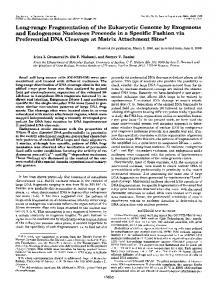
was measured as follows. 10 pl of the pure peptide (10 pg) were incubated in a microtiter plate with 100 pl of the 0.5% red blood cells. As a positive control (100% lysis), a solution of 1% detergent (Triton X-100) A was used instead of the molecule for analysis. After 1h of incubation at 37 "C, the plate was centrifuged (288 x g, at 10 "C, for 5 min), and the absorbance of 50 pl of the supernatant was measured at 405 nm. Proteolysis-Purified peptide was treated with S. aureus V8 protease (Takara, Kyoto), trypsin, chymotrypsin, elastase, and thermolysin (all from Boehringer Mannheim), in the conditions recommended by the supplier with an enzymdprotein ratio of 25% (w/w),except for endoproteinase Glu-C (S. aureus V8 protease) where the ratio was 1%.For pepsin (Boehringer Mannheim) we used 0.1 M Tris-HC1buffer at pH 3.5, and incubation was performed over 16 h at 37 "C. All reactions were stopped directly by injection on reversed-phase HPLC. Reduction and Alkylation-An aliquot of the purified peptide (2 nmol) was dissolved in 40 pl of 0.5 M Tris-HC1, 2 mM EDTA, pH 7.5, 10 20 30 4a Time (min) containing 6 M guanidine hydrochloride to which 2 pl of 2.2 M dithiothreitol were added. The sample was flushed with nitrogen and incubated for 1h a t 45 "C in thedark. Freshly distilled 4-vinylpyridine (2 pl) was added, and the sample was incubated for 10 min at 45 "C in the PIxl dark. The pyridylethylated peptide was separated by reversed-phase 2 I HPLC prior to sequencing. Microsequence Analysis-Automated Edman degradation of the native and of the pyridylethylated peptide and detection of phenylthiohydantoin derivatives were performed on a pulse liquid automatic sequenator (Applied Biosystems, model 473A). Mass Spectrometry-The purified peptide was dissolved in water/ methanol (50:50, v/v) containing 1%acetic acid and analyzed on a VG BioTech BioQ mass spectrometer (Manchester). This instrument consists of an electrostatic ion spray source operating at atmospheric pressure, followedby a quadruple mass analyzer with a mass range of 14,000. The extraction cone voltage value was 55 V. Scanning was performed from mlz 1,500 in 10 s with the resolution adjusted so that the mlz 998 peak from horse heart myoglobin was 1.5-1.7 Da at its r base. The data system was operated as a multichannel analyzer, and 10 20 30 40 several scans were summed to obtain the final spectrum. Each molecTime (min) ular species produced a seriesof multiply charged protonated molecular FIG.1. Reversed-phaseHPLC separation of extracts of naive ions from a separate introduction of horse heart myoglobin (16,951.4 material Da). Molecular masses are given as average values based on the atomic (A) and challenged adult malesof Drosophila ( B ) .The contained in fraction F-30% (see "Results") was loaded on an Aquapore weights of the elements (C = 12.011, H = 1.00794, N = 14.0067, 0 = OD300C1, column and eluted with a linear gradient (dotted line) of 15.9994, and S = 32.06); only average masses were measured. acetonitrile in 25 mM ammonium acetate buffer pH 5.2. Absorbance was monitored at 225 nm (solid line). Note the appearance of an absorption RESULTS peak P-30 in the extract of challenged insects.
Isolation of a 44-Residue Peptide from Bacteria-challenged 1nsects-2,000 1-day-old adult males of Drosophila were challenged as described under "Material and Methods." They were kept for 24 hat room temperature before extraction undermild acidic conditions (pH 5.2). In parallel, 2,000 untreated adult males were subjected to extraction. The supernatants of the two crude extracts were prepurified by solid phase extraction on Sep-Pak C,, cartridges. The elution was successively performed with a 10, 30, and 80% solution of acetonitrile in ammonium acetate buffer at pH 5.2, yielding three fractions (referred to as F-lo%,F-30%, and F-80%) which were separately analyzed by reversed-phase HPLC using various gradients of acetonitrile in ammonium acetate buffer. When the HPLC chromatograms of fraction F-10% from extracts of bacteria-challenged and untreated Drosophila were compared, no clear-cut difference could be observed, neither for the numberof absorption peaks nor for their relative intensities (data not shown). A similar result wasobtained for fraction F-80%. In markedcontrast, the analysis of fraction F-30% showed the presence of a strong absorption peak in bacteria-challenged insects which could not be detected in control insects (Fig. 1,peak P-30). This experiment was repeated five times with similar results. No other obvious difference was noted in the HPLC chromatograms obtained from extracts of bacteria-challenged or untreated insects. Peak P-30 was purified to homogeneity by reversed-phase HPLC on a C, column with a linear gradient of acetonitrile in acidified water, which also served as a desalting step. Two nanomoles of pure compound P-30 were then subjected t o automated Edmandegradation and gave a sequence of 43 residues withseven blanks. To check whether these blanks
I
10
20
30
40
44
DCLSGRYKGP CAVWDNETCR RVCKEEGRSS GHCSFSLKCW CEGC
FIG.2. Amino acid sequence of the bacteria-induced 44-residue peptide.
could correspond to cysteine residues, 2 nmol of P-30 were submitted to reduction and alkylation with 4-vinylpyridine, followed by Edman degradation. A sequence of 44 residues was obtained comprising 8 cysteine residues (Fig. 2). One of these cysteine corresponds to the C-terminal residue. This residue could not be detected in the absence of derivatization, thus explaining that we initially obtained a sequence of only 43 residues. We next submitted 250 pmol of P-30 to electrospray mass spectrometry in theconditions described in Bulet et al. (4) and obtained a molecular mass of 4889.1 Da. The mass calculated for the 44 residues presented inFig. 2 is 4897.5, i.e. in excess of 8 Da t o that measuredby mass spectrometry. This difference of 8 Da is attributable to the arrangement of the 8 cysteine residues intofour intramolecular disulfide bridges, a process which eliminates 8 hydrogen atoms. We repeatedly analyzed extracts from unchallenged larvae and adults andfailed to detect the44-residue peptide. In adult males, which we investigated in more detail, the 44-residue peptide was clearly evidenced as early as 1 h after bacterial challenge. The amounts recovered from challenged insects increased for a period of up t o 16 h after which they reached a plateau. Inone experiment we collected the hemolymph of 100
Peptide Antifungal Drosophila
33161
GATAATTCAAACAGAAATCATTTACCAAGCTCCGTGAGAACCTTTTCCAAT
ATG ATG CAG ATC
M
M
Q
i?
I
1
> - X TAC TTG TTC GCC CTC TTC GCT GTC CTG ATG CTG GTG GTC CTG GGA GCC114 X Y L F A L F A V L M L V V L G A 20
10
M C GAG GCC GAT GCC GAC TGC CTG TCC GGA AGA TAC AAG GGT CCC TGT GCC 165 K
K
A
D
A
D
C
L
S
G
R
Y
K
G
P
C
A
30
GTC TGG GAC AAC GAGA CC TGT CGT CGT GTG TGC AAG GAG GAG GGA CGC TCC 2 1 6 V
W 40
D
N
E
T
C
R
R
V
C
K 50
E
E
G
R
S
AGT GGC CAC TGC AGC CCC AGT CTG AAG TGC TGG TGC GAA GGA TAATGC ATCC S
G
H
C
S 60
P
S
L
K
C
W
C
E
G
268
C 70
CATGAGCAATTAGCATGAACGTTCTGAAAAGCGCGTTTAGCTCTCCACTACTTACGACATATTCTATG CTSCAATATTGAAAATCTAATAAACAAAATGTACATT(A)38
335 313
FIG. 3. Nucleotide sequence of a cDNA clone and deduced amino acid sequence correspondingto the precursor of the "residue peptide. A size-selectedcDNA library was screened using a standard protocol (as in Bulet et al.(4) with a degenerate probe pool (5'-GT(Cm)TC(A/ GjTT(A/G)TCCCA(C/G!AC-3') complementary to residues 13-18 of the newly characterized peptide. The polyadenylation consensus signal is underlined. The construction of the size-selected hgt22 cDNA and all conditions were as in Buletet al. (4).
-"
bacteria-treated adults 24 h after challenge and observed the adults Male Pupae Larvae presence of the 44-residue peptide (data not shown). We ascertained its identity by partial peptide sequencing and electro0 4 0 7 0 1.5 7 24 spray mass spectrometry. The peptide was absent from the hemolymph of untreated adults. Isolation of cDNA Clones Encoding a 70-Residue Precursor Peptide-From the peptide sequence established above, we devised a degenerate oligonucleotide probe pool corresponding to residues 13-18. The probe pool was used to screen a size-selected cDNA library preparedfrom bacteria-challenged larvae. 10,000 plaque-forming units were plated out and 120 hybridization-positive clones wereobtained,4 of which weresequenced. They were all 373 nucleotides long and contained an open reading frame of 70 codons starting with two Met codons and ending witha stop codon (Fig. 3). Therewere no sequence FIG. 4. Northern blot analysis of the precursor of the 44-resivariations between these four cDNA clones. The amino acid due peptide. 20 pg of total RNAfrom naive ( 0 )and bacteria-challenged sequence deduced from the cDNA clones obviously corresponds third instar larvae, young pupae, and adults (times after pricking inby denaturing electrophoresis in 1% to a precursor peptide containing the 44-residue sequence of dicated in hours) were fractionated gels with MOPS buffer. Transfer to nylon memthe newly isolated peptide. The deduced sequence of the pep- agarose-formaldehyde branes (HybondT", Amersham Corp.) was performed essentially a s detide is in perfect agreement with that obtained by Edman deg- scribed in Thomas (18).The membrane was hybridized overnight a t radation. The 44-residue peptide is N-terminally flanked by a 42 "C with nick-translated"P-labeled cDNAprobe ([a-"'PldCTP, [U-'~PI putative strongly apolar signal peptide up to residue Ala-21, dATP, 3000 Ci/mmol). Hybridization was performed under high stringency conditions. The membrane was first hybridized with the cDNA which is a good candidate cleavage site for signal peptidase. encoding the precursor of the 44-residue peptide (A), then with a riboAlternatively, residues Ala-24 or Ala-26 could serve as cleavage somal protein (rp49) cDNA probe as a control ( B ) . sites. If the signal peptidase acts at the level of Ala-21, a n additional cleavage by dipeptidylaminopeptidases would be necessary to yield the 44-residue peptide, as is the case for erythrocytes. In marked contrast,however, in antifungal tests, the peptide showed a strong activity againsteight fungal other bacteria-induced peptides in insects (8-10). Analysis of Dunscripts Encoding the 70-Residue Precursor strains tested (Fig. 6): N. crassa (IC5o0.6 PM),B. cinerea (IC5o Peptide-One of the cDNA clones was used after nick transla- 1.2 PM),I? culmorum (IC5o1.0 PM), A. brassicola (IC5o0.9 PM),A. N. haematococca (IC5o1.8 PM),l? oxytion to detectby Northern blotting experiments thepresence of longipes (IC5o 1.4 p ~ ) , sporum (IC5o4.2 PM), and A. pisi (IC5o3.2 PM).No or minimal transcripts in untreated and bacteria-challenged Drosophila. As shown in Fig. 4, a faint signalis present innaive insects and activity was found against two Dichoderma species (I: viride, I: hamatum). Fungal growthinhibitionwas also monitored is noticeably increased in bacteria-challenged larvae, pupae, microscopically. High concentrations of the 44-residue peptide and adults. In situ hybridization was performed on paraffin-embedded (10 J ~ Mand above) completely inhibited spore germination and naive and bacteria-challenged larvae and adults using a digoxi- no hyphae were observed. Lower concentrations of the peptide caused delayed growth genin-labeled cDNA probe. A marked reaction was observed in the fat body cells of challenged insects, as shown in Fig. 5. A of hyphae with abnormal morphology. More than 50% of the fainter, but distinct,reaction was also seen in the fat body in observed hyphae of B.cinerea treated with the 44-residue peptide at a concentrationof 1.2 PM,extruded cytoplasmic material absence of challenge (data not shown). Antifungal Activity of the 44-Residue Peptide-The purified along the hyphae (Fig. 7), indicating that the protein causes peptide was tested ina variety of biological assays for antibac- partial lysis. The antifungal activity was shown in similar tests to be terial, antifungal, or hemolytic activities. No antibacterial activity against a large variety of Gram-positive and Gram-neg- resistant to variationsof pH from 2 to 10 and toa 30-min heat ative bacteria(B. megaterium, M.luteus, S. aureus, S. sanguis, treatment at 100 "C (data not shown). Finally, spores of the highly sensitive N. crassa were cultured S. agalatae, I? cepacia, E. cloacae, E. coli) was detected in standard conditions at concentrations ranging from 1to 20 p ~ . in thepresence of concentrations of 0.1-10 PMof the 44-residue The peptide did not exhibit hemolytic activity against bovine peptide. After 48 h, the medium containing the peptide was
Drosophila Antifungal Peptide
33 162
-
-~
"~ ~
FIG.5. Detection of transcripts encoding the 44-residue peptide in 6-h bacteria-challenged third instar larvae by in situ hybridization. Insects were fixed, embedded, sectioned, and hybridized with a digoxigenin-labeled cDNA probe encoding the precursor of the 44-residue peptide, using a DIG-DNA labeling and detection kit (Boehringer Mannheim). The protocol of Tautz and Pfeiflle (19)was modified a s follows. Fixation was in Carnoy's fix; prehybridation and hybridization were carried out at 48"C in 50% formamide. cu, cuticle; fa, fat body.As a control, the same experiment was performed after RNase treatment of slices, and they showed no staining (data not shown).
FIG.7. Inhibition of fungal growth bythe 44-residue peptide. Photomicrographs were taken after 24 h of incubation of a B. cinerea spore suspension in half-strength potato dextrose broth supplemented with 1mM CaCI, and 20 mM KC1 in theabsence of the 44-residue peptide (control, Panel A), or in the presence of 1.25 PM peptide (Panel B ) . Arrowheads indicate sitesof hyphal lysis.
by fresh medium. This result demonstrates that 44-residue the peptide is fungicidal on N. crassa. DISCUSSION
1
10
100 pM
Concentration
A/. brassicola AI. lmgipes T. viride T. hamatum AS. pis;
Concentration
FIG.6. Dose-response curves for different fungal strains. Growth inhibition was measured at various concentrations of the 44residue peptide. Percent growth inhibition was recorded after 48 (IC5o, peptide concentration giving50% of growth inhibition).
removed and replaced with fresh medium. Forty-eight hours later the cultureswere examined spectrometrically and microscopically. No growth recovery had occurred after replacement
The present studyis the first report on an inducible antifungal peptide in aninsect. As stated in the Introduction, insects have been shown to produce a variety of potent antibacterial peptides within hours following septic injury or a bacterial challenge. However, it had not been documented so far that this type of challenge can also induce the synthesisof an antifungal peptide. Natori's group (11)has recently isolated from larvae of the dipteran species Sarcophaga peregrina a 67-residue histidine-rich antifungal protein devoid of cysteines. This protein is constitutively present in the hemolymph, in contrast to the inducible peptidewhich we have now characterized in Drosophila. I n situ hybridization points to the fat body as the major site of synthesis of this novel peptide in larvae and adultsof Drosophila. The Northern blot and in situ hybridization experiments indicate that transcripts encoding the antifungal peptide are constitutively present in larvae and adults and that their level is significantly enhanced upon septic injury. This constrasts with our chemical isolation studies which failed to detect the presence of the antifungal peptide prior to immune challenge. A few hours after challenge, however, a dramatic induction of the peptide can be detected. This high level of production is illustratedby the fact that we could extract up to 10 pmol of the peptide from one insect (this corresponds of an overall concentrations of 50 pg/g and could account for a blood concentration of up to 0.1 mM; note that theIC,, values are in the low micromolar range). Our presentworking hypothesis is that theexpression of the antifungal peptide is subjected both to transcriptional and translational control mechanisms in contrast to the majority of the inducible antibacterial peptides, the expression of which is primarily controlled at the transcriptional level. Our cDNA cloning studies show that the44-residue peptide is processed from a 70-residue precursor molecule. The exact h mechanism of the processing remains to be elucidated. The inducible antifungal peptide of Drosophila is a small sized, slightly cationic molecule (calculated PI = 7.81, which is particularly resistant to heat treatment, toaction the of various proteases, and to pH variations. It has four intramolecular disulfide bridges, and we may assume that it adopts a very
Peptide
AntifungalDrosophila
33163
most of the fungi tested, theIC,, values were in thelow micromolar ranges and even below (0.6 p ~ in) thecase of N. crassa. Experiments with the highly sensitive N. crassa point to a FIG.8. Sequencecomparison of the "residue antifungal fungicidal activity. peptide from Drosophila with an antifungal peptide (Rs-AFP2) In conclusion, we show that Drosophila larvae and adults isolate from the seeds of R. satiuus. Identical amino acids and conservative replacements are boxed. Bars indicate gaps to optimize se- can be induced by a septic injury, such as pricking with a bacteria-soaked needle,t o produce remarkably high amounts of quence alignments. a potent antifungal peptide. Thestructuraland functional compact tridimensional arrangement. Of major interest is the characteristics indicate that this peptide is a homologue of a fact that the Drosophila peptide has a significant sequence family of antifungal peptides recently isolatedfrom the seeds of homology with plant antifungal peptides recently isolatedfrom Brassicaceae,where theyparticipate t o theplant defense seeds of Brassicaceae which play a role in plant defense (6). against microorganisms. To our knowledge, this is the first Indeed, apart from the already well known plant antifungal report showing that insects and plants can rely on similar proteins (chitinases, glucanases, thionins, chitin-binding lec- molecules in theirhost defense. We proposed the nameof drosotins, ribosome-inactivatingproteins) (12-14) anincreasing mycin for this new antifungal peptide from Drosophila. number of new plant proteins capable of inhibiting fungal growth in vitro is emerging (see Refs. 6, 15, and 16).In parREFERENCES ticular, several 5-kDa cationic antifungal peptides which con1. Hoffmann,J. A,, Hetru, C., and Reiehhart, J-M.(1993) FEBS Lett. 3 2 5 , 6 3 4 6 2. Cocianeich, S., Bulet, P., Hetru, C., and Hoffmann, J . A. (1994a) Parasitol. tain disulfide bridges were isolated from seeds (AFPs, antiToday 10, 132-139 fungal peptides). Fig. 8 compares the peptide sequence of the 3. Hultmark, D. (1993) Dends Genet. 9, 178-183 Drosophila antifungal peptide with Rs-AFP2 (from seeds of 4. Bulet, P., Dimarcq, J.-L., Hetm, C., Laguew, M., Charlet, M., Hegy, G., Van Dorsselaer, A., and H o h a n n , J. A. (1993) J. Biol. Chem. 268,14893-14897 radish Raphanus satiuus) (17). Both peptideshave 8 cysteines 5. Dimareq, J.-L., Hoffmann, D., Meister, M., Bulet, P., Lanot, R., Reichhart, which are engaged in intramoleculardisulfide bridges. One of J.-M., and Hoffmann, J.A. (1994) Eul: J. Biochem. 221,201-209 6. Terras, F. R. G., Torrekens, S., Van Leuven, F., Osborn, R. W., Vanderleyden, J., these cysteines is positioned C-terminally inboth molecules. Cammue, B. P. A,, and Broekaert, W. F. (1993) FEBS Lett. 316,233-240 Allowing for several minor gaps in both sequences, is it ap7. Broekaert, W. F., Terras, F. R. G., Cammue, B. P. A,, and Vanderleyden, J. parent that the eightcysteines are arranged in a similar pat(1990) FEMS Microbiol. Lett. 69, 5 5 4 0 8. Kreil G., (1990) l k n d s Biochem. Sci. 15,23-25 tern. Theoverall homology between the Drosophila peptide and 9. Wicker, C., Reichhart, J.-M.,Hoffmann, D., Hultmark, D., Samakovlis, C., and Rs-AFP2 is 38%, takinginto accountconservative replaceH o h a n n , J. A. (1990)J. B i d . Chem. 265,22493-22498 ments. It will be of great interest t o work out the tridimen- 10. Boman, H. G., Faye, I., Gudmundsson, G. H., Lee, J. Y.,and Lidholm, D. A. (1991)E m J. Biochem. 201,23-31 sional structure of the Drosophila peptide and compare it t o the 11. Ijima, R., Kurata, S., and Natori, S. (1993) J. Biol. Chem. 268, 12055-12061 structure of the plant antifungal peptides in the future. It is 12. Roberts, W. K., and Selitrennikoff, C. P. (1986) Biochim. Biophys. Acta 880, 161-170 also interesting tonote that theproduction of Rs-AFP2 and its 13. Bowles, D. J. (1990) Annn. Reu. Biochem. 59,873-907 homologue in radishis not restricted to seeds but also occurs in 14. Raikhel, N.V., Lee, H. I., and Broekaert,W. F. (1993)Annu. Reu. Plant Physiol. Plant Mol. Biol. 44, 5 9 1 4 1 5 leaves after challenge with fungal pathogens.' W. E , Marien, W., Terras, F. R. G . ,De Bolle, M. F. C., Proost, P., Van As is the case for Rs-AF'P2, the Drosophila peptide is active 15. Broekaert, Damme, J., Dillen, L., Claeys, M., Rees, S . B., Vanderleyden, J.,and against a relatively broad spectrum of filamentous fungi. It Cammue, B. P. A. (1992) Biochemistry 31,43084314 inhibits spore germination at high concentrations and, at lower 16. Cammue, B. P. A,, De Bolle, M. F. C., Terras, F. R. G., Proost, P., Van Damme, J., Rees, S. B., Vanderleyden, J., and Broekaert, W. F. (1992) J. Biol. Chem. concentrations, delays growth of hyphae which subsequently 267,2228-2233 exhibit abnormal morphology. The IC,, values indicate an ex- 17. Terras, F. R. G . , Schoofs, H. M. E., De Bolle, M. F. C., Van Leuven, F., Rees, S . B., Vanderleyden, J., Cammue, B. P. A,, and Broekaert, W. F. (1992) J. Biol. ceptionally high potency of the Drosophila peptide. Indeed, for Chem. 267, 15301-15309 F. R. G . Terras and W. F. Broekaert, unpublished results.
18. Thomas, P. (1980)Proc. Natl. Acad. Sci. U.S. A. 77,5201-5205 19. Tautz, D., and Pfeifle, C. (1989) Chromosoma (Berl.)98, 80-85