Induction of c-fos, c-jun and p33 mRNAs after growth factor treatment of rat-1 parental cells and p2lHras-transformed cell lines. Subconfluent cultures of different ...
The EMBO Journal vol. 10 no. 5 pp. 1 103 - 1109, 1991
Insulin stimulation of p21 ras activation
gene
expression mediated by
Boudewijn M.Th.Burgering1 4, Rene H.Medema' 4, J.Antonie Maassen2 Marcus L.van de Wetering1, Alex J.van der Eb', Frank McCormick3 and Johannes L.Bos1'4 'Laboratory for Molecular Carcinogenesis and 2Laboratory of Protein Synthesis and Hormone Regulation, Sylvius Laboratory, PO Box 9503, 2300 RA Leiden, The Netherlands and 3Department of Molecular Biology, Cetus Corporation, 1400, 53rd Street, Emeryville, CA 94608, USA 4Present address: Laboratory for Physiological Chemistry, University of Utrecht, Vondellaan 24A, 3521 GG Utrecht, The Netherlands. Communicated by A.J.van der Eb
In fibroblasts, insulin is a weak mitogen and does not induce expression of c-fos, c-jun or p33. However, increasing the expression levels of either normal p21Hras or the insulin receptor, but not mutant p2lHras, enables insulin to induce the expression of these genes. In cells expressing elevated levels of insulin receptor, this process involves a rapid increase in p2lrasGTP levels (from 20% to 70% GTP as a percentage of total guanine nucleotides). No increase in p2lrasGTP levels was observed after PDGF and EGF stimulation of cells expressing high levels of the cognate receptor, stressing the specificity of the insulin-induced increase. We conclude that in fibroblasts, p2lras is an intermediate of the insulin signal transduction pathway involved in the regulation of gene expression and mitogenicity. Key words: GDP-GTP exchange/GTPase/insulin/phosphatidylinositol-3-kinase/signal transduction
Introduction Proteins encoded by members of the p2lras gene family bind guanine nucleotides and have low intrinsic GTPase activity (Barbacid, 1987). p2lras GTPase activity is strongly increased by a cytosolic protein of 120 kd called GTPase activating protein (GAP) (Trahey and McCormick, 1987). Specific point mutations at positions 12, 13 or 61 of p2 Iras result in proteins that have lost their intrinsic GTPase activity (Gibbs et al., 1984; Sweet et al., 1984) as well as regulation by GAP (Trahey and McCormick, 1987). These mutant p21ras proteins are capable of transforming immortalized cells in vitro, indicating that the GTP form of p2 Iras is a positive signal in cell proliferation (Gibbs et al., 1987; Hoshino et al., 1988; Satoh et al., 1988; Trahey and McCormick, 1987). Moreover, microinjection of p21 rasGTP but not p2lrasGDP induces the expression of c-fos, showing that also in the regulation of gene expression p2lrasGTP is the active state (Stacey et al., 1987). By analogy to the heterotrimeric G proteins, it has been postulated that GTP binding to p21ras is under the control of growth factors. Oxford University Press
Thus, a specific growth factor may constitute or induce a messenger upstream of p21ras and drive p21ras into an active, signal generating state (Hall, 1990). Recently, Downward et al. (1990a) strengthened this concept, by showing that upon stimulation of T cells with phytohaemagglutinin, the GTP form of p2lras accumulates rapidly. In most cell types the signal that activates p2iras is still unknown, although several candidates have been put forward (see also Hall, 1990). Korn et al. (1987) showed that p2lras mediates insulin-induced maturation of Xenopus oocytes and we recently obtained evidence that in rat fibroblasts, p2lras might mediate insulin- or insulin-like growth factor I-induced processes as well (Burgering et al., 1989). For the neuronal PC12 cells it has been suggested that p21ras might be involved in nerve growth factor-induced neurite outgrowth (Bar-Sagi and Feramisco, 1985; Hagag et al., 1986; Noda et al., 1985). Furthermore, using interfering p21ras mutants (Asn17), a role for p2lras has been suggested in EGF and TPA signal transduction (Cai et al., 1990). Also, a close linkage between p2 lras and PDGF-stimulated responses has been implicated (Kaplan et al., 1990; Kazlauskas et al., 1990; Molley et al., 1989). The fact that microinjection of antibodies against p2iras can block the action of a variety of growth factors, suggests a more general role for p21ras in growth control, and that, for instance, p2lras might be a connecting point for several growth factor mediated signalling pathways (Mulcahy et al., 1985; Yu et al., 1988). We have studied whether p2 lras can mediate signals induced by stimulation of cells with insulin. For that purpose we investigated the effects of expression of mutant p2 lHras or overexpression of normal p2 lHras and of insulin receptor on insulin-induced gene expression. Furthermore, we have measured the effect of insulin treatment on the activation state of p2lras. Our results show that insulin stimulation results in a rapid conversion of p2 lrasGDP into p2 lrasGTP, which provides evidence that the activation of p2 Iras may be part of the insulin signal transduction pathway leading to the induction of gene expression.
Results Overexpression of insulin receptor and overexpression of normal p2lHras enhance insulin-induced gene
expression We have previously proposed a role for normal p21ras in the mitogenic effect of insulin (Burgering et al., 1989). To extend our initial observations, we analysed the effect of overexpression of normal p2 lHras and mutant p21 Hras on insulin-induced expression of c-fos, c-jun and p33 [p33 is an insulin-inducible gene isolated from rat hepatocytes (Messina et al., 1985)]. The different cells lines were serum starved for 24 h, insulin or serum was added for various lengths of time, and RNA was isolated and probed for the expression of c-fos, c-jun and p33 mRNA. A clear induction by insulin of c-fos, c-jun and p33 gene expression was 1 103
B.M.T.Burgering et al.
.KQ
-
FOS
_w
up'
*_L2
-JUN
D.33
w JUN
-. 6
62(
.M,mww,
qAP
*0 HE F
"':::":
'101 01.1111"IM
":
:.,
f3
ul1l
HEF
_
_
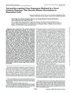
Fig. 1. Induction of c-fos, c-jun and p33 mRNAs after growth factor treatment of rat-1 parental cells and p2lHras-transformed cell lines. Subconfluent cultures of different cell lines were serum-arrested for
24 h and stimulated with insulin (ins, 10 yg/ml) or fetal calf serum (FCS, 10%). Total RNA was isolated at the indicated time points (minutes) and probed for the expression of the genes indicated. Hybridizations with human elongation factor 1 (HEF) cDNA were performed to indicate equal amounts of RNA. Control inductions with the solution in which insulin is dissolved (1% BSA, 4 mM HCI) did not show any increase in the expression of the genes analysed (not shown). Results shown were obtained with the H13 cell line, overexpressing normal p2lras (Downward et al., 1988) and RR3, a mutant p2lras expressing cell line. Similar results were obtained with two other p2lHras overexpressing cell lines, H9 (Downward et al., 1988) and HE+ (Burgering et al., 1989) and with two other mutant p2lHras expressing cells, RR2 and RR7.
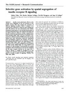
observed in cells overexpressing normal p2lHras (Figure 1). In contrast, we observed only a small, hardly detectable, insulin-induced increase in the expression of these genes in the untransformed, parental rat-I cells or in the mutant p21 Hras-transformed cells. As in the normal p21 Hras overexpressing cell lines, insulin also stimulated expression of c-jun and p33 in the insulin receptor-overexpressing A14 cell line (Figure 2). These results confirm those obtained by us (Burgering et al., 1989) and others (Stumpo and Blackshear, 1986) for insulin induced c-fos expression. Taken together these results show that both increased expression of insulin receptor as well as increased expression of p2lHras result in enhanced insulin signalling, as reflected by the ability of insulin to stimulate gene expression. Overexpression of p21Hras does not enhance all cellular responses to insulin In cell lines expressing high levels of insulin receptor, several other processes are induced rapidly by insulin, including an increase in tyrosine phosphate-containing phos-
phatidylinositol-3-kinase (PI-3-K) activity (Endemann et al., 1990; Ruderman et al., 1990). We have investigated whether
1104
e
ee: -. >
Fig. 2. Induction of c-jun and p33 expression after insulin stimulation of insulin receptor overexpressing cells. Subconfluent cultures of A14 cells, overexpressing the insulin receptor, were serum-arrested for 24 h and stimulated with insulin (ins, 10 tiglml). Total RNA was isolated at the indicated time points (minutes) and probed for the expression of the genes indicated. Hybridizations with human elongation factor 1 (HEF) cDNA were performed to indicate equal amounts of RNA.
overexpression of p2 lHras potentiates this insulin response in a way similar to the induction of gene expression. For this purpose we measured insulin-induced PI-3-K activity in anti-phosphotyrosine immunoprecipitates. The various cell lines were serum starved for 24 h and stimulated with insulin. Cells were lysed 10 min after stimulation and phosphotyrosine-containing proteins were immunoprecipitated with a polyclonal antibody. PI-3-K activity was measured in the immunoprecipitate collected on protein A - Sepharose beads. As shown in Figure 3, PI-3-K activity was strongly induced in the cells overexpressing the insulin receptor, but not in the cell lines overexpressing normal p21Hras or in any of the other cell lines. In the cell lines overexpressing normal p21Hras and mutant p2lHras, PI-3-K is not defective, since in these cells PDGF can induce PI-3-K activity normally (B.M.Th.Burgering, A.M.M.de Vries Smits, F. McCormick, J.L. Bos, manuscript in preparation). These results show that overexpression of p2lHras does not augment all cellular responses to insulin and that overexpression of p21 Hras specifically contributes to the response pathway of insulin involved in the regulation of gene expression. Insulin stimulates increase of p21rasGTP levels The simplest model deduced from the experiments described above would assume that p21 ras is an intermediate in insulin signalling. To demonstrate this more directly, we have analysed the effect of growth factor treatment, in particular insulin, on the relative levels of GTP bound to p2 lras. To this end, cells were labelled in vivo with [32P]orthophosphate for 3 h and after growth factor stimulation for an additional 5 min, cells were lysed and p21 ras was collected by immunoprecipitation with the monoclonal antibody Y13-259. Bound nucleotides were eluted and separated by thin layer chromatography. Using this protocol, we observed in untransformed cells a low amount of GTP bound to p2 Iras as a proportion of total nucleotides (20% GTP; Figure 4A), compared with the level of GTP bound
Insulin activates p2l ras
A. -
B.
F~~DGF
'iS.
-
r,,
!P0D0GF
*9
0
pip ?pip
0
,*'0 rat-1
*
0
mutant
: e-ex
H- r
,
ori
*
4
ori
p ress r'i
AJ~tp eH-
-s-
r ece-cto r
Fig. 3. Activation of P1-3-K after growth factor treatment. Cells were serum-arrested for 24 h and stimulated with insulin (ins, 10 pugIml) or PDGF (40 nglml) for 15 mmi. Cells were lysed and 400 itg of protein was precipitated with polyclonal anti-phosphotyrosine serum. The precipitates bound to protein A -Sepharose beads were assayed for PI-3-K activity using phosphatidylinositol as substrate. Lipids were separated by thin layer chromatography and detected by autoradiography. -, no growth factor added. Cell lines used are (A) rat-l, RR2 (mutant p2lHras), H9 and H13 (overexpressing normal p2lHras); (B) NIH 3T3 and A14 (overexpressing insulin receptor). The position of the different phosphatidylinositols (PI, PIP and PIP2) were determined by running standards and staining with iodine vapour.
B
.-DP
II
t
I
'S..
GT-P
F DGI
I GDP
eI
Ln~
K>. -
C
.i
KT>
to p2lIras. Autoradiographs after chromatographic separation of GTP and GDP eluted from p2lIras. Cells were labelled with [3p]orthophosphate for 3 h and stimulated with growth factors for 5 min. Cells were lysed and p21 ras was collected by immunoprecipitation with the ras-specific monoclonal Y13-259 or, when indicated, with the p2lras-specific monoclonal Y13-238 or the control monoclonals KT3 and 9CI0. GDP/GTP was eluted and separated by thin layer chromatography. A. NIH/3T3 cells, unstimulated (-) and stimulated with insulin (ins, 10 [tg/ml), PDGF (20 ng/m-l), TPA (100 ng/m-l) and EFG (20 ng/m-l). B. RR3 cells expressing mutant Hras, unstimulated (-) and stimulated with insulin. C. H13 cells, overexpressing normal p2lHras, unstimulated (-) and stimulated with insulin or PDGF. D. A14 cells, overexpressing the insulin receptor, unstimulated (-) and stimulated with insulin. E. HER 14 cells, overexpressing the EGF receptor, unstimulated (-) or stimulated with insulin or EGF.
Fig. 4. GTP/GDP bound
p2lras in mutant p2lHras-expressing cells (70% GTP; Figure 4B). Treatment of normal fibroblasts-NIH 3T3 cells (Figure 4A) and rat- I cells (not shown)-with various growth
to
factors did not result in a detectable shift in the GTP/GDP balance on p2lras. Next, we analysed the effect of insulin stimulation on the cells expressing increased levels of 1105
B.M.T.Burgering et at.
A
31-
i ',' ll'
p2lras,
1'
B .1
---
NW'..
:1
iV
Fig. 5. Time course of insulin-induced increase in GTP bound to p21ras and increase in insulin receptor phosphorylation. A. Graphic presentation of the p21rasGTP/GDP ratio in A14 cells after stimulation with insulin for the indicated times. The insert shows the autoradiograph of the thin layer separation of GTP and GDP bound to p21ras (for details see legend to Figure 4) c; control unstimulated with antibody KT3. B. Graphic presentation of the phosphorylation of the 3-chain of the insulin receptor. In vivo phosphorylated insulin receptor was immunoprecipitated and the ,8-chain was separated by SDS-polyacrylamide gel electrophoresis. After autoradiography (see insert), the phosphorylated bands corresponding to the insulin receptor 13-chain were cut out and the 32p content was quantified by liquid scintillation counting.
p21Hras. Insulin did not significantly stimulate a shift in the balance of GTP/GDP bound to p2 lras in the normal p2 lHras overexpressing cells, although in some experiments a small increase was observed (from 3 to 5 % GTP; Figure 4C). This insulin-induced increase was within the same order as that observed after stimulation of p21 Hras overexpressing cell lines with PGDF (Figure 4C) or EGF (data not shown; see also Satoh et al., 1990a,b). However, insulin stimulation of cells expressing increased levels of insulin receptor resulted in drastic accumulation of GTP on p2lras (20% to 70% GTP; Figure 4D). The insulin-induced activation of p21ras in the insulin receptor overexpressing cells was analysed using the Triton XI 14 phase-split method (Bordier, 1981; Hancock et al., 1990). In this method palmitoylated/ farnesylated p21ras is partitioned in the detergent phase, 1106
whereas the non-modified p21 ras is retained in the aqueous phase. It demonstrates that the activation of p21 ras occurred within the fraction of processed p2lras. To substantiate the specificity of the observed activation of p2lras, we performed several control experiments. First, precipitation with another anti-ras monoclonal antibody (Y13-238) yielded similar results, whereas precipitation with two unrelated antibodies [KT3 (anti-SV40 LT) and 9C 1O (anti-adenovirus ElB)] were negative, underscoring that the GTP/GDP binding was to p2lras. Secondly, we analysed the time course of the insulin-induced increase of GTP bound to p2 l ras (Figure 5A). After 1 min, the accumulation of GTP on p2 lras was clearly visible and almost maximal, indicating that the activation of p2lras is a very rapid process. The increased GTP/GDP ratio on p21 ras remained elevated for the at least 15 min. Thirdly, after precipitation of 32P-labelled lysates used for the time course experiment were re-used to immunoprecipitate the insulin receptor with a polyclonal anti-insulin receptor serum. After SDS -PAGE and autoradiography, the phosphorylated bands corresponding to the insulin receptor fl-chain were cut out and 32p content was quantified by liquid scintillation counting. The kinetics of insulin-stimulated phosphorylation of the insulin receptor (Figure SB) were rapid and almost identical to the kinetics of insulin stimulated accumulation of GTP on p2 lras (Figure 5A), indicating a tight link between receptor activation and p2lras activation. Finally, we analysed whether EGF can stimulate the increase of GTP on p21ras in cells overexpressing the EGF receptor [HER14 cells, 3 x 10⁵ EGF receptors (Honegger et al., 1987)]. As shown in Figure 4E, EGF stimulation of HER14 cells did not result in a significant increase in the levels of GTP bound to p2 I ras.
Discussion Insulin induced gene expression In this paper we have studied the effects of insulin on gene expression, PI-3-K activity and the GTP/GDP balance on p21ras in cell lines expressing different levels of insulin receptor, normal p2lHras and mutant p2lHras. The data presented provide evidence that insulin-induced activation of p2 lras is an intermediate in the route leading from insulin receptor activation to the induction of gene expression. The premise that normal p2lras proteins are mediators of growth factor-induced signals to downstream targets implies that overexpression of normal p21 Hras should result in a qualitative and/or quantitative change in growth factor response. In this respect there is no conceptual difference between the effect of overexpression of a growth factor receptor or a key intermediate in the signal transduction pathway. Indeed, we do not observe a difference between the effect of overexpression of the insulin receptor and the effect of overexpression of p21 Hras in insulin-induced gene expression. Both in cells overexpressing the insulin receptor and in cells overexpressing normal p21Hras, but not in the parental cells, insulin can induce the expression of genes like c-fos, c-jun or p33. This therefore indicates the p2 lras may be part of the insulin signalling pathway to induce gene expression. The observation that in mutant p21 Hras expressing cells insulin did not induce gene expression is consistent with this conclusion. In these cells, p2lHras is
Insulin activates p21 ras
postulated to be active constitutively and is likely to be insensitive to the upstream regulatory mechanisms controlling normal p2 lHras function. The elevated level of p33 expression might be due to the constitutive activation of the downstream part of the p21 ras pathway. The expression of c-fos and c-jun is not elevated in mutant p2lHras transformed cells, presumably because these genes are expressed only transiently after growth factor stimulation. Insulin induced activation of p2 1ras The direct link between p2 Iras and insulin-induced receptor activation was shown in cells expressing increased amounts of the insulin receptor. In these cells the level of GTP bound to p21 ras increases dramatically within 1 min after insulin stimulation (Figure 4). Furthermore, the time course of insulin-induced phosphorylation of the insulin receptor 3chain, a measure for insulin receptor activation, and of insulin-induced activation of p2iras is identical (Figure 5). We do not observe insulin-induced stimulation of p2 lras in normal fibroblasts, presumably due to the low number of insulin receptors (2 x 10³ receptors/cell compared with 3 x 10⁵ receptors/cell in the insulin receptor overexpressing cells). This does not imply that insulin-induced activation of p21ras does not occur in these cells. A small shift in the GTP/GDP balance might be brought about after insulin stimulation, yet remain undetected. In this respect it is interesting to note that other growth factors tested, including serum, do not cause a significant shift of the GTP/GDP balance on p21ras in NIH 3T3 cells and rat-I cells (Figure lA; J.Downward, personal communication), while blocking p2lras, by micro-injection of Y13-259 in these cells, inhibits mitogenic signalling of these growth factors (Mulcahy et al., 1985; Yu et al., 1988). Similarly, in cells overexpressing normal p2lHras, hardly any increase in the level of GTP bound on p21ras after insulin stimulation was observed. It should be noted, however, that in these cells, which overexpress p2lHras 100-fold, an increase of 0.5 % GTP bound to p2l ras represents an absolute increase of GTP bound to p2lras that is similar to the increase observed after insulin stimulation of the cells expressing increased levels of insulin receptor but normal levels of p2 lras. Such an increase, although observed by Satoh et al. (1990a,b) in p2lHras overexpressing cells after PDGF and EGF stimulation, would be undetectable in our assay. An effect on the nucleotide balance of p21ras similar to that found in the cells expressing increased levels of the insulin receptor with insulin, has been shown for T cells, where stimulation of the T cell receptor or treatment with the phorbol-ester TPA results in a rapid and strong shift in the GTP/GDP balance on p2 lras (Downward et al., 1990a).
p2 lras is part of the insulin signal transduction pathway From our results we conclude that insulin-induced activation of p2 Iras is an intermediate step in the insulin signal transduction pathway leading to the activation of gene expression. However, one could argue that increased expression of the insulin receptor results in an improper linkage to other pathways and, thus, is permissive in the activation of p21ras. Although this possibility cannot be formally excluded, it would imply that overexpression of p2lHras is also pennissive for stimulation of gene expression by insulin. We
therefore consider a role for p21 ras as an intermediate in the insulin signal transduction more likely than a mechanism involving some kind of interchangeable permissiveness. Insulin has many effects on the cell and a variety of pathways has been proposed (Rosen, 1987). Our results do not imply that p21ras mediates all these insulin-induced processes. Indeed, the analysis of insulin-induced PI-3-K activity suggests that this process is not mediated by p2lras. Apparently, p2 lras mediates only part of the insulin-induced signal transduction pathway. This notion is further strengthened by observations that insulin-induced hexose uptake and 1,2- and 1,3-diacylgylcerol increase are also unaffected by overexpression of p2lHras (unpublished observations). The involvement of p2 lras in only a specific branch of the insulin signal transduction pathway may also explain the observation that under certain experimental conditions insulin can synergize with p21ras elicited signals (Morris et al., 1989), maybe through the increase in glucose and/or metabolite uptake. p2lras: mediator of multiple signal transduction pathways? The fact that p21ras is not involved in all insulin-induced cellular responses, but only in a part that is common to many different growth factors, i.e. the induction of gene expression, suggests that p2 lras may also be involved in the signal transduction pathway of these growth factors. Indeed, activation of p2 Iras appears to be obligatory for the induction of mitogenesis by several growth factors and the induction of c-fos by serum (Cai et al., 1990; Mulcahy et al., 1985; Yu et al., 1988). However, whether it is the relative or absolute level of p2lrasGTP that is required to achieve stimulation of the downstream part of the 'ras-pathway', is still unknown. The situation is further complicated by the possibility that partial activation of p2iras may synergize with other intracellular routes activated by the same growth factor. Our results show that p2lras is more sensitive to activation by insulin than by other growth factors. For instance, normal fibroblasts express an amount of PDGF-B receptors within the same order of magnitude as insulin receptors on the A14 insulin receptor overexpressing cell line (W.H.Moolenaar, personal communication). PDGF, however, although a strong mitogen, does not activate p21 ras significantly. Also after EGF stimulation of NIH 3T3 cells expressing increased levels of the EGF receptor, no increase in GTP bound to p2 lras was observed. These observations do not rule out the involvement of p2 lras in PDGF and EGF signal transduction, but they indicate that insulin increases p2l rasGTP levels more effectively than do PDGF and EGF. Clearly, the involvement of p21ras activation in insulininduced mitogenesis and gene expression differs from its involvement in PDGF and EGF signalling. In fibroblasts p21 ras may be the main mediator in insulin-induced mitogenesis and gene induction, whereas for other growth factors activation of p21ras, although necessary, may synergize with other activated pathways. Activation of p2 lras may be a main crossroad in growth factor-induced signal transduction and it will be of interest to study the relative contributions of the various growth factors in the activation of p2 Iras and the different mechanisms employed. In this respect insulin-induced activation of p21 ras may prove to be an important paradigm for
1107
B.M.T.Burgering et al.
study of the mechanism by which p2 lrasGTP/GDP cycling is regulated. At present we do not know what precise mechanism is employed by insulin to increase the level of GTP on p2iras. In analogy to the results obtained with T lymphocytes, inactivation of GAP activity via protein kinase C (Downward et al., 1990a), is a possibility we are currently investigating. On the other hand, activation of an exchange factor remains a possibility as well. The existence of multiple GAP activities, such as the recently identified NF1 gene product (Ballester et al., 1990; Martin et al., 1990; Xu et al., 1990) and different exchange activities [membranebound (West et al., 1990) and cytoplasmic (Downward et al., 1990b; Wolfman and Macara, 1900)] indicates the complexity of p2 Iras regulation.
Materials and methods Materials and cells Tissue culture media and sera were from Gibco Laboratories. Insulin, protease inhibitors, phosphatidylinositol and lipid standards were from Sigma Chemical Co. Platelet-derived growth factor (PDGF) BB homodimer was from Amersham. Polyclonal anti-PY was prepared as described (Pang et al., 1985), polyclonal anti-insulin receptor has been described (Maassen et al., 1987). The overexpressing H-ras cell lines (H9 and H13) have been described (Downward et al., 1988) and were kindly provided by J.de Gunzburg (Paris). Mutant p21Hras cell lines (RR2, RR3, and RR7) were made by transfecting a human mutant H-ras gene (pEJ 6.6, G12V) and kindly provided by R.Offringa (Leiden). An insulin receptor-overexpressing cell line (A14) was made by transfection of NIH 3T3 cells with a plasmid expressing a full length human insulin receptor cDNA under control of the SV40 early promoter, in combination with a dihydrofolate reductase gene for amplification and a neomycin resistance gene for selection. The cells contain 3 x 10⁵ high affinity insulin receptors (KD S l0-9 M) per cell (J.A.Maassen, manuscript in preparation). HER14 cells overexpressing 3 x 10⁵ EGF receptors per cell (Honegger et al., 1987) were kindly obtained from J.Schlessinger. Cells were cultured routinely in DMEM supplemented with 8% fetal calf serum (FCS). For serum starvation subconfluent cultures were cultured in DMEM plus 0.5% FCS and 10 Mug/ml transferrin. After 24 h the cells were stimulated with the indicated growth factor. RNA analysis RNA was isolated by LiCl -urea lysis and prepared for Northern blotting as described (Schrier et al., 1983). The probes used, HEF and c-fos, have been described previously (Burgering et al., 1989). Mouse c-jun cDNA was kindly provided by R.Bernards (Boston, MA) and the p33 cDNA (Messina et al., 1985) was kindly provided by D.K.Granner (Nashville, TN). Determination of GTP/GDP ratio The determination of the GTP/GDP ratio of p21ras was essentially as described (Downward et al., 1990a). Cells were serum arrested for 18 h and subsequently labelled for 3 h with 400 MCi [32P]orthophosphate per 9 cm dish in phosphate-free/serum-free medium (Gibco). Cells were stimulated with insulin (10 Mg/ml) for the times indicated. Cells were put on ice and rapidly washed with ice-cold Tris-buffered saline and lysed in 50 mM HEPES buffer, pH 7.4, 1% Triton X-1 14, 100 mM NaCl, 5 mM MgCl2, 1 mg/nil BSA, 10 mM benzamidine, 10 Mg/ml leupeptin, 10 Mg/ml aprotinin, 10 Mg/ml soybean trypsin inhibitor. 100 MM GTP, 100 MtM GDP, 1 mM ATP and 1 mM sodium phosphate pH 7.4 was included to prevent post-lysis labelling of p2 lras. Nuclei were removed by centrifugation and the Triton X-1 14 and aqueous phases were split at 37°C for 2 min followed by a brief spin (Bordier, 1981). The detergent phase was deluted 10-fold with lysis buffer without Triton X-1 14. The lysate was precleared for 5 min with protein G-Sepharose beads and further incubated for 40 min with anti-p2lras monoclonal Y13-259 or Y13-238 (Oncogene Science) or with a control monoclonal KT3 (anti-SV40 large T) or 9C10, a rat antibody directed against adenovirus E1B protein (kindly provided by A.Zantema), all bound to protein G -Sepharose. Immunoprecipitates were collected and washed eight times with 50 mM HEPES buffer, pH 7.4, 500 mM NaCl, 5 mM MgCI2, 0.1% Triton X-100, 0.005% SDS. GTP/GDP was eluted in 2 mM EDTA, 2 mM DTT, 0.2% SDS, 0.5 mM GDP, 0.5 mM GTP at 68°C for 20 min and separated on PEI-cellulose plates (Merck) developed in 1.2 M ammonium formate, 0.8 M HCI. Plates were autoradiographed and the GTP/GDP ratio was determined by scintillation counting.
1108
Phosphatidylinositol-3-kinase assay Cells were washed twice with cold PBS containing 1 mM sodium vanadate and lysed by scraping in 1 ml of lysis buffer containing 2 mM Tris-CI (pH 7.8), 137 mM NaCl, 1% NP40, 10% glycerol, 2 mM sodium EDTA, 1 mM sodium vanadate, 1 mM phenylmethylsulphonyl fluoride, 0.15 U/nil aprotinin and 20 AM leupeptin. After 5 min on ice the nuclei were removed by an Eppendorf spin for 10 min at 4°C. Supernatant was transferred to a clean tube and protein content was measured by the Bradford method (BioRad). 400 Mg of protein was incubated with polyclonal antiphosphotyrosine serum for at least 3 h at 4°C. Protein A -Sepharose beads were used to collect the antigen-antibody complexes. The complexes were washed twice with lysis buffer in the presence of I M LiCl, twice with lysis buffer and once with 10 mM Tris-Cl (pH 7.5), 1 mM sodium vanadate. PI-3-K activity was determined essentially as described by Kaplan et al. (1990). In brief, anti-phosphotyrosine immunoprecipitates collected on protein A-Sepharose were incubated in 30 mM HEPES (pH 7.5), 200 AM adenosine and 0.2 mg/ml sonicated phosphatidylinositol for 15 min at 25°C in a total volume of 50 ,1u. Adenosine was included to inhibit any contaminating PI-4-K activity (Whitman et al., 1987). The reaction was started by adding 30 mM MgCl2, 40 MM ATP and 10 MCi [-y-32P]ATP (final concentrations) and incubation continued for another 25 min at 250C. The reaction was terminated by the addition of 100 M1 1 M HCI and quickly mixing. Lipids were extracted by addition of 200 Mi1 chloroform-methanol (1: 1). The organic phase was washed once more with methanol - I M HCI (1:1). An aliquot of the organic phase of 50 M1 was applied to a silica gel G plate and developed in chloroform-methanol -4 M NH40H (45:35: 10).
Acknowledaements We thank Matty Verlaan-de Vries and Jan-Paul Medema for excellent technical support during this project. We also thank Julian Downward (ICRF, London) and Wouter Moolenaar (NKI, Amsterdam) for sharing unpublished data and Aart-Gerrit Jochemsen for discussions. This project was supported by grants from the Dutch Cancer Society.
References Ballester,R., Marchuk,D., Boguski,M., Saulino,A., Letcher,R., Wigler,M. and Collins,F. (1990) Cell, 63, 851-859. Bar-Sagi,D. and Feramisco,J.R. (1985) Cell, 42, 841-848. Barbacid,M. (1987) Annu. Rev. Biochem, 56, 779-827. Bordier,C. (1981) J. Biol. Chem., 256, 1604-1607. Burgering,B.M.T., Snijders,A.J., Maassen,J.A., Van der Eb,A.J. and Bos,J.L. (1989) Mol. Cell. Biol., 9, 4312-4322. Cai,H., Szeberenyi,J. and Cooper,G.M. (1990) Mol. Cell. Biol., 10, 5314-5323. Downward,J., De Gunzburg,J., Riehl,R. and Weinberg,R.A. (1988) Proc. Natl. Acad. Sci., 85, 5774-5778. Downward,J., Graves,J.D., Warne,P.H., Rayter,S. and Cantrell,D.A. (1990a) Nature, 346, 719-723. Downward,J., Riehl,R., Wu,L. and Weinberg,R.A. (1990b) Proc. NatI. Acad. Sci. USA, 87, 5998-6002. Endemann,G., Yonezama,K. and Roth,R.A. (1990) J. Biol. Chem., 265, 396-400. Gibbs,J.B., Sigal,I.S., Poe,M. and Scolnick,E.M. (1984) Proc. Natl. Acad. Sci. USA, 81, 5704-5708. Gibbs,J.B., Schaber,M.D., Marshall,M.S., Scolnick,E.M. and Sigal,I.S. (1987) J. Biol. Chem., 262, 10426-10429. Hagag,N., Halegoua,S. and Viola,M. (1986) Nature, 319, 680-682. Hall,A., (1990) Science, 249, 635-640. Hancock,J.F., Magee,A.I., Childs,J.E. and Marshall,C.J. (1990) Cell, 57, 1167-1177. Honegger,A.M., Dull,T.J., Felder,S., Van Obberghen,E., Bellot,F., Szapary,D., Schmidt,A., Ullrich,A. and Schlessinger,J. (1987) Cell, 51, 199-209. Hoshino,M., Kawakita,M. and Hatorri,S. (1988) Mol. Cell. Biol, 8, 4169-4173. Kaplan,D.R., Morrison,D.K., Wong,G., McCormick,F. and Williams,L.T. (1990) Cell, 61, 125-133. Kazlauskas,A., Ellis,C., Pawson,T. and Cooper,J.A. (1990) Science, 247, 1578- 1581. Korn,L.J., Siebel,C.W., McCormick,F. and Roth,R.A. (1987) Science, 236, 840-843. Maassen,J.A., Krans,H.M.J. and Moller,W. (1987) Biochim. Biophys. Acta, 930, 72-78. Martin,G.A., Viskochil,D., Bollag,G., McCabe,P.C., Crosier,W.J.,
Insulin activates p21 ras Haubruck,H., Conroy,L., Clark,R., O'Connell,P., Cawthon,R.M., Innes,M.A. and McCormick,F. (1990) Cell, 63, 843-849. Messina,J.L., Hamlin,J. and Larner,J. (1985) J. Biol. Chem., 260, 16418- 16423. Molloy,C.J., Bottaro,D.P., Fleming,T.P., Marshall,M.S., Gibbs,J.B. and Aaronson,S.A. (1989) Nature, 342, 711 -714. Morris,J.D.H., Price,B., Lloyd,A.C., Self,A.J., Marshall,C.J. and Hall,A. (1989) Oncogene, 4, 27-31. Mulcahy,L.S., Smith,M.R. and Stacey,D.W. (1985) Nature, 313, 241-243. Noda,M., Ko,M., Ogura,A., Liu,D., Amano,A., Takano,T. and Ikawa,Y. (1985) Nature, 318, 73-75. Pang,D.T., Sharma,B.R. and Shafer,J.A. (1985) Arch. Biochem. Biophys., 242, 176-186. Rosen,O.M. (1987) Science, 237, 1452 - 1458. Ruderman,N.B., Kapeller,R., White,M.F. and Cantley,L.C. (1990) Proc. Natl. Acad. Sci. USA, 87, 1411-1415. Satoh,T., Endo,M., Nakamura,S. and Kaziro,Y. (1988) FEBS Lett., 236, 185-189. Satoh,T., Endo,M., Akiyama,T., Yamamoto,T. and Kaziro,Y. (1990a) Proc. Natl. Acad. Sci. USA, 87, 7926-7929. Satoh,T., Endo,M., Nakafuku,M., Nakamura,S. and Kaziro,Y. (1990b) Proc. Natl. Acad. Sci. USA, 87, 5993-5997. Schrier,P.I., Bernards,R., Vaessen,R.T.J.M., Houweling,A. and Van der Eb,A.J. (1983) Nature, 305, 771-775. Stacey,D.W., Watson,T., Kung,H.-F. and Curran,T. (1987) Mol. Cell. Biol., 7, 523-527. Stumpo,D.J. and Blackshear,P.J. (1986) Proc. Natl. Acad. Sci. USA, 83,
9453-9457. Sweet,R.W., Yokoyama,S., Kamata,T., Feramisco,J.R., Rosenberg,M. and Gross,M. (1984) Nature, 311, 273-275. Trahey,M. and McCormick,F. (1987) Science, 238, 542-545. West,M., Kung,H. and Kamata,T. (1990) FEBS Lett., 259, 245-248. Whitman,M., Kaplan,D.R., Schaffhausen,B., Roberts,T. and Cantley,L. (1987) Biochem. J., 247, 165-174. Wolfman,A. and Macara,I.G. (1990) Science, 248, 67-69. Xu,G., O'Connell,P., Viskochil,D., Cawthon,R., Robertson,M., Culver,M., Dunn,D., Stevens,J., Gesteland,R., White,R. and Weiss,R. (1990) Cell, 62, 599-608. Yu,C.-L., Tsai,M.-H. and Stacey,D.W. (1988) Cell, 52, 63-71. Received on December 10, 1990
1109