Oct 30, 1978 - activation in mice infected with ectromelia or lympho- cytic choriomeningitis viruses. Aust. J. Exp. Biol. Med. Sci. 51:393-398. 2. Boyum, A.1968.
Vol. 23, No. 2
INFECTION AND IMMUNITY, Feb. 1979, p. 320-324 0019-9567/79/02-0320/05$02.00/0
Interactions Between Immune Cells and Antibody in Protection from Fatal Sindbis Virus Encephalitis ROBERT L. HIRSCH,l* DIANE E. GRIFFIN,"'2 AND RICHARD T. JOHNSON13 Departments of Neurology,' Medicine,2 and Microbiology,3 The Johns Hopkins University School of Medicine, Baltimore, Maryland 21205 Received for publication 30 October 1978
Transfer of anti-Sindbis virus serum, obtained from peripherally inoculated donors, protected mice from an otherwise fatal intracerebral infection with neuroadapted Sindbis virus (NSV). F(ab)'2 preparations of serum were not protective, indicating that the Fc piece of immunoglobulin G was important. Complement-depleted animals were protected with anti-NSV serum, ruling out as essential the complement-fixing function of the Fc piece. The presence of protective antibody correlated with the ability of serum to inhibit T-cell cytotoxicity. However, experiments using athymic nude mice showed that T cells played no role in killing the mice since the 50% lethal dose was the same as that in normal BALB/c mice, and that T cells were not required for protection since athymic nude mice were protected with antibody alone. Cyclophosphamide treatment of NSV-infected mice ablated the protective capacity of anti-NSV serum. Therefore, a non-T cell, cyclophosphamide-sensitive cell was required for antibody-mediated protection.
Several components of the immune system are involved in the host response to acute viral infections. The activation of macrophages (1), natural killer cells (22), lymphocytes (6), and the production of interferon (17), and anti-viral antibody (18) all appear to play a role in the response. The significance of in vitro correlates of the immune response to viral infections as well as how the components of the immune system interact to clear viral infections is not clear. Studies in this laboratory have been aimed at understanding the immune response to Sindbis virus infection in mice. Sindbis virus, an alphavirus of the togavirus group, causes an acute encephalitis in mice, in which mortality is age dependent (7, 11, 19). A mononuclear cell inflammatory response is observed 4 to 8 days after infection. This response appears to be specific, since Sindbis virus-sensitized lymphocytes, but not immune serum, can transfer this response to immunosuppressed, infected hosts. Bone marrow cells enhance the inflammatory response (16). To investigate the role of the immune response in recovery from Sindbis virus encephalitis a neuroadapted strain of Sindbis virus (NSV) was developed (9). Unlike tissue culturegrown virus, NSV causes fatal encephalitis in weanling mice. Animals can be protected from a fatal NSV infection by passive transfer of immune serum 24 h after intracerebral infection.
Sensitized lymphocytes are not protective. The protective factor in immune serum is immunoglobulin G (IgG) and protection is not solely a function of transfer of neutralizing antibody: serum-treated animals did not have significantly lowered amounts of brain virus, certain fractions of serum with high viral neutralization activity were not protective, and cyclophosphamide treatment of animals abolished the protective capacity of serum, indicating that a host cell was also required for antibody protection (9). The studies presented here further explored the characteristics of the protective serum, and the effect of immune serum on lymphocyte effector functions in vitro and the host factors required for its effect. MATERIALS AND METHODS Animals. BALB/c mice (Charles River Animal Breeding Laboratories, Wilmington, Mass.) or athymic nude mice bred on a BALB/c background (SpragueDawley, Madison, Wis.) were used in this study. Fourweek-old mice served as recipients in adoptive transfer experiments and 6- to 8-week-old mice served as serum and lymphocyte donors and as the source of lymphocytes for in vitro studies. Virus. NSV was prepared as described previously (9). Stock virus contained 109 plaque-forming units (PFU)/g of brain on BHK-21 cells and 3 x 10⁶ 50% lethal doses (LD5o)/g of brain after intracerebral (i.c.) inoculation of 4-week-old BALB/c (or nude) mice. Virus titrations were performed as described previously (7), except that BHK-21 cells were used as 320
VOL. 23, 1979
PROTECTION FROM SINDBIS VIRUS ENCEPHALITIS
indicator cells in place of chick embryo fibroblasts. Virus inoculation and passive transfer studies. Mice were inoculated i.c. with 10 LD50 of NSV in 0.03 ml of Hanks balanced salt solution (HBSS) containing 5% fetal calf serum (FCS). Anti-Sindbis serum, obtained at various times from donors after footpad inoculation of 105 PFU of NSV, was given intraperitoneally (i.p.) to animals 24 h after i.c. injection. In some experiments animals were immunosuppressed by cyclophosphamide treatment (Cytoxan, MeadeJohnson), 150 mg/kg i.p., 8 h after virus inoculation. Lymph node cell (LNC) suspensions for cell transfer experiments were prepared by passage of nodes through stainless steel grids (32 mesh/cm). A portion (5 X 10⁷ LNC/0.2 ml of HBSS) was administered i.p. 24 h after virus. In experiments in which the role of complement was evaluated, complement depletion was accomplished by 2 i.p. injections of cobra venom factor (CoVF) 4 h before and 2 days after serum transfer. CoVF was purified as previously described (20). Complement depletion of CoVF-treated mice was confirmed by assessment of serum C3 levels (10). Preparation of lymphocytes for cytotoxicity testing. Spleens and axillary, brachial, and popliteal lymph nodes from 6- to 8-week old mice were removed aseptically and placed in RPMI-1640 (Gibco, Grand Island, N.Y.) medium containing 10 mM HEPES (N2-hydroxyethylpiperazine-N'-2-ethanesulfonic acid), and supplemented with 5% FCS (Microbiological Associates, Walkersville, Md.). Lymph nodes were teased apart with 26-guage needles, and the released cells were washed three times with RPMI before in vitro culture. Spleen cells were teased apart with 26-gauge needles, and lymphocytes were obtained from the suspension by separation on Ficoll-Hypaque (2). Modification of effector cell populations. Adherent cells were removed from LNC suspensions by the method of Vassar et al. (21). A total of 10 to 15% of LNC suspensions were adherent by this method. Treatment of LNC suspensions with anti-Thy 1.2 serum and complement was performed as described previously (8). Modification of serum. Heat-killed, Formalinfixed Staphylococcus aureus containing protein A was prepared by the method of Jonsson and Kronvall (12), and absorption of mouse serum with this preparation was performed as previously described (13). Levels of immunoglobulin before and after absorption were quantitated by radial immunodiffusion by using monospecific antiserum to mouse IgG subclasses (Miles Labs, Elkhardt, Ind.). IgG2a, -2b, and -3 were no longer detectable after absorption. F(ab)'2 fragments of mouse immunoglobulin were prepared as described previously (5) with the exception that pepsin digestion was performed on 50% (NH4)2SO4 precipitates of mouse serum, and the reaction was performed at pH 4.9. The antigen binding capacity of F(ab)'2 fragments was intact as shown by the ability to inhibit cell-mediated cytotoxicity (CMC). In addition, F(ab)'2 preparations did not fix complement in complement fixation assays with Sindbis virus antigen. CMC assay. Previous studies indicated that LNC from Sindbis virus-infected mice were cytotoxic for
321
virus-infected primary murine fibroblasts (15). In the current study, BALB/c 3T3 clone A-31 cells were used as targets. A portion of 104 cells on flat-bottom microtiter plates (Falcon no. 3042) was infected with NSV for 1 h at 370C. The multiplicity of infection was 2. At 3 h after infection 1 LCi of 51Cr (Na25"CrO4, Amersham/Searle, specific activity 1 Ci/mM) in 0.2 ml of HBSS containing 5% FCS was added to each well for 1 h at 370C. It was found that LNC and splenic lymphocytes obtained from animals 6 days after footpad inoculation with 105 PFU of NSV were cytotoxic towards Sindbisinfected 3T3 cells (effector-to-target-cell ratio, E/T = 50). (Approximately 90% of target cells showed viral antigen on the cell surface as determined by indirect fluorescent microscopy of live cells 7 h after infection). Cytotoxic activity was maximum 18 h after addition of LNC (22 h after infection of target cells). Due to the lytic nature of Sindbis virus infection, low levels of cytotoxicity were observed at 24 to 26 h after infection as a result of the increase in "spontaneous" 61Cr release. Thus, all subsequent assays were evaluated 22 h after target cell infection. In experiments in which the effects of anti-NSV serum were tested, 0.2 ml of heat-inactivated serum was added for 1 h. Serum was diluted in HBSS, and all dilutions of serum were corrected to 10% serum with heat-inactivated FCS. After 1 h at 37CC, serum was removed and 5 x 10⁵ lymphocytes were added to each well (E/T = 50). After 16 h medium from each well was aspirated and counted in a Nuclear Chicago gamma counter (model 1185). The percentage of 51Cr release was calculated as described previously (3), except that spontaneous release was not subtracted from total 5"Cr incorporation in the denominator.
RESULTS Serum effects in vivo. Approximately 90% mortality was observed in animals inoculated with 10 LD50 of NSV i.c. (Table 1). As previously reported (9), high levels of protection from fatal encephalitis were conferred by transfer of antiserum collected 12 days after extraneural injection of mice. To further characterize the factors necessary for this protective effect, the same TABLE 1. Effect of anti-Sindbis Ab preparations on course of NSV infection No. Treatment
dead/total no.
None (virus only) 53/61 Mouse anti-NSV serum 5/49 (12 days p.i.) Protein A absorbed se- 18/22
% Mortal- Mean day of death ity
86.9 10.0
7.0 8.2
81.8
7.8
rum 75.0 8.1 F(ab)2' 24/32 9.1 5.0 Anti-NSV serum + 1/11 CoVF aMice were infected with 10 LD50 of NSV i.c. (approximately 60 PFU) and given serum preparations i.p. 24 h after virus challenge.
322
HIRSCH, GRIFFIN, AND JOHNSON
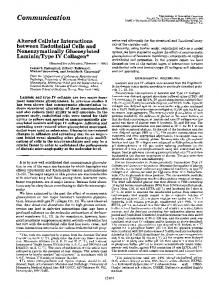
antiserum was absorbed with protein A or was pepsin digested to prepare F(ab)'2 fragments. Serum after protein A absorption was no longer protective, indicating that IgG-1 alone is not protective. Treatment of animals with F(ab)'2 fragments did not protect animals from fatal encephalitis. This indicated that an Fc piece function is necessary for protection. However, animals depleted of C3 by CoVF treatment were protected by antiserum treatment as well as normocomplementemic animals (Table 1). Effect of anti-Sindbis serum on CMC. To test the hypothesis (9) that antibody (Ab)-mediated protection may be mediated by the inhibition of T-cell cytotoxicity, anti-NSV serum was evaluated for effects on lymphoid cell effector function and ability to protect in vitro and in vivo. Mouse LNC or splenic lymphocytes obtained from mice 6 days after footpad inoculation of NSV were cytotoxic for Sindbis virusinfected 3T3 cells. Cytotoxicity was abolished by treatment with anti-Thy 1.2 serum and complement; removal of adherent cells did not affect cytotoxicity. Sera obtained from mice at various times after footpad inoculation of NSV were investigated for effects on lysis of infected target cells. Antiserum collected 6 or more days after NSV infection significantly inhibited lysis of infected cells by sensitized spleen cells or LNC (Fig. 1). Antiserum taken at 8 and 12 days after infection was markedly inhibitory and protective. The inhibition was dose dependent, and at 1/500 dilution no inhibition was observed. Treatment of cells with normal mouse serum did not inhibit cytotoxicity. When nonsensitized splenic lymphocytes or LNC were applied to serumtreated, infected targets, no enhancement of cytotoxicity was observed (Table 2). Furthermore, 2-ME treatment of serum did not decrease the inhibitory capacity of serum, indicating that IgM was not responsible for inhibition. Host cell requirements. Previous studies (9) showed that cyclophosphamide-treated, infected hosts could not be protected with Ab. Animals treated with cyclophosphamide were not protected by 12-day serum, even if serum was given repeatedly (every 2 days) (Table 3). To evaluate the host cellular component required for protection, cell transfer studies were carried out in cyclophosphamide-immunosuppressed hosts. LNC obtained from donors 6 days after footpad inoculation, or from uninoculated mice, were injected i.p. along with 12-day anti-NSV serum, 24 h after virus inoculation. LNC did not restore the cyclophosphamide-sensitive cell population required for Ab-mediated protection (Table 3). Furthermore, nude mice were equally sensitive to NSV infection (same LD50) and could be protected by Ab alone (Table 3).
INFECT. IMMUN.
~5O 0
50i
~~~~~~~~
I
400
240
0
30
30
Mortali0ty2 0-20 %
20
O/
/
10
0
O
_10
2 3 4 5 6 7 8 9 10 11 12 DAY OF INFECTED DONOR SERUM COLLECTION
FIG. 1. Protection from fatal NSV encephalitis and inhibition of CMC by anti-NSV antisera. Solid line indicates mortality of animals infected i.c. with 10 LD5o of NSV and treated with 0.2 ml of anti-NSV serum ip. 24 h after infection. Broken line represents antiserum inhibition of CMC by the same serum used in vivo. Target cells were pretreated with a 1:10 dilution of serum prior to addition of LNC.
DISCUSSION confirm and extend previous studies These results which showed that anti-NSV IgG protects animals from fatal NSV encephalitis. We have shown that the inhibition of T-cell-mediated cytotoxicity by anti-NSV Ab, although correlating with protection, does not relate to the mechanism by which Ab exerts its effect in vivo. Nude mice, which were equally sensitive to NSV encephalitis, could be protected by Ab even though lacking T cells. This indicates that the phenomenon of Ab inhibition of T-cell cytotoxicity in vitro does not explain the in vivo protection in this system. Furthermore, the fact that 5 x 10⁷ sensitized or normal LNC could not restore the cyclophosphamide-sensitive cell population, required along with Ab for protection, indicates that another cell population is necessary for protection and possible interaction with Ab. Studies with non-neuroadapted Sindbis virus have shown that the presence of the mononuclear cell inflammatory response in the brains of Sindbis virus-infected mice correlates with clear-
VOL. 23, 1979
PROTECTION FROM SINDBIS VIRUS ENCEPHALITIS
323
TABLE 2. Cytotoxic activity of mouse lymphoid cell populations toward Sindbis virus-infected cells % 5"Cr Release ± SE' Effector cell
Normal mouse serum Infected 56.8 ± 6.7 43.8 ± 2.4 22.9 ± 3.2 24.9 ± 0.6
Uninfected -6.6 ± 1.6 -3.9 ± 4.8 -5.3 ± 3.9 -7.6 ± 1.1
6-day serum Infected 25.3 ± 10.5 14.5 ± 4.2 22.7 ± 4.2 18.8 ± 3.6
12-day serum
Uninfected -7.5 ± 1.8 -4.8 ± 1.3 -7.5 ± 2.6 -9.5 ± 4.9
Infected 9.0 ± 3.0 4.9 ± 1.5 18 ± 2.9 21.3 ± 3.2
Uninfected -8.6 ± 2.5 -4.1 ± 0.7 -5.5 ± 5.0 -8.2 ± 1.0
Sensitized LNC Sensitized spleen Normal LNC Normal spleen aPercentage of 51Cr release was calculated as described in the text. Each value represents data from at least three different experiments. 10% serum was added to target cells in HBSS for 1 h before the addition of lymphocytes. SE, Standard error of the mean.
TABLE 3. Effect of cyclophosphamide immunosuppression and ceU transfer on the course ofNSV infection No. % Mortal- Mean day Treatment Dead/total ity of death no. 7.0 None (virus only) 86.9 53/61 88 6.8 45/51 Cyclophosphamide
(Cy)na
60 6.3 CY + anti-NSV serum 6/10 80 11.0 CY + anti-NSV serum 8/10 every 2 days 80 6.6 CY + sensitized LNCb 16/20 60 6.6 CY + sensitized LNC + 24/40 anti-NSV serum 87.5 CY + normal LNC + 7/8 8.3 anti-NSV serum 100 7.6 Nude mice (virus only) 5/5 0 Nude mice + anti-NSV 0/6 serum a A 150-mg dose of cyclophosphamide (Cytoxan) per kg was given ip. 8 h after virus challenge. b LNC (5 x 10') obtained from animals 6 days after foot-pad inoculation with NSV were given i.p. 24 h after virus challenge.
ance of virus, and that the transfer of this inflammatory response to cyclophosphamide-treated
mice by specifically sensitized lymphocytes could be enhanced with bone marrow cells (16). Perhaps a population of cells within the marrow is required for the Ab-mediated protection observed in this system. Our studies with F(ab)'2 preparations of antiNSV Ab indicate that an Fc-dependent IgG function is necessary at some point for protection. Complement-mediated mechanisms seem unlikely since depletion of C3 by CoVF treatment of infected animals did not change the protective capacity of Ab. Alternatively, the Fc piece of IgG may be required for transfer of immunoglobulin across the blood-brain barrier or for efficient processing of opsonized virus. Antibody-dependent cellular cytotoxicity is another possible Fc-dependent IgG-mediated
mechanism which could operate in protection. The murine macrophage has been shown to mediate antibody-dependent cellular cytotoxicity against cells infected with Semliki Forest virus (14), another alpha-togavirus. Although we have not yet been able to demonstrate antibodydependent cellular cytotoxicity against Sindbisinfected cells, macrophage-enriched populations have not been tested. The transfer of anti-NSV IgG to infected hosts allows for the presence of specific anti-viral Ab 2 to 3 days before detection of Ab in untreated, infected animals. The presence of this Ab could mediate protection by coating infected cells in the brain and, in the presence of the mononuclear cell infiltrate, infected cells could be eliminated. The elimination of infected cells early in the infection could limit viral replication in the brain and resolve an otherwise fatal infection. ACKNOWLEDGMENTS This work was supported in part by Public Health Service research grants NS-10920 and NS-07000 from the National Institute of Neurological and Communicative Disorders and Stroke. D.E.G. is an investigator of the Howard Hughes Medical Institute. We thank Jerry A. Winkelstein for supplying the purified CoVF and for measuring serum complement levels.
LITERATURE CITED 1. Blanden, R. V., and C. A. Mims. 1973. Macrophage activation in mice infected with ectromelia or lymphocytic choriomeningitis viruses. Aust. J. Exp. Biol. Med.
Sci. 51:393-398. 2. Boyum, A. 1968. Separation of leucocytes from blood and bone marrow. Scand. J. Lab. Invest. Supply. 97, 31-50. 3. Ennis, F. A., W. J. Martin, M. W. Verbonitz, and G. M. Butchko. 1977. Specificity studies on cytotoxic thymus-derived lymphocytes reactive with influenza virus infected cells: evidence for dual recognition of H-2 and viral hemagglutinin antigens. Proc. Natl. Acad. Sci. U.S.A. 74:3006-3010. 4. Fuson, E. W., H. D. Whitten, R. D. Ayers, and E. W. Lamon. 1978. Antibody dependent cell-mediated cytotoxicity by human lymphocytes. L Comparison of IgMand IgG-induced cytotoxicity. J. Immunol. 120: 1726-1732. 5. Gorini, G., G. A. Medgyesi, and G. Doria. 1969. Heterogeneity of mouse myeloma G globulins as revealed by enzymatic proteolysis. J. Immunol. 103:1132-1142.
324
HIRSCH, GRIFFIN, AND JOHNSON
6. Greenberg, S. G., B. S. Criswell, H. R. Six, and R. B. Couch. 1978. Lymphocyte cytotoxicity to influenza virus-infected cells: response to vaccination and virus infection. Infect. Immun. 20:640-645. 7. Griffin, D. E. 1976. Role of the immune response in agedependent resistance of mice to encephalitis due to Sindbis virus. J. Infect. Dis. 133:456-464. 8. Griffin, D. E., and R. T. Johnson. 1973. Cellular immune response to viral infection: in vitro studies of lymphocytes from mice infected with Sindbis virus. Cell. Immunol. 9:426-434. 9. Griffin, D. E., and R. T. Johnson. 1977. Role of the immune response in recovery from Sindbis virus encephalitis in mice. J. Immunol. 118:1070-1075. 10. Hirsch, R. L., D. E. Griffin, and J. A. Winkelstein. 1978. The effect of complement depletion on the course of Sindbis virus infection in mice. J. Immunol., 121: 1276-1278. 11. Johnson, R. T., H. F. McFarland, and S. E. Levy. 1972. Age dependent resistance to viral encephalitis: studies of infections due to Sindbis virus in mice. J. Infect. Dis. 125:257-262. 12. Jonsson, S., G. Kronvall. 1974. The use of protein Acontaining Staphylococcus aureus as a solid phase antiIgG reagent in radioimmunoassay as exemplified in the quantitation of a-fetoprotein in normal human adult serum. Eur. J. Immunol. 4:29-33. 13. Kronvall, G., H. M. Grey, and R. C. Williams. 1970. Protein A reactivity with mouse immunoglobulins: structural relationship between some mouse and human immunoglobulins. J. Immunol. 105:1116-1123. 14. Macfarlan, R. I., W. H. Burns, and D. 0. White. 1977.
INFECT. LIMMUN.
15. 16. 17.
18. 19.
20.
21. 22.
Two cytotoxic cells in peritoneal cavity of virus-infected mice: antibody-dependent macrophages and non-specific killer cells. J. Immunol. 119:1569-1574. McFarland, H. F. 1974. In vitro studies of cell-mediated immunity in an acute viral infection. J. Immunol. 113: 173-180. McFarland, H. F., D. E. Griffin, and R. T. Johnson. 1972. Specificity of the inflammatory response in viral encephalitis. J. Exp. Med. 136:216-226. Notkins, A. (ed.). 1975. Interferon as a mediator of cellular immunity to virus infections, p. 149. In Viral Immunology and Immunopathology. Academic Press, Inc., New York. Oldstone, M. B. A. 1975. Virus neutralization and virusinduced immune complex disease. Prog. Med. Virol. 19: 84-119. Reinarz, A. B. G., M. G. Broome, and B. P. Sagik. 1971. Age dependent resistance of mice to Sindbis virus: viral replication as a function of host age. Infect. Immun. 3:268-273. Shin, H. S., H. Gewurz, and R. Synderman. 1969. Reaction of a cobra venom factor with guinea pig complement and generation of an activity chemotactic for polymorphonuclear leukocytes. Proc. Soc. Exp. Biol. Med. 131:203-207. Vassar, P. S., E. M. Levy, and D. E. Brooks. 1976. Studies on the electrophoretic separability of B and T human lymphocytes. Cell. Immunol. 21:257-271. Welsh, R. M. 1978. Cytotoxic cells induced during lymphocytic choriomeningitis virus infection of mice. I. Characterization of natural killer cell induction. J. Exp. Med. 148:163-181.