May 6, 1993 - reproduce the respiratory epithelial cytopathology that is the hallmark of pertussis (18). We have monitored the biological effects of TCT by ...
Vol. 61, No. 8
INFECTION AND IMMUNrrY, Aug. 1993, p. 3123-3128
0019-9567/93/083123-06$02.00/0 Copyright © 1993, American Society for Microbiology
Interleukin-1 Is Linked to the Respiratory Epithelial Cytopathology of Pertussis LINDA NIXON HEISS,1 STEPHEN A. MOSER,2t EMIL R. UNANUE,2 AND WILLIAM E. GOLDMAN'* Department of Molecular Microbiology' and Department ofPathology, 2 Washington University School ofMedicine, St. Louis, Missouri 63110 Received 3 March 1993/Accepted 6 May 1993
BordeteUla pertussis, the causative agent of whooping cough, releases a muramyl peptide known as tracheal cytotoxin (TCT) that is responsible for destruction of ciliated epithelial cells lining the large airways. In vitro, TCT has been shown to cause this specific pathology in human or hamster respiratory epithelium and to inhibit the proliferation of cultured hamster trachea epithelial cells. The diverse biological actions of muramyl peptides, including adjuvanticity, somnogenicity, and pyrogenicity, have been correlated with the production and release of the inflammatory mediator interleukin-1 (IL-1). Consistent with its ability to reproduce other muramyl peptide actions, recombinant IL-1 caused TCT-like damage to the respiratory epithelium. In the nanogram-per-milliliter range, exogenous IL-1 inhibited DNA synthesis in hamster trachea epithelial cells and reproduced the pathology of TCT in hamster tracheal organ culture. Tumor necrosis factor alpha and IL-6, cytokines also associated with inflammation, were unable to reproduce TCT cytopathology. Furthermore, exposure of respiratory epithelial cells to TCT stimulated production of cell-associated IL-la, which could be detected within 2 h of TCT treatment. In contrast, there was no evidence of TCT-triggered release of IL-i. Previous studies have suggested that intracellular IL-la, as well as exogenous IL-la and IL-113, can inhibit cell proliferation. Our results therefore implicate IL-la, produced by epithelial cells in response to TCT, as a potential intracellular mediator of the primary respiratory cytopathology of pertussis. suggest that some muramyl peptide activities are the result of IL-1 induction. In vitro, muramyl peptides stimulate IL-1 production by monocytes and macrophages (13, 35), and in vivo production of IL-1 is seen when muramyl peptides are injected into experimental animals (36). The pyrogenic activities of muramyl peptides correspond to their ability to stimulate host production of endogenous pyrogens, including IL-1 (12). A correlation has also been demonstrated between the adjuvant activity of muramyl peptides and their ability to induce production of macrophage membraneassociated IL-1 (3). In addition, muramyl peptides may elicit their somnogenic effects via cytokines such as IL-1. The time courses of sleep effects stimulated by recombinant IL-1 or muramyl peptides in rabbits are consistent with the hypothesis that muramyl peptides modulate sleep by triggering the production of IL-1 (see reference 28). Given the correlations between IL-1 and muramyl peptide biological activities, we hypothesized that TCT might induce respiratory epithelial cells to produce IL-1, which in turn may be responsible for mediating TCT toxicity.
Whooping cough (pertussis) is classically characterized in infants and children by violent, debilitating coughing episodes, a result of respiratory tract colonization by Bordetella pertussis (34). Although B. pertussis produces a variety of toxins, tracheal cytotoxin (TCT) is the only one able to reproduce the respiratory epithelial cytopathology that is the hallmark of pertussis (18). We have monitored the biological effects of TCT by using two in vitro models of the respiratory epithelium: hamster trachea epithelial (HTE) cells and hamster tracheal organ culture. HTE cells are a nontransformed, homogeneous epithelial cell population isolated from excised tracheal tissue (17). TCT inhibits DNA synthesis by HTE cells in a dose-dependent manner but has little effect on overall RNA or protein synthesis (18). Sectioned hamster tracheas serve as a parallel organ culture model to reproduce the dramatic ciliated cell pathology of human infection (6). When treated with purified TCT, the epithelium of tracheal rings exhibits pathological changes indistinguishable from those observed in human pertussis (7). Exposure of human nasal tissue explants to TCT has also been shown to result in the destruction of ciliated cells (40). TCT is a 921-D disaccharide-tetrapeptide (8) that is a member of a large family of molecules known as muramyl peptides, which are fragments of bacterial cell wall peptidoglycan. Muramyl peptides have been shown to have a variety of biological activities in vivo, including somnogenicity, pyrogenicity, arthritogenicity, and adjuvanticity (1, 5, 27). There are many similarities between the biological effects of muramyl peptides and those of the inflammatory mediator interleukin-1 (IL-1), and several lines of evidence
MATERIALS AND METHODS
Reagents. TCT was purified from B. pertussis Tohama I or Tohama III culture supernatant as described previously (7). Recombinant murine IL-1,8 and the anti-IL-lo monoclonal antibody (MAb) 161.1-ALF were gifts from K. Hogquist and D. Chaplin (Washington University School of Medicine). Recombinant murine IL-6 and recombinant murine tumor necrosis factor alpha (TNF-a) were purchased from Genzyme (Cambridge, Mass.). Escherichia coli 026:B6 lipopolysaccharide (LPS) was purchased from Sigma Chemical Co. (St. Louis, Mo.). Cell culture. HTE cells were isolated from a Golden Syrian hamster as described previously (17) and cultured in F-12
* Corresponding author. Electronic mail address: goldman@ borcim.wustl.edu. t Present address: Department of Pathology, University of Alabama-Birmingham, Birmingham, AL 35294.
3123
3124
INFECT. IMMUN.
HEISS ET AL.
lo-A
tissue culture medium (GIBCO, Grand Island, N.Y.) con-
taining 10% fetal bovine serum (Hyclone Sterile Systems, Logan, Utah), 100 U of penicillin per ml, 100 jig of streptomycin per ml, and 2 mM L-glutamine. In certain experiments (e.g., those whose results are shown in Fig. 1 and Table 2), 3 ng of epidermal growth factor (EGF; Sigma) per ml was included during culture. EGF is not required for TCT toxicity, but growth rate and longevity of HTE cells may be improved by culture with EGF. Cells were maintained in 95% air-5% CO2 at 37°C. HTE cell toxicity assay. Inhibition of DNA synthesis by HTE cells was assessed in a modification of the microtiter assay described previously (18). Briefly, HTE cells resuspended in minimal essential medium (GIBCO) containing 2.5% fetal bovine serum were plated in 96-well culture plates at a density of 12,000 cells per well. In experiments where cells were cultured with EGF, 0.1 ng of EGF per ml was included. Incubation for 24 h synchronized these cells in the nonproliferative phase of the cell cycle. The nonproliferative cultures were incubated for 4 h with the test compound in serum-free minimal essential medium containing 5 mM HEPES (N-2-hydroxyethylpiperazine-N'-2-ethanesulfonic acid; pH 7.3; U.S. Biochemicals, Cleveland, Ohio). Cells were then stimulated with 15% fetal bovine serum, pulsed with [3H]thymidine, and allowed to incubate for 26 h. The cells were harvested on cotton-tipped applicators, and the amount of DNA synthesis was measured as trichloroacetic acid-precipitable 3H counts. The relative inhibition of DNA synthesis was calculated by comparing incorporated counts per minute of treated cells to untreated controls. The data shown are the means and the standard deviations of quadru-
plicate samples. E. coli LPS was included at a concentration of 10 ng/ml as a cofactor for TCT toxicity. Toxicity of purified TCT for HTE cells was previously shown to require 1 ,uM bovine serum albumin (BSA), which was thought to act as a protectant or carrier protein (4). Recent experiments determined that it is actually low-level endotoxin contamination of BSA that is responsible for this enhanced TCT toxicity (manuscript in preparation). Approximately 10 ng of E. coli LPS or B. pertussis lipooligosaccharide per ml can substitute for BSA in toxicity assays with HTE cells; this corresponds to lipooligosaccharide levels that we have measured in B. pertussis culture supernatant (7). However, even high levels (10 i,g/ml) of LPS alone do not produce significant toxicity. In the tracheal ring assays described below, there is no corresponding requirement for BSA, LPS, or lipooligosaccharide. Hamster tracheal ring toxicity assay. Damage to hamster tracheal epithelium was assessed as described previously (18). Tracheas from male Golden Syrian hamsters (Charles Rivers Laboratories, Wilmington, Mass.) at least 90 days old were dissected and sectioned into rings. Tracheal rings were cultured in F-12 medium containing the compound to be tested under an atmosphere of 95% air-5% CO2 at 37°C. Tracheal rings were screened for ciliated cell damage daily by light microscopy over the course of 102 h. Toxicity was defined as ciliostasis and extrusion of ciliated cells from the tracheal epithelium after exposure to the test compound for 4 to 5 days. Transmission electron microscopy. Tracheal rings were fixed overnight at 4°C in Tyrode's buffer containing 2% glutaraldehyde and then postfixed for 1 to 2 h in 1% osmium tetroxide at 4°C. Fixed samples were embedded with Poly/ Bed 812 (Polysciences, Inc., Warrington, Pa.) after passage through 70, 95, and 100% ethanol and propylene oxide. Thin
100
-u
80 60 CO (Ia 4)
40 20
m
zco
. ...
V
.001
....
.1
...
.
II
1
110
Concentration of TCT (gM)
0
100.0
.
.01
B
80 .
60
40 20 ,
.01
.. ...... .1 100 1. 0 Concentration of IL-1 (ng/ml) .
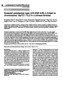
FIG. 1. Inhibitory effects of TCT and IL-1 on the proliferation of HTE cells. HTE cells were exposed to the indicated concentrations of TCT (A) or recombinant IL-lp (B) for 30 h. The relative inhibition of DNA synthesis was calculated by comparing incorporated counts per minute of treated cells with those of untreated controls. [3H]thymidine incorporation for untreated controls was 70,471 ± 1365 cpm. The data shown are the means and standard deviations for quadruplicate samples.
sections were cut with a glass knife, stained with 2% uranyl acetate and 0.4% lead citrate, and then examined on a Philips model 400 transmission electron microscope. Induction of IL-1 and IL-1 bioassay. HTE cells were seeded for synchronization and treated with 3 ,M TCT in the presence of 10 ng of LPS per ml. To assess IL-1 production, culture supernatants were collected, and the cell monolayers were washed with pH 3.0 medium for 2 min (acidic eluate) or subjected to three cycles of freeze-thawing in minimal essential medium-HEPES. Cell culture supernatants, acidic eluates, and cell lysates were tested for IL-1 activity as described previously (29) by using the D1O.G4.1 T-cell clone as an indicator (25). Samples were added to 2 x 10⁴ D1O.G4.1 cells plus 2.5 ,ug of concanavalin A per ml in microtiter wells. The indicator cells were pulsed with 0.2 ,uCi of [3H]thymidine per well during the last 24 h of a 72-h incubation at 37°C.
RESULTS Toxic effects of TCT and IL-1 for respiratory epithelial cells. HTE cells were treated with TCT or IL-1 for 30 h to determine whether exogenous IL-1 could reproduce the ability of TCT to inhibit proliferation of HTE cells. At concentrations as low as 50 pg/ml, IL-1 inhibited DNA synthesis in HTE cells in a concentration-dependent manner (Fig. 1). The toxicity curve for IL-1 was similar to that obtained for TCT. IL-1 was also tested for the ability to generate pertussislike destruction of ciliated respiratory epithelium. Hamster
VOL. 61, 1993
IL-1 IS LINKED TO PERTUSSIS CYTOPATHOLOGY
3125
A
C
0 0