Eur Respir J 2006; 28: 1005–1012 DOI: 10.1183/09031936.06.00038806 CopyrightßERS Journals Ltd 2006
Interstitial lung diseases associated with amyopathic dermatomyositis T. Suda*, T. Fujisawa*, N. Enomoto*, Y. Nakamura*, N. Inui*, T. Naito*, D. Hashimoto*, J. Sato*, M. Toyoshima*, H. Hashizume# and K. Chida*
ABSTRACT: The aim of the present study was to clarify the clinical characteristics and prognosis of patients with interstitial lung disease (ILD) associated with amyopathic dermatomyositis (ILD-ADM). The study consisted of 14 consecutive patients with ILD-ADM. Patients were classified into two categories, acute/subacute and chronic forms, according to the clinical presentation of ILD. The clinical features, responsiveness to therapy, and prognosis between the two forms were compared. Nine ILD-ADM patients were categorised as the acute/subacute form, and five as the chronic form. Arterial oxygen tension was significantly lower in the acute/subacute ILD than chronic ILD patients. On high-resolution computed tomography, ground-glass opacities were frequently found in the two forms, but consolidation was more common in acute/subacute ILD than chronic ILD. Bronchoalveolar lavage analysis showed higher numbers of total cells and lymphocytes in acute/subacute ILD than chronic ILD. Histologically, the most common finding was nonspecific interstitial pneumonia in the two forms, while diffuse alveolar damage was only found in acute/ subacute ILD. Acute/subacute ILD was generally resistant to therapy, while chronic ILD responded well. Notably, the mortality of acute/subacute ILD was much higher than that of chronic ILD (67 versus 0%, respectively). In conclusion, interstitial lung disease associated with amyopathic dermatomyositis includes two different forms, the acute/subacute and chronic forms, with distinct prognoses.
AFFILIATIONS *Second Division, Dept of Internal Medicine, and # Dermatology, Hamamatsu University School of Medicine, Hamamatsu, Japan. CORRESPONDENCE T. Suda 1-20-1 Handayama Hamamatsu 431-3192 Japan Fax: 81 534352354 E-mail:
[email protected] Received: March 18 2006 Accepted after revision: June 28 2006
KEYWORDS: Amyopathic dermatomyositis, interstitial lung disease
myopathic dermatomyositis (ADM) is recognised as a distinct subgroup of dermatomyositis (DM) with the typical skin rash of classic DM, but without muscle involvement [1–6]. Several studies have demonstrated that rapidly progressive interstitial lung disease (ILD) with a poor prognosis occurs in patients with ADM [7–12]. These patients were often resistant to intensive therapy, such as highdose corticosteroids plus immunosuppressive agents, resulting in fatal respiratory failure. In contrast, a recent report from Europe emphasised a favourable prognosis of ILD associated with ADM (ILD-ADM) among ILD associated with ADM, DM, and polymyositis (PM) [13]. As ILDADM is a rare condition, its characteristics have not been fully clarified. In the present study, therefore, the current authors examined a series of patients with ILD-ADM and attempted to determine its clinical features and prognosis.
A
METHODS Patient selection The study consisted of 14 consecutive patients (one male, 13 female) diagnosed with ILD-ADM. The diagnosis of ADM was confirmed based on modified Euwer’s criteria as follows: 1) characteristic dermatological manifestations of classic DM, including a heliotrope rash and Gottoron’s papules; 2) no muscle weakness; and 3) no increases in serum muscle enzymes during the observation period. All the subjects were seen as inpatients or outpatients at institutions between 1985 and 2005.
For editorial comments see page 893.
ILD presentations ILD was diagnosed based on the presence of radiological abnormalities with respiratory symptoms. Patients were classified into two categories according to clinical presentation: acute/subacute or chronic ILD-ADM. The acute/subacute form was defined as a rapidly progressive ILD showing deterioration within 3 months. According to the International Consensus
EUROPEAN RESPIRATORY JOURNAL
VOLUME 28 NUMBER 5
European Respiratory Journal Print ISSN 0903-1936 Online ISSN 1399-3003
c 1005
LUNG DISEASE OF AMYOPATHIC DERMATOMYOSITIS
T. SUDA ET AL.
Statement of idiopathic pulmonary fibrosis of the American Thoracic Society (ATS) with modification [14], the deterioration was defined by two or more of the following: 1) symptomatic exacerbation (dyspnoea on exertion); 2) an increase in parenchymal abnormality on high-resolution computed tomography (HRCT) scan; and 3) physiological change defined by one of the following, .10% decrease in vital capacity (VC) or .1.33 kPa decrease in arterial oxygen tension (Pa,O2). The chronic form was defined as a slowly progressive ILD that gradually deteriorated over .3 months. Regarding the temporal relationship between the onset of ADM and that of ILD, the interval between them within 3 months was defined as concomitant onset.
Treatment and outcome During the course of treatment, respiratory symptoms, chest radiograph/computed tomography findings, VC and Pa,O2 were assessed. According to the International Consensus Statement of idiopathic pulmonary fibrosis by the ATS with a slight modification [16], ‘‘improvement’’, or ‘‘favourable (or good) response’’ was defined by two or more of the following: 1) a decrease in symptoms (dyspnoea on exertion); 2) reduction in parenchymal abnormalities on chest radiographs or HRCT scans; and 3) physiological improvement defined as one of the following, .10% increase in VC or total lung capacity, .1.33 kPa increase in Pa,O2.
Data collection Clinical data, including history, treatment and laboratory findings, were obtained from patients’ medical records at the first encounter, which eventually led to a diagnosis of ILD. Signs and symptoms were also recorded. The following pulmonary function test parameters were assessed: VC and forced expiratory volume in one second.
Statistical analysis For two group comparisons involving binary data, either the Chi-squared test or Fisher’s exact test were used, depending on the sample size. Comparisons involving continuous data were made using the Mann–Whitney U-test. The cumulative survival rate was calculated using the Kaplan–Meier test. A p-value ,0.05 was considered significant. All data are expressed as mean¡SD.
HRCT HRCT examinations of the lung were performed on 1.0- or 1.5-mm thick sections to evaluate radiographic abnormalities. The HRCT images were reviewed for the presence of each of the following signs: consolidation, ground-glass opacities, traction bronchiectasis, irregular linear opacities, bronchovascular bundle thickening, honeycombing, and pleural effusion.
RESULTS Clinical features and laboratory findings Clinical characteristics of patients with acute/subacute ILDADM and chronic ILD-ADM are shown in table 1. Nine patients (all female) and five patients (one male and four female) were diagnosed as having acute/subacute ILD-ADM and chronic ILD-ADM, respectively. None of the patients had been given any drugs that might have caused ILD. Age and sex did not differ between the two groups. The observation period and duration of respiratory symptoms were shorter in acute/ subacute than chronic ILD-ADM. In most patients (89%) with acute/subacute ILD-ADM, ILD onset was concomitant with a diagnosis of ADM. In contrast, 60% of patients with chronic ILD-ADM developed ILD after an initial diagnosis of ADM (range 6–24 months). There were no patients in whom ILD onset preceded the initial diagnosis of ADM. None of the patients had any malignancies. Dyspnoea on effort, fever and arthralgia were more common in acute/subacute than chronic ILD-ADM. Chest auscultation revealed fine crackles in most patients with acute/subacute ILD-ADM as well as in chronic ILD-ADM.
Bronchoalveolar lavage Bronchoalveolar lavage (BAL) was performed as described previously [15]. Briefly, a fibreoptic bronchoscope was passed transorally and wedged in a segmental or subsegmental bronchus of the middle lobe. Three 50-mL aliquots of sterile 0.9% saline were instilled and the returns gently aspirated through the side channel of the bronchoscope. BAL fluid (BALF) was centrifuged at 8006g for 10 min to obtain the cellular components. The total cell count was determined using a haemocytometer and a differential cell count was taken on Giemsa-stained cytocentrifuged preparations. To characterise the phenotype of the lymphocytes in the BALF, flow cytometric analysis was performed in a flow cytometer (EPICS Profile; Coulter Electronics, Hialeath, France) using monoclonal antibody OKT3 (anti-CD3; Coulter Electronics), OKT4 (anti-CD4; Coulter Electronics) and OKT8 (anti-CD8; Coulter Electronics). Lung biopsy Surgical lung biopsy was not performed in patients with severe respiratory failure. In patients without severe respiratory failure, six (three acute/subacute ILD, three chronic ILD) underwent surgical lung biopsy, and two patients were autopsied. Lung specimens were obtained from at least two lobes. The specimens were categorised using the following abnormalities consistent with ILD: usual interstitial pneumonia (UIP), nonspecific interstitial pneumonia (NSIP), bronchiolitis obliterans organising pneumonia (BOOP), and diffuse alveolar damage (DAD), according to the current classification of interstitial pneumonias [16]. 1006
VOLUME 28 NUMBER 5
Laboratory findings are presented in table 2. Serum levels of KL-6, a marker of interstitial pneumonia, were elevated in the two forms, but no difference was found between them. Only one patient with chronic ILD-ADM had the anti-Jo-1 antibody. Pa,O2 was significantly lower in acute/subacute than chronic ILD-ADM. In addition, %VC tended to be lower in acute/ subacute than chronic ILD-ADM. HRCT findings HRCT images of the lung were available for 13 patients (eight acute/subacute and five chronic ILD) and the findings are summarised in table 3. Representative HRCT scans are shown in figure 1. All patients showed ground-glass opacities. Consolidation was more common in acute/subacute than chronic ILD-ADM, while the frequency of traction bronchiectasis was higher in chronic than acute/subacute ILD-ADM. In acute/subacute ILD-ADM, consolidation was a main finding in patients having this abnormality. Ground-glass opacity was EUROPEAN RESPIRATORY JOURNAL
T. SUDA ET AL.
TABLE 1
LUNG DISEASE OF AMYOPATHIC DERMATOMYOSITIS
Comparison of clinical characteristics between acute/subacute and chronic interstitial lung disease associated with amyopathic dermatomyositis (ILD-ADM) Acute/subacute ILD-ADM
Subjects n Age yrs Sex M/F
Chronic ILD-ADM
9
5
55.1¡10.4
53.8¡8.2
0/9
1/4
Observation period months
18.8¡30.0
43.3¡40.4
Duration of respiratory symptoms months
1.2¡0.7*
9.0¡2.6
Time of ILD diagnosis Before ADM diagnosis
0
0
Concomitant with ADM diagnosis
89
40
After ADM diagnosis
11
60
Malignancy
0
5
Dyspnoea on effort
89
20
Cough
78
60
Fever
78*
0
Arthralgia
56
20
Raynaud’s phenomenon
0
20
Fine crackles
89
100
Data are presented as mean¡SD, n or %. M: male; F: female.*: p,0.05.
a main finding in those showing no consolidation. No patients had ground-glass opacities and/or consolidation superimposing on honeycombing. In terms of histological patterns, consolidation was more frequently seen in DAD than in NSIP (100 versus 60%, respectively). Honeycombing was found in only one patient with UIP.
TABLE 2
Comparison of laboratory findings between acute/subacute and chronic interstitial lung disease associated with amyopathic dermatomyositis (ILD-ADM) Acute/subacute ILD-ADM
Chronic ILD-ADM
Subjects n
9
5
WBC?mm-3
6436¡2092
7286¡2090
ESR mm?h-1 LDH IU?L-1
45¡28
39¡9
450¡211
300¡84
CPK IU?L-1
73¡44
96¡48
Aldolase IU?L-1
6.1¡3.1
5.2¡4.0
KL-6 U?mL-1
851¡543
1035¡801
IgG mg?dL-1
1611¡390
1755¡480
Positive ANA %
38
60
Positive Jo-1 %
0
20
8.8¡0.8*
11.1¡1.6
VC %
65¡16
81¡17
FEV1 %
85¡12
82¡8
Pa,O2 kPa
Data are presented as mean¡SD or %, unless otherwise stated. WBC: white blood cell count; ESR: erythrocyte sedimentation rate; LDH: lactate dehydrogenase; CPK: creatine phosphakinase; Ig: immunoglobulin; ANA: antinuclear antibody; Pa,O2: arterial oxygen tension; VC: vital capacity; FEV1, forced expiratory volume in one second. *: p,0.05.
EUROPEAN RESPIRATORY JOURNAL
BAL analysis BAL was performed in seven patients (four acute/subacute and three chronic ILD). Patients with acute/subacute ILDADM had a significantly higher number of total cell counts than those with chronic ILD-ADM (table 3). Higher percentages of lymphocytes and neutrophils were found in acute/ subacute ILD-ADM than chronic ILD-ADM. The ratio of CD4+/CD8+ lymphocytes was higher in acute/subacute ILDADM than chronic ILD-ADM, but the difference was not statistically significant. Pulmonary pathology Specimens obtained from surgical lung biopsy (three acute/ subacute ILD and three chronic ILD) and autopsy (two acute/ subacute ILD) were reviewed. The most common histological pattern was NSIP (five fibrotic NSIP) in ILD-ADM (table 3). DAD was seen only in acute/subacute ILD-ADM at autopsy, but not in chronic ILD-ADM. UIP was found in one patient with chronic ILD-ADM. Treatment All patients, except one with chronic ILD-ADM, received corticosteroids in the form of oral prednisolone (0.75– 1.0 mg?kg-1?day-1), but patients with respiratory failure were treated with i.v. methylprednisolone pulse therapy (1 g?day-1 for 3 days; table 4). Immunosuppressive agents such as cyclosporine (2–3 mg?kg-1?day-1), cyclophosphamide (daily oral treatment 1–2 mg?kg-1?day-1, or monthly i.v. treatment 500–700 mg?month-1), azathioprine (1–2 mg?kg-1?day-1), were added to corticosteroid therapy in eight patients in whom there was not a favourable response to corticosteroids. Four patients received immunosuppressive agents initially, together with corticosteroids. Intravenous immunogloblins were administered to one patient with acute/subacute ILD-ADM that did not respond to corticosteroids plus immunosuppressive agents. VOLUME 28 NUMBER 5
1007
c
LUNG DISEASE OF AMYOPATHIC DERMATOMYOSITIS
T. SUDA ET AL.
Comparison of high-resolution computed tomography (HRCT), bronchoalveolar lavage (BAL) and histological findings between acute/subacute and chronic interstitial lung disease associated with amyopathic dermatomyositis (ILD-ADM)
TABLE 3
Acute/subacute ADM-ILD
Chronic ADM-ILD
HRCT findings n
8
5
Consolidation %
75
40
Ground glass opacities %
100
100
Traction bronchiectasis %
38
80
Irregular linear opacities %
50
60
Bronchovascular bundle thickening %
20
40
Honeycombing %
0
20
Pleural effusion % BAL findings n
0
0
4
3
Total cell 6105 mL
3.8¡1.1*
1.6¡1.4
Macrophages %
76.4¡18.1
90.7¡9.5
Lymphocytes %
12.5¡3.8*
3.3¡0.8
Neutrophils %
10.4¡14.3
5.0¡8.5
Eosinophils %
0.2¡0.2
0.9¡0.5
CD4/CD8 ratio
3.8¡2.7
0.7¡0.5
Histological findings n
5
3
UIP
0
1
NSIP
3
2
DAD
2
0
Data are presented as mean¡SD, unless otherwise stated. UIP: usual interstitial pneumonia; NSIP: nonspecific interstitial pneumonia; DAD: diffuse alveolar damage. *: p,0.05.
In acute/subacute ILD-ADM, corticosteroids alone did not achieve a significant improvement. Cyclophosphamide had no therapeutic effect in any of the seven patients receiving it, and it was replaced with cyclosporine in five, although four subsequently died of respiratory failure. Cyclosporine was given to eight patients, but five died. Several recent studies reported that early administration of cyclosporine might improve the prognosis of acute ILD-ADM [11, 17]. Thus, the duration between the start of corticosteroids and addition of cyclosporine, and the duration between onset of respiratory
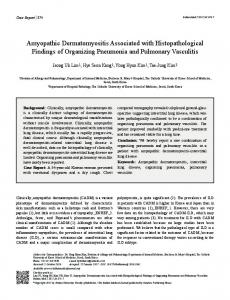
a)
b)
symptoms and the start of cyclosporine among survivors and nonsurvivors was compared. However, no significant differences were found between survivors and nonsurvivors (duration between the start of corticosteroids and addition of cyclosporine 26.7¡29.0 versus 19.0¡20.1 days; duration between onset of respiratory symptoms and the start of cyclosporine 66.7¡55.3 versus 42.5¡47.6 days, respectively). Three patients with acute/subacute ILD-ADM were treated with high-dose corticosteroids plus cyclosporine very early in the course of ILD, but two died of respiratory failure. Intravenous immunoglobulin therapy was effective in one patient that showed resistance to corticosteroids plus cyclosporine. In chronic ILD-ADM, corticosteroids alone were given to one patient, who showed improvement. Cyclosporine was given to three patients. Two of them received cyclosporine plus corticosteroids initially with a favourable response, while the other patient was given cyclosporine as a corticosteroidsparing agent after the start of corticosteroid therapy. The duration of therapy was longer in chronic than acute/subacute ILD-ADM.
FIGURE 1.
High-resolution computed tomography (HRCT) scans of patients
with acute/subacute and chronic interstitial lung disease associated with amyopathic dermatomyositis (ILD-ADM). a) HRCT scan of acute/subacute ILD-ADM shows consolidation, ground-glass opacities and irregular peribronchovascular thickening. b) HRCT scan of chronic ILD-ADM shows the areas of irregular linear and ground-glass opacities predominantly in the subpleural region.
1008
VOLUME 28 NUMBER 5
Mortality and survival During the observation period, six (67%) of the nine patients with acute/subacute ILD-ADM died (table 4). Of these, all died from respiratory failure due to progression of ILD. In contrast, no patients with chronic ILD-ADM died. A comparison of survival curves in the two groups is shown in figure 2. Patients with acute/subacute ILD-ADM had a much lower survival rate than those with chronic ILD-ADM (5-yr survival 35 versus 100%, respectively). Interestingly, four out of the five EUROPEAN RESPIRATORY JOURNAL
T. SUDA ET AL.
TABLE 4
LUNG DISEASE OF AMYOPATHIC DERMATOMYOSITIS
Comparison of treatment and outcome between acute/subacute and chronic interstitial lung disease associated with amyopathic dermatomyositis (ILD-ADM) Acute/subacute ADM-ILD
Chronic ADM-ILD
9
5
0 (0)
1 (20)
9 (100)
3 (60)
Subjects n Treatment Corticosteroids alone Corticosteroids + immunosuppressive agents Cyclophosphamide
7
0
Cyclosporine
8
3
Azathioprine
1
0
Intravenous Igs
1
0
Duration of therapy months
20¡31
46¡59
Death due to respiratory failure
6 (67)
0 (0)
Data are presented as mean¡SD or n (%). Ig: immunoglobulin.
deaths of patients with acute/subacute ILD-ADM occurred within 2 months, suggesting that the failure of the initial treatment was associated with early death in those patients. DISCUSSION In the present study, the authors retrospectively reviewed consecutive cases of ILD-ADM and attempted to elucidate its clinical characteristics and prognosis. It was found that ILDADM includes two different forms, acute/subacute and chronic, each with distinct prognoses. Acute/subacute ILDADM, which rapidly progressed, showed poor response to therapy and had high mortality. In contrast, chronic ILD-ADM responded well to therapy with a favourable prognosis. As ADM is a rare disease, previous studies of ILD-ADM included only one, or a few patients [7–13]. The present study investigated the highest number of patients with ILD-ADM so far. To date, contradictory data have been reported in the prognosis of ILD-ADM. Previous studies, mainly from Asia, have demonstrated that ILD-ADM generally runs an aggressive course, leading to fatal respiratory failure [7–12]. In contrast, COTTIN et al. [13] recently described a benign form of
ILD-ADM. They reported three patients with chronic ILDADM, and corticosteroids alone achieved a favourable response in two. The other one responded well to corticosteroids plus immunosuppressive agents. None of these three patients died during the observation period, leading to the conclusion that ILD-ADM has a good prognosis. In the present study, it was demonstrated that ILD-ADM consisted of at least two different forms with distinct outcomes. Acute/subacute ILD, which constituted about two thirds of the ILD-ADM patients, showed a rapid progression that was poorly responsive to therapy, resulting in severe respiratory failure. Conversely, chronic ILD-ADM showed a mild course and responded well to therapy. Notably, the outcome was completely different between these two forms. The mortality was much higher in patients with acute/subacute ILD-ADM (67%) than those with chronic ILD-ADM (0%). In acute/ subacute ILD-ADM, no patients had chronic respiratory symptoms before ILD diagnosis. Additionally, in patients developing acute/subacute ILD after ADM diagnosis, no abnormality was found on HRCT at the initial ADM diagnosis. These data suggest that chronic ILD is unlikely to pre-exist in the study patients with acute/subacute ILD. Taken together, these results may account for the contradictory data previously reported in the prognosis of ILD-ADM. Namely, the fatal progressive ILD-ADM described is likely to correspond to the acute/subacute ILD-ADM in the present study, while the ILDADM with a favourable prognosis reported by COTTIN et al. [13] may be equivalent to chronic ILD-ADM. Although ethnic differences may affect the clinical manifestations of ILD-ADM, it should be noted that ILD-ADM has these two different forms.
interstitial lung disease associated with amyopathic dermatomyositis.
In a comparison of the clinical characteristics between acute/ subacute ILD-ADM and chronic ILD-ADM, ILD onset was concomitant with ADM in all the patients with acute/subacute ILD-ADM except one, while more than half of patients with chronic ILD-ADM developed ILD after an ADM diagnosis. In addition, extrapulmonary symptoms, including fever and arthralgia, were more common in acute/subacute ILD-ADM than chronic ILD-ADM. Pa,O2 and %VC were significantly lower in acute/subacute ILD-ADM than chronic ILD-ADM. These data suggest that patients with acute/subacute ILD-
EUROPEAN RESPIRATORY JOURNAL
VOLUME 28 NUMBER 5
FIGURE 2.
Survival curves of acute/subacute (– – –) and chronic (–––)
1009
c
LUNG DISEASE OF AMYOPATHIC DERMATOMYOSITIS
ADM, which commonly occur simultaneously with ADM onset, have more severe ILD with systemic symptoms at the initial examination than those with chronic ILD. Regarding the anti-Jo-1 antibody, only one patient with chronic ILD-ADM had this antibody among the present ILD-ADM study. To date, several studies have reported a low incidence of anti-Jo-1 antibody in ILD-ADM patients [11–13, 17–19]. The current results were consistent with those studies. There have been few reports of the HRCT findings of ILDADM [7, 13, 20]. According to these reports, ground-glass opacities, consolidation and irregular linear opacities were often seen in ILD-ADM. Consistently, the study patients with ILD-ADM had a high frequency of these findings on HRCT. Notably, ground-glass opacities were found in all the patients. Between the two forms, consolidation was more common in acute/subacute ILD-ADM, while traction bronchiectasis was more frequently found in chronic ILD-ADM. Honeycombing was seen in only one patient with chronic ILD-ADM, who was histologically proven to have UIP. Overall, 60% of ILD-ADM patients with NSIP histology had consolidation. Originally, consolidation was reported not to be a common finding in idiopathic NSIP [21, 22]. In ILD-PM/DM, however, much higher prevalence of consolidation was demonstrated in NSIP [23, 24]. A recent study showed that consolidation was found in 42.9% of NSIP patients associated with PM/DM [23]. In addition, a previous study indicated that 86% of NSIP patients associated with PM/DM showed consolidation [24]. These data suggest that consolidation is more common in NSIP associated with PM/DM/ADM than in idiopathic NSIP. To date, little is known about the BAL findings of ILD-ADM. Two reports from Japan demonstrated an increase of BALF lymphocytes and neutrophils in acute/subacute ILD-ADM [11, 12]. The present study showed that the BAL findings differed between the two forms. Patients with acute/subacute ILD-ADM had an increase in percentages of BALF lymphocytes, as well as neutrophils, while those with chronic ILDADM showed only a moderate increase in percentages of BALF neutrophils. In addition, the total cell counts were significantly higher in acute/subacute than chronic ILD-ADM. Interestingly, the CD4/CD8 ratio of BALF T-lymphocytes tended to be higher in acute/subacute ILD-ADM than chronic ILD-ADM. Inconsistent with the current data, YOKOYAMA et al. [12] recently reported a case of fatal acute ILD-ADM with a low CD4/CD8 ratio (0.2) of BALF T-lymphocytes. However, none of the present patients with acute/subacute ILD-ADM exhibited ,1.0 CD4/CD8 ratio; the reason for this discrepancy is unknown. Further studies including larger numbers of patients may elucidate this point. Several studies on the histopathology of ILD-PM/DM have demonstrated various histological patterns, such as UIP, NSIP, BOOP and DAD, and emphasised their prognostic significance [13, 25–27]. However, limited data are available on the histopathology of ILD-ADM. LEE et al. [7] recently reported three cases of acute ILD-ADM with a histological finding of DAD, which was associated with poor outcome; whilst COTTIN et al. [13] described three patients with chronic ILD-ADM who showed NSIP with a good prognosis [13]. Based on these observations, acute/subacute ILD-ADM and chronic ILD-ADM might histologically correspond to DAD and NSIP, 1010
VOLUME 28 NUMBER 5
T. SUDA ET AL.
respectively. In the present study, however, NSIP was found in three out of five patients with acute/subacute ILD-ADM, of which two responded to therapy and survived, while the third patient died despite intensive therapy. The remaining two with acute/subacute ILD-ADM had DAD, and all died within 2 months. More recently, MIYAZAKI et al. [11] reported three patients with rapidly progressive ILD-ADM, and of these, NSIP was found in two and DAD in one. In addition, SAKAMOTO et al. [10] described a case of fatal ILD-ADM with a histological finding of NSIP. Taking the current data together with recent studies, the histological patterns of acute/subacute ILD-ADM include NSIP in addition to DAD. Conversely, the study patients with chronic ILD-ADM showed UIP and NSIP, but not DAD. Possibly, the favourable prognosis of chronic ILD-ADM was partially associated with the fact that no DAD was found in those patients. The optimal treatment for patients with ILD-ADM has not been established because of its rarity. Recent studies of rapidly progressive ILD-ADM have highlighted the effectiveness of cyclosporine combined with corticosteroids in the early course of ILD [11, 17]. Among the current acute/subacute ILD-ADM study, eight out of the nine patients received cyclosporine, although five of whom subsequently died of respiratory failure with a poor response to therapy. Between survivors and nonsurvivors, no difference was found in the duration between the start of corticosteroids and addition of cyclosporine, or the duration between onset of respiratory symptoms and the start of cyclosporine. In addition, two acute/subacute ILD-ADM patients receiving corticosteroids plus cyclosporine together during the very early course of ILD failed to respond. Collectively, these results suggest that early administration of cyclosporine may be beneficial in certain patients with acute/ subacute ILD-ADM, but not in all. Interestingly, intravenous immunoglobulin therapy was effective in one patient with acute/subacute ILD that was resistant to corticosteroids plus cyclosporine. More recently, TSUKAMOTO et al. [28] reported the efficacy of autologous peripheral blood stem cell transplantation in a patient with ILD-ADM that was unresponsive to corticosteroids plus cyclosporine. To date, however, there is no concrete evidence of treatment for ILD-ADM. Thus, future investigations are needed to elucidate an effective therapy for acute/subacute ILD-ADM. In chronic ILD-ADM, all four of the study patients had good outcomes. Interestingly, corticosteroids alone achieved a favourable response in one patient. Three patients received cyclosporine plus corticosteroids, which proved to be effective. However, it remains to be determined whether immunosuppressive therapy is actually required for chronic ILD-ADM patients. Recently, the current authors reported the characteristics of patients with ILD-PM/DM and highlighted the differences in clinical features and prognosis between ILD-PM and ILD-DM [24]. ILD-DM was shown to be more refractory to therapy, resulting in poorer prognosis than ILD-PM. Compared with ILD-DM/PM, the survival curve of overall ILD-ADM patients tended to be worse than that of ILD-DM patients without a significant difference, and significantly worse than that of ILDPM patients (data not shown). In terms of clinical presentation, acute/subacute forms were found in 35 and 47% of ILD-PM and ILD-DM patients, respectively. Thus, the proportion of acute/subacute form was highest in ILD-ADM patients (64%). EUROPEAN RESPIRATORY JOURNAL
T. SUDA ET AL.
LUNG DISEASE OF AMYOPATHIC DERMATOMYOSITIS
REFERENCES 1 Euwer RL, Sontheimer RD. Amyopathic dermatomyositis: a review. J Invest Dermatol 1993; 100: 124S–127S. 2 Kagen LJ. Amyopathic dermatomyositis. Arch Dermatol 1995; 131: 1458–1459. 3 Erel A, Toros P, Tokcaer AB, Gurer MA. Amyopathic dermatomyositis. Int J Dermatol 2000; 39: 771–773. 4 Olsen NJ, Park JH, King LE Jr. Amyopathic dermatomyositis. Curr Rheumatol Rep 2001; 3: 346–351. 5 Caproni M, Cardinali C, Parodi A, et al. Amyopathic dermatomyositis: a review by the Italian Group of Immunodermatology. Arch Dermatol 2002; 138: 23–27. 6 Dalakas MC, Hohlfeld R. Polymyositis and dermatomyositis. Lancet 2003; 362: 971–982. 7 Lee CS, Chen TL, Tzen CY, et al. Idiopathic inflammatory myopathy with diffuse alveolar damage. Clin Rheumatol 2002; 21: 391–396. 8 High WA, Cohen JB, Murphy BA, Costner MI. Fatal interstitial pulmonary fibrosis in anti-Jo-1-negative amyopathic dermatomyositis. J Am Acad Dermatol 2003; 49: 295–298. 9 Sontheimer RD, Miyagawa S. Potentially fatal interstitial lung disease can occur in clinically amyopathic dermatomyositis. J Am Acad Dermatol 2003; 48: 797–798. 10 Sakamoto N, Mukae H, Fujii T, et al. Nonspecific interstitial pneumonia with poor prognosis associated with amyopathic dermatomyositis. Intern Med 2004; 43: 838–842. 11 Miyazaki E, Ando M, Muramatsu T, et al. Early assessment of rapidly progressive interstitial pneumonia associated with amyopathic dermatomyositis. Clin Rheumatol 2006; [Epub ahead of print PMID: 16365687].
12 Yokoyama T, Sakamoto T, Shida N, et al. Fatal rapidly progressive interstitial pneumonitis associated with amyopathic dermatomyositis and CD8 T lymphocytes. J Intensive Care Med 2005; 20: 160–163. 13 Cottin V, Thivolet-Bejui F, Reynaud-Gaubert M, et al. Interstitial lung disease in amyopathic dermatomyositis, dermatomyositis and polymyositis. Eur Respir J 2003; 22: 245–250. 14 American Thoracic Society. Idiopathic pulmonary fibrosis: diagnosis and treatment. International consensus statement. American Thoracic Society (ATS), and the European Respiratory Society (ERS). Am J Respir Crit Care Med 2000; 161: 646–664. 15 Suda T, Sato A, Ida M, Gemma H, Hayakawa H, Chida K. Hypersensitivity pneumonitis associated with home ultrasonic humidifiers. Chest 1995; 107: 711–717. 16 American Thoracic Society/European Respiratory Society International Multidisciplinary Consensus Classification of the Idiopathic Interstitial Pneumonias. This joint statement of the American Thoracic Society (ATS), and the European Respiratory Society (ERS) was adopted by the ATS board of directors, June 2001 and by the ERS Executive Committee, June 2001. Am J Respir Crit Care Med 2002; 165: 277–304. 17 Shimojima Y, Ishii W, Kato T, et al. Intractable skin necrosis and interstitial pneumonia in amyopathic dermatomyositis, successfully treated with cyclosporin A. Intern Med 2003; 42: 1253–1258. 18 Santiago MB, Chalhoub M, Pereira ST. Amyopathic dermatomyositis complicated by interstitial pulmonary disease and pneumomediastinum. J Rheumatol 1998; 25: 2042–2043. 19 Chow SK, Yeap SS. Amyopathic dermatomyositis and pulmonary fibrosis. Clin Rheumatol 2000; 19: 484–485. 20 Kang EH, Lee EB, Shin KC, et al. Interstitial lung disease in patients with polymyositis, dermatomyositis and amyopathic dermatomyositis. Rheumatology (Oxford) 2005; 44: 1282–1286. 21 Hartman TE, Swensen SJ, Hansell DM, et al. Nonspecific interstitial pneumonia: variable appearance at highresolution chest CT. Radiology 2000; 217: 701–705. 22 MacDonald SL, Rubens MB, Hansell DM, et al. Nonspecific interstitial pneumonia and usual interstitial pneumonia: comparative appearances at and diagnostic accuracy of thin-section CT. Radiology 2001; 221: 600–605. 23 Arakawa H, Yamada H, Kurihara Y, et al. Nonspecific interstitial pneumonia associated with polymyositis and dermatomyositis: serial high-resolution CT findings and functional correlation. Chest 2003; 123: 1096–1103. 24 Fujisawa T, Suda T, Nakamura Y, et al. Differences in clinical features and prognosis of interstitial lung diseases between polymyositis and dermatomyositis. J Rheumatol 2005; 32: 58–64. 25 Tazelaar HD, Viggiano RW, Pickersgill J, Colby TV. Interstitial lung disease in polymyositis and dermatomyositis. Clinical features and prognosis as correlated with histologic findings. Am Rev Respir Dis 1990; 141: 727–733. 26 Nakamura Y, Chida K, Suda T, et al. Nonspecific interstitial pneumonia in collagen vascular diseases: comparison of the clinical characteristics and prognostic significance with usual interstitial pneumonia. Sarcoidosis Vasc Diffuse Lung Dis 2003; 20: 235–241.
EUROPEAN RESPIRATORY JOURNAL
VOLUME 28 NUMBER 5
Interestingly, none of the study patients developed ILD before ADM onset, regardless of acute/subacute or chronic form. To date, only two cases have been reported in which ILD onset preceded ADM [13], whilst, in all other cases, reported ILD onset was concomitant with, or followed ADM [7–12]. In ILDPM/DM, however, 20–30% of patients have been reported to develop ILD before PM/DM diagnosis [27, 29]. Indeed, a previous study of ILD-DM/PM by the current authors demonstrated that ILD onset preceded diagnosis of PM/DM in 19% of ILD-PM patients and in 33% of ILD-DM patients [24]. Thus, a low proportion of patients in which ILD onset precedes collagen vascular diseases may be one of the clinical characteristics of ILD-ADM. Collectively, these data suggest that ILD-ADM, which is more likely to take an acute/subacute course and not to precede ADM, has the poorest prognosis among ILD associated with DM, PM and ADM. In conclusion, the present study demonstrated two different forms of interstitial lung disease associated with amyopathic dermatomyositis, acute/subacute and chronic forms, which were closely related to outcome. In order to appropriately care for patients with interstitial lung disease associated with amyopathic dermatomyositis, these two conditions should be taken into account. Further studies will provide information regarding the optimal treatment for patients with acute/ subacute and chronic interstitial lung disease associated with amyopathic dermatomyositis.
1011
c
LUNG DISEASE OF AMYOPATHIC DERMATOMYOSITIS
27 Douglas WW, Tazelaar HD, Hartman TE, et al. Polymyositis-dermatomyositis-associated interstitial lung disease. Am J Respir Crit Care Med 2001; 164: 1182–1185. 28 Tsukamoto H, Nagafuji K, Horiuchi T, et al. A phase I-II trial of autologous peripheral blood stem cell transplantation
1012
VOLUME 28 NUMBER 5
T. SUDA ET AL.
in the treatment of refractory autoimmune disease. Ann Rheum Dis 2006; 65: 508–514. 29 Marie I, Hachulla E, Cherin P, et al. Interstitial lung disease in polymyositis and dermatomyositis. Arthritis Rheum 2002; 47: 614–622.
EUROPEAN RESPIRATORY JOURNAL